1204
Brain glutathione levels decrease with age and correlate with cognitive function1Department of Radiology, Shandong Provincial Hospital Affiliated to Shandong First Medical University, Jinan, China, 2Philips Healthcare, Shanghai, China
Synopsis
Keywords: Nerves, Aging
Cognitive impairment and the improvement of oxidative stress are common in the aged. GSH is a key player to defend oxidative stress and avoid ferroptosis. We aim to explore the variation of brain GSH levels with age including anterior cingulate cortex, posterior cingulate cortex and occipital cortex, and to test whether GSH levels in these regions are associated with cognitive function. Our findings indicate that oxidative stress and ferroptosis abnormality caused by the decreased GSH levels may contribute to cognitive decline of the aged without a regional specificity manner.Purpose
Oxidative stress increases with aging 1. Accumulating evidence suggest that the accumulated oxidative stress plays an important role in cognitive aging and neurodegenerative diseases 2. As the most abundant endogenous antioxidant in the brain 3, glutathione (GSH) is considered as key players in oxidative stress defending. Therefore, we aim to explore how human brain GSH levels vary with aging and whether brain GSH levels are associated with cognition function.Method
253 healthy volunteers (20-70 years) underwent magnetic resonance spectroscopy (MRS) on a 3.0 T scanner to measure GSH levels in the anterior cingulate cortex (ACC), posterior cingulate cortex (PCC) and occipital cortex (OC) using PRESS and HERMES, and underwent neuropsychological tests (MOCA, SDMT, Stroop, TMT and RCFT) to assess cognitive function. MRS data were processed and quantified using the LCModel software package (version 6.3-1 M) and Gannet (version 3.1.5). The relationships between age and GSH level and between cognitive function scores and GSH level were analyzed using Pearson correlation coefficients.Result
Aging is significantly correlated with the decreased PRESS GSH levels in ACC (r = -0.252, p<0.001), PCC (r = -0.358, p < 0.001) and OC (r = -0.163, p = 0.010) and decreased HERMES GSH levels in ACC (r = -0.168, p = 0.008) and PCC (r = -0.244, p < 0.001). Cognitive impairment is significantly correlated with the decreased PRESS GSH levels in ACC (r SDMT = 0.163, p = 0.011; r Stroop = -0.167, p = 0.010; r TMT A = -0.221, p = 0.001; r TMT B = -0.155, p = 0.018; r RCFT1 = 0.223, p = 0.001; r RCFT2 = 0.200, p = 0.002; r RCFT3 = 0.164, p = 0.014), PCC (r SDMT = 0.228, p < 0.001; r Stroop = -0.143, p = 0.027; r TMT A = -0.194, p = 0.003; r TMT B = -0.186, p = 0.004; r RCFT1 = 0.170, p = 0.009; r RCFT2 = 0.162, p = 0.013; r RCFT3 = 0.158, p = 0.016) and OC (r SDMT = 0.138, p = 0.030; r TMT A = -0.173, p = 0.007; r RCFT1 = 0.229, p < 0.001; r RCFT2 = 0.182, p = 0.005; r RCFT3 = 0.187, p = 0.004) and the decreased HERMES GSH levels in OC (r SDMT = 0.129, p = 0.045; r RCFT1 = 0.153, p = 0.020; r RCFT2 = 0.173, p = 0.009; r RCFT3 = 0.156, p = 0.020).Discussion
GSH is consumed in anti-oxidative stress 4.The absence of GSH causes damage to the function of antioxidative defense system, which further leads to ferroptosis 5. And the essence of ferroptosis is correlated with an increased ratio of Fe(iron) to S(sulfhydryl) with aging 6. Consequently, it might be reasonable to conclude that the reduction of GSH suggests the oxidative stress and ferroptosis abnormality in the brain of elderly adults, and the mechanisms were not regional specificity in the ACC, PCC and OC.Oxidative stress and ferroptosis 7 are key players in cognitive dysfunction. We find that cognitive impairment is correlated with the reduction of GSH levels in ACC, PCC and OC, suggest that the reduction of GSH causes the oxidative stress abnormality and ferroptosis in brain, which further lead to cognitive impairment. It reminds us that the changes of GSH levels in ACC, PCC and OC may be predictive biological markers to detect the early changes of cognitive performance without a regional specificity manner.Conclusion
Our findings indicate that oxidative stress and ferroptosis abnormality caused by the decreased GSH may contribute to cognitive decline of the aged without a regional specificity manner.Acknowledgements
This work was supported by the National Natural Science Foundation of China for Young Scholars (No. 81601479), Taishan Scholars Project (No. tsqn201812147), Shandong Provincial Natural Science Foundation of China (Nos. ZR2021MH030, ZR2021MH355), Jinan Science and Technology Development Program of China (No. 202019098).
References
1 Martinez de Toda, I., Vida, C., Garrido, A. & De la Fuente, M. Redox Parameters as Markers of the Rate of Aging and Predictors of Life Span. J Gerontol A Biol Sci Med Sci 75, 613-620, doi:10.1093/gerona/glz033 (2020).
2 Ionescu-Tucker, A. & Cotman, C. W. Emerging roles of oxidative stress in brain aging and Alzheimer's disease. Neurobiol Aging 107, 86-95, doi:10.1016/j.neurobiolaging.2021.07.014 (2021).
3 Mandal, P. K., Saharan, S., Tripathi, M. & Murari, G. Brain glutathione levels--a novel biomarker for mild cognitive impairment and Alzheimer's disease. Biol Psychiatry 78, 702-710, doi:10.1016/j.biopsych.2015.04.005 (2015).
4 Rae, C. D. & Williams, S. R. Glutathione in the human brain: Review of its roles and measurement by magnetic resonance spectroscopy. Anal Biochem 529, 127-143, doi:10.1016/j.ab.2016.12.022 (2017).
5 Dixon, S. J. et al. Ferroptosis: an iron-dependent form of nonapoptotic cell death. Cell 149, 1060-1072, doi:10.1016/j.cell.2012.03.042 (2012).
6 Toyokuni, S. et al. Ferroptosis at the crossroads of infection, aging and cancer. Cancer Sci 111, 2665-2671, doi:10.1111/cas.14496 (2020).
7 Hao, L. et al. SLC40A1 Mediates Ferroptosis and Cognitive Dysfunction in Type 1 Diabetes. Neuroscience 463, 216-226, doi:10.1016/j.neuroscience.2021.03.009 (2021).
Figures
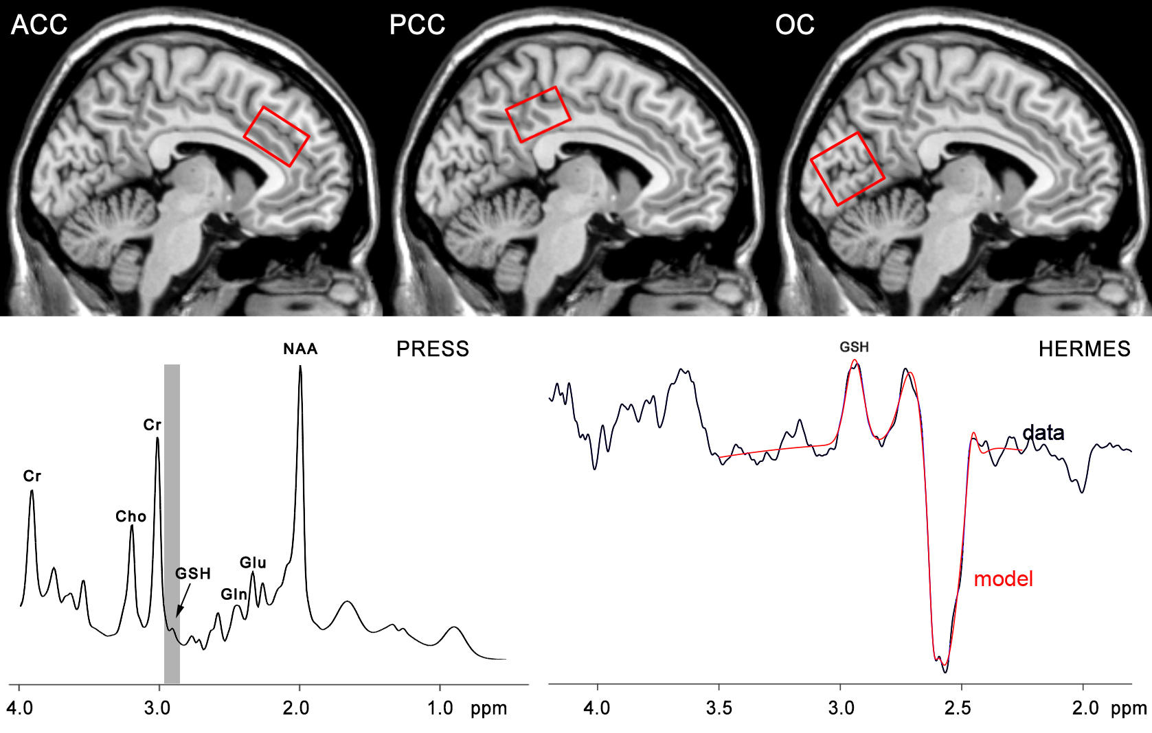
Figure 1. Positions of VOIs in ACC, PCC and OC, corresponding GSH spectra obtained by PRESS sequence and HERMES sequence. Cho, choline; Cr, creatine; Glu, glutamate; Gln, glutamine; GSH, glutathione; NAA, N-acetylaspartate.
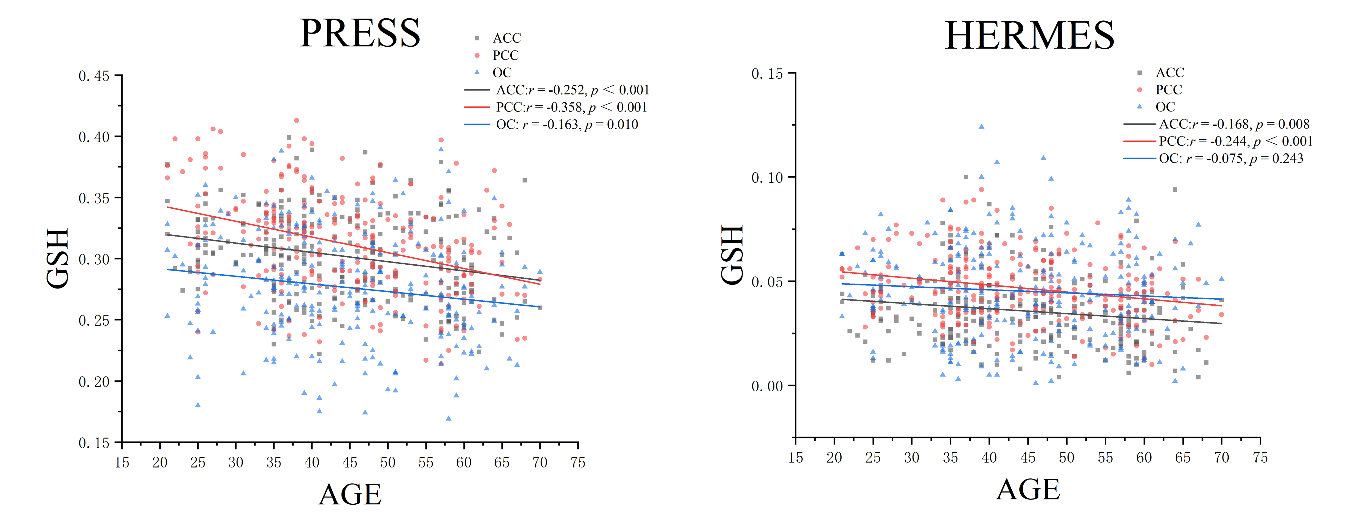
Figure 2. GSH levels correlations with age. Significant correlations are observed between age and PRESS GSH levels in ACC, PCC and OC and between age and HERMES GSH levels in ACC and PCC. r indicates the Pearson correlation coefficient.
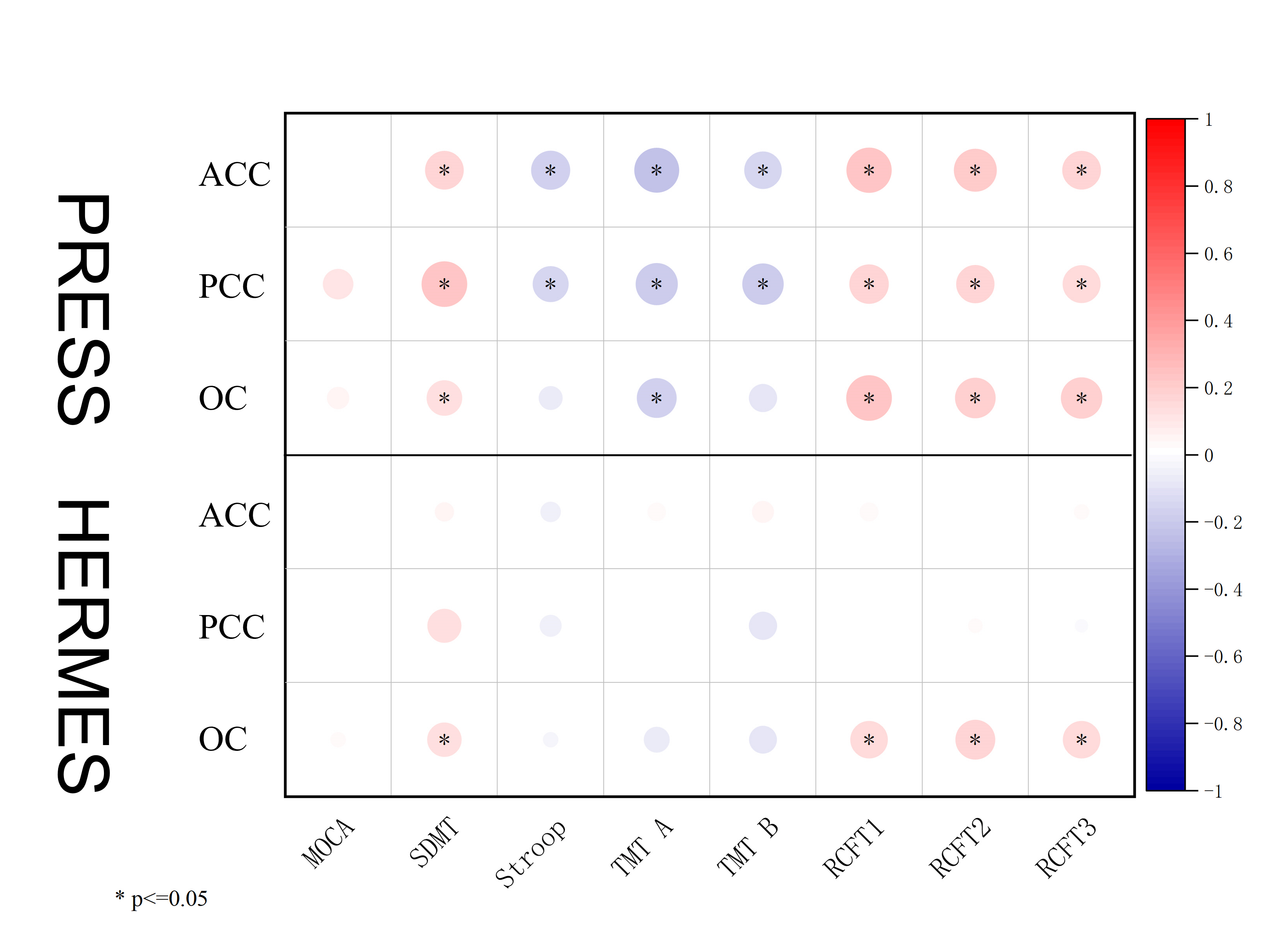
Figure 3. The GSH levels in ACC, PCC and OC detected by PRESS and HERMES MRS sequence correlates with cognitive function scores. Dot radius and color indicate correlation strength, while color indicates correlation directionality. MOCA, The Montreal Cognitive Assessment;SDMT, Symbol Digit Modalities Test; Stroop, Stroop test; TMT, Trail making Test; RCFT, Rey Complex Figure Test and Recognition Trial.