1159
Clinical DIADEM diffusion-weighted-imaging (DWI) of the brain: Comparison with commercially available DWI techniques.1Department of Radiology, Mayo Clinic, Rochester, MN, United States
Synopsis
Keywords: Artifacts, Diffusion Tensor Imaging
A novel distortion-free multi-shot diffusion-weighted-imaging (DWI), termed DIADEM (Distortion-free Imaging: A Double Encoding Method), was compared with commercially available state-of-the-art DWI techniques for clinical brain imaging. High-resolution distortion-free DWI is feasible within clinically acceptable acquisition times using the DIADEM technique, and demonstrated better performance than current commercially available DWI techniques.Introduction
Recently, a novel multi-shot diffusion-weighted imaging technique, termed DIADEM DWI, was developed to enable distortion-free imaging1. DIADEM DWI was initially optimized for brain imaging on a high-performance compact 3T scanner2 and subsequently translated for use on conventional 510(k)-cleared clinical whole-body 3T scanners3. In this study, DIADEM DWI of the brain obtained on clinical 3T MRI scanners was compared directly with commercially available DWI techniques to evaluate its relative merits for evaluation of both normal brain structures and detected pathology.Methods and Materials
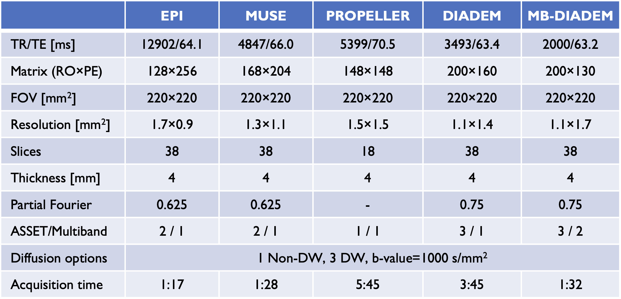
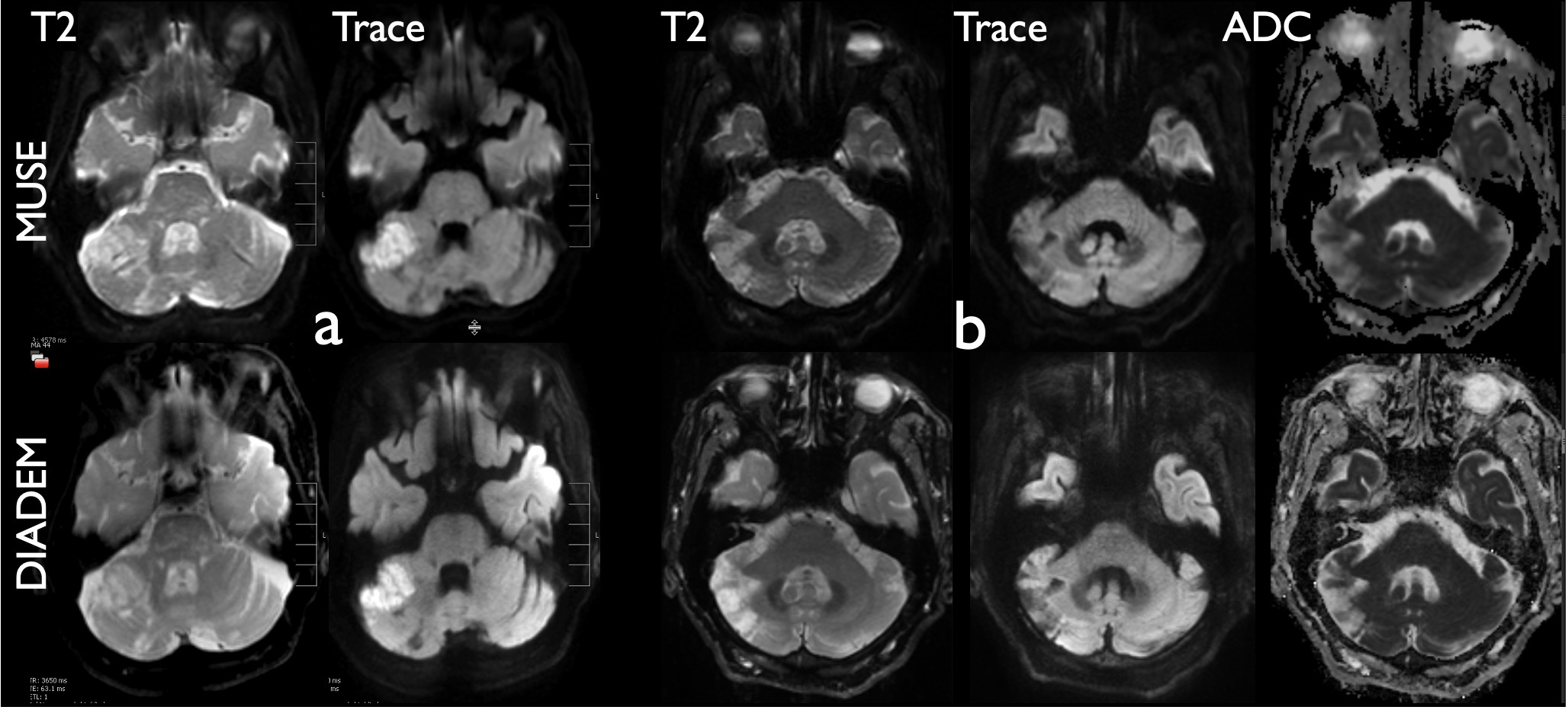
Four healthy subjects and four patients were scanned under IRB-approved protocols on clinical whole-body 3T MRI scanners (GE750, Waukesha, WI). DIADEM DWI imaging of the brain was compared with various vendor DWI techniques including single-shot EPI (echo-planar-imaging), MUSE (multiplexed sensitivity-encoding)4, and PROPELLER (periodically rotated overlapping parallel lines with enhanced reconstruction)5 in four healthy subjects. The default clinical DWI protocols at our institution are listed in the table in Fig 1. A vendor-provided distortion correction, termed PROGRES (Polarity Reversed On Gradients to Reduce Susceptibility), was applied to EPI and MUSE DWI. In PROPELLER DWI, only 18 slices rather than 38 in others were obtained due to the prolonged acquisition time. DIADEM DWI was acquired with and without multiband6. MUSE and DIADEM DWI were further assessed in four patients; two with acute brain infracts, and two others for which no specific diffusion abnormality was identified.Results and discussion
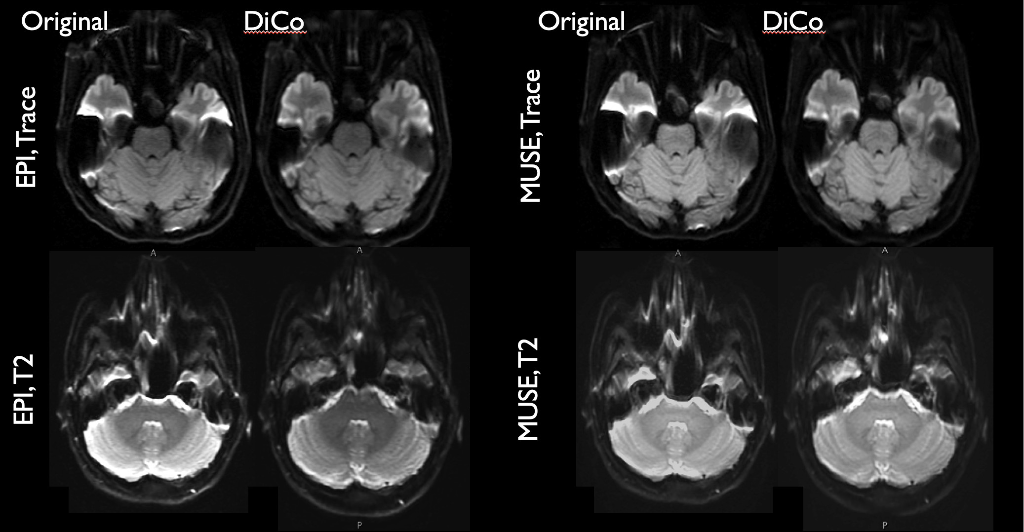
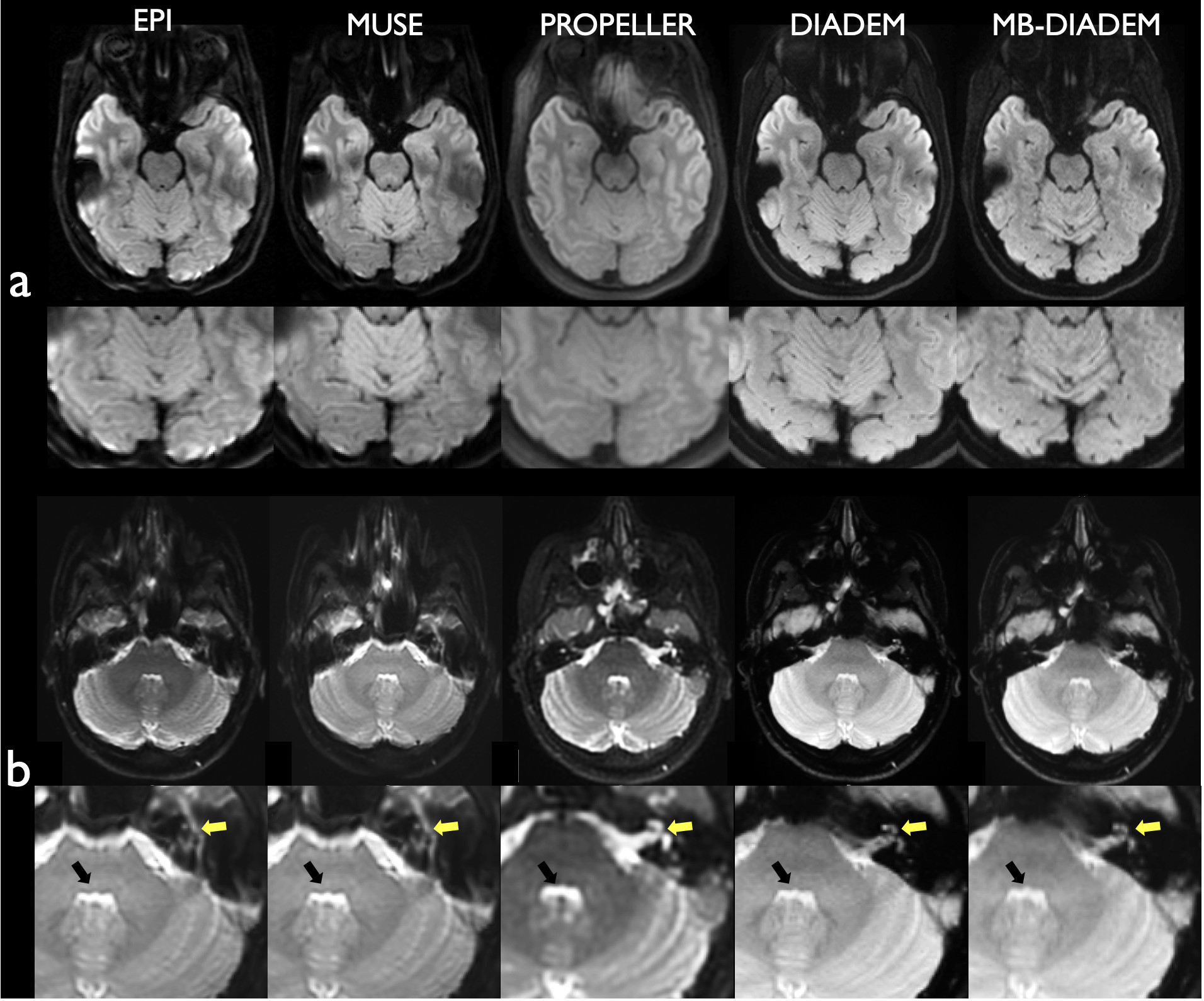
While the acquisition times in EPI and MUSE DWI were relatively short (see Fig. 1), these techniques suffered from stronger geometric distortion compared to DIADEM. Although distortion was generally well mitigated with PROGRES, it introduced image blurring and there were still noticeable residuals, especially in areas near air-tissue interfaces such as internal auditory canal (Fig. 2). PROPELLER DWI was robust to geometric distortion and superior in minimizing signal dropouts, but at the costs of severe image blurring, decreased resolution and prolonged scan time (Fig. 3). With DIADEM DWI, high-resolution imaging was achievable without any visible distortion within clinically feasible scan time, which could be further reduced with multiband. In patient exams, DIADEM DWI was deemed superior to MUSE DWI due to the decreased artifacts, improved resolution, decreased anatomic distortion, and increased confidence in correctly attributing diffusion changes (Fig. 4).Conclusion
High-resolution distortion-free DIADEM DWI of the brain is feasible within clinically acceptable scan times. Early clinical experience demonstrated superior performance of DIADEM DWI compared to current commercially available state of the art DWI techniques.Acknowledgements
This work was supported by NIH U01 EB024450, NHI U01 EB026979, and Mayo Clinic Imaging Biomarker Discovery Program.References
1. In MH, Posnansky O, Speck O.
High-resolution distortion-free diffusion imaging using hybrid spin-warp and
echo-planar PSF-encoding approach. NeuroImage
2017; 148: 20-30.
2. In MH, Tan ET, Trzasko JD, et al.
Distortion-free imaging: A double encoding method (DIADEM) combined with
multiband imaging for rapid distortion-free high-resolution diffusion imaging
on a compact 3T with high-performance gradients. J Magn Reson Imaging 2020; 51(1):
296-310.
3. In MH, Campeau NG, Trzasko J, Kang D,
Welker KM, Huston III J. Clinical DIADEM: Distortion-free high-resolution
diffusion-weighted imaging of the brain.
ISMRM 2022. p. 4662.
4. Chen N-K, Guidon A, Chang H-C, Song AW.
A robust multi-shot scan strategy for high-resolution diffusion weighted MRI
enabled by multiplexed sensitivity-encoding (MUSE). Neuroimage 2013; 72:
41-7.
5. Forbes KP, Pipe JG, Karis JP, Heiserman
JE. Improved image quality and detection of acute cerebral infarction with
PROPELLER diffusion-weighted MR imaging. Radiology
2002; 225(2): 551-5.
6. Setsompop K, Gagoski BA, Polimeni JR,
Witzel T, Wedeen VJ, Wald LL. Blipped‐controlled aliasing in parallel imaging
for simultaneous multislice echo planar imaging with reduced g‐factor penalty. Magnetic resonance in medicine 2012; 67(5): 1210-24.
Figures