1157
Dynamic distortion correction using blip-rewound EPI and joint multi-echo reconstruction1Wellcome Centre for Integrative Neuroimaging, FMRIB, Nuffield Department of Clinical Neurosciences, University of Oxford, Oxford, United Kingdom
Synopsis
Keywords: Artifacts, Data Acquisition, EPI distortion
In this work, we propose a new method for dynamic distortion correction by integrating a tailored EPI trajectory and a joint multi-echo reconstruction, which permits robust dynamic field mapping and distortion correction without compromising spatial resolution. The performance of the proposed method is demonstrated using in vivo experiments.Introduction
Echo planar imaging (EPI) is the workhorse for most functional MRI studies. However, given its low bandwidth along the phase encoding direction, EPI is prone to B0 inhomogeneity that results in image distortions. Static distortions can be corrected in postprocessing using a separately acquired field map1, which however cannot capture dynamic B0 variations due to subject motion, respiration, gradient heating, and eddy currents. These uncompensated field fluctuations result in temporally varying image distortions that might obscure subtle activation signals in functional MRI.Dynamic distortion correction can be achieved using a multi-echo EPI acquisition, which acquires multiple EPI volumes from a single readout and estimates a dynamic field map from the phase evolution between two echoes2. However, the sequential acquisition of multiple EPI volumes in each TR limits the achievable spatial resolution of this method, particularly at higher fields with short T2* values.
In this work, we propose a new method for dynamic distortion correction by integrating a tailored EPI trajectory and a joint multi-echo reconstruction, which permits robust dynamic field mapping and distortion correction without compromising spatial resolution. The performance of the proposed method is demonstrated using in vivo experiments.
Methods
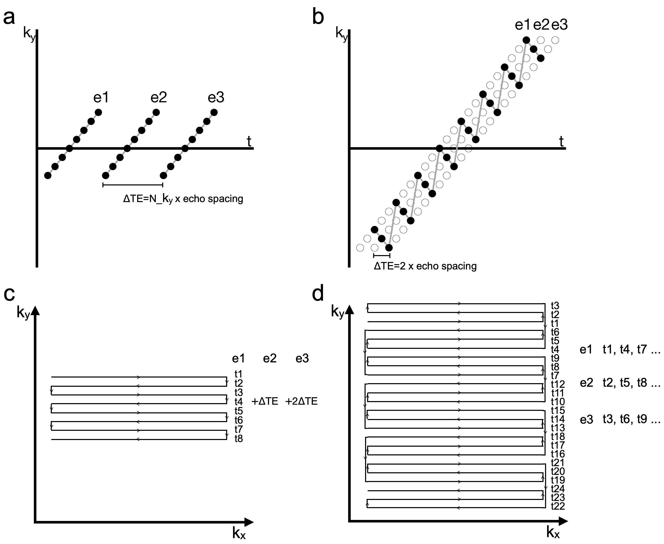
Blip-rewound EPIConventional multi-echo EPI acquires multiple EPI volumes sequentially, corresponding to several straight lines in the ky-t space (Fig. 1a shows an example with 3 echoes) that are separated in t. We developed a blip-rewound EPI (rEPI) trajectory to acquire multiple EPI volumes in an interleaved fashion, which leads to a zig-zag trajectory in the ky-t space (Fig.1b) that covers three temporally adjacent lines. In k-space, the rEPI trajectory traverses along the phase encoding direction with a periodic rewinding pattern (Fig. 1d). With the interleaved acquisition, rEPI de-couples ky coverage with TEs of latter echoes, achieving a higher spatial resolution than conventional multi-echo EPI using the same amount of data. The TE difference between two adjacent EPI volumes is very small in rEPI (i.e., twice the echo spacing, Fig. 1b), which is a desirable property for robust image reconstruction and field map estimation. For reconstruction, a small TE difference leads to high similarities between different EPI volumes, which can be leveraged using a structured low rank based reconstruction3 to improve the reconstruction of each EPI volume that is highly undersampled. A small TE difference also facilitates robust field map estimation by preventing extreme phase wrapping that might be hard to resolve. Finally, rEPI based field map estimation is considerably robust against motion and respiration as each field map is generated from a single-shot readout with a short temporal duration (~50ms).
Experiments
In vivo data from one subject was acquired using gradient-echo rEPI with three echoes on a Siemens 3T scanner. Acquisition parameters: FOV 220mmx220mmx100mm, resolution 1.7mmx1.7mmx2mm, TR=3.6s, echo spacing 0.76ms. Two datasets with (R=2) and without (R=1) GRAPPA acceleration were acquired, and TE were 31ms and 55ms, respectively. To investigate the efficacy of dynamic field mapping in the presence of motion, an rEPI time series was acquired and the subject was instructed to perform head movement during the scan. In addition, a conventional field map was also acquired using a multi-echo gradient echo (GRE) sequence.
Reconstruction and distortion correction
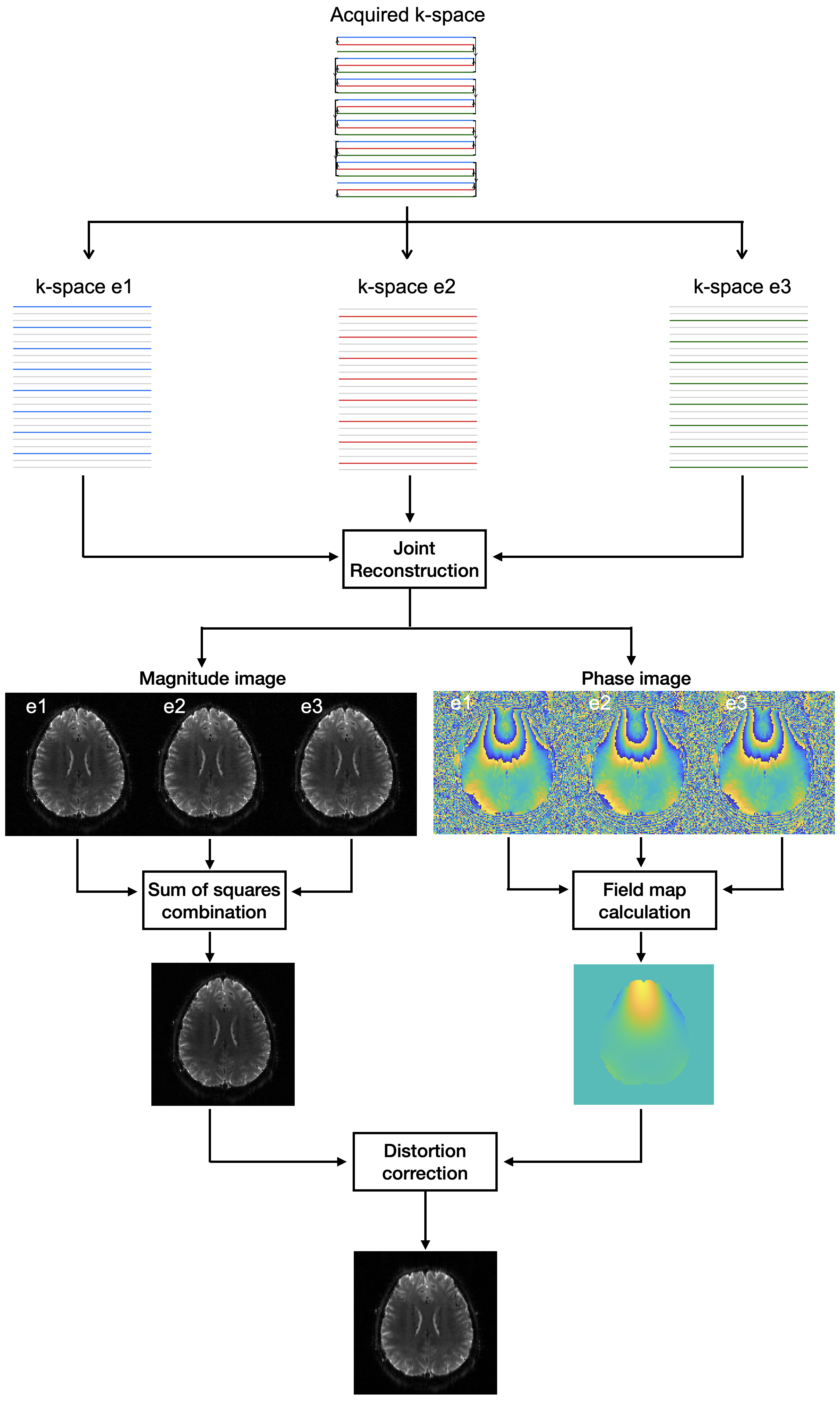
The pipeline for image reconstruction and distortion correction is shown in Fig.2. All echoes were jointly reconstructed using a structured low rank based reconstruction to leverage the information redundancy across echoes. The reconstructed individual echo images were sum-of-squares combined to produce the final magnitude reconstruction. The phase images were used to estimate a B0 map.
For comparison, distortion correction was performed with both rEPI derived field maps and GRE based field maps. All field maps were smoothed with a fourth-order polynomial fitting. Distortion correction was performed using SPM124. The rEPI time series were co-registered using FSL’s MCFLIRT5 after distortion correction.
Results
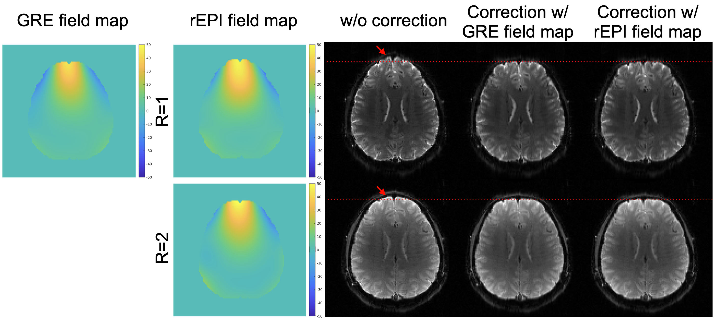
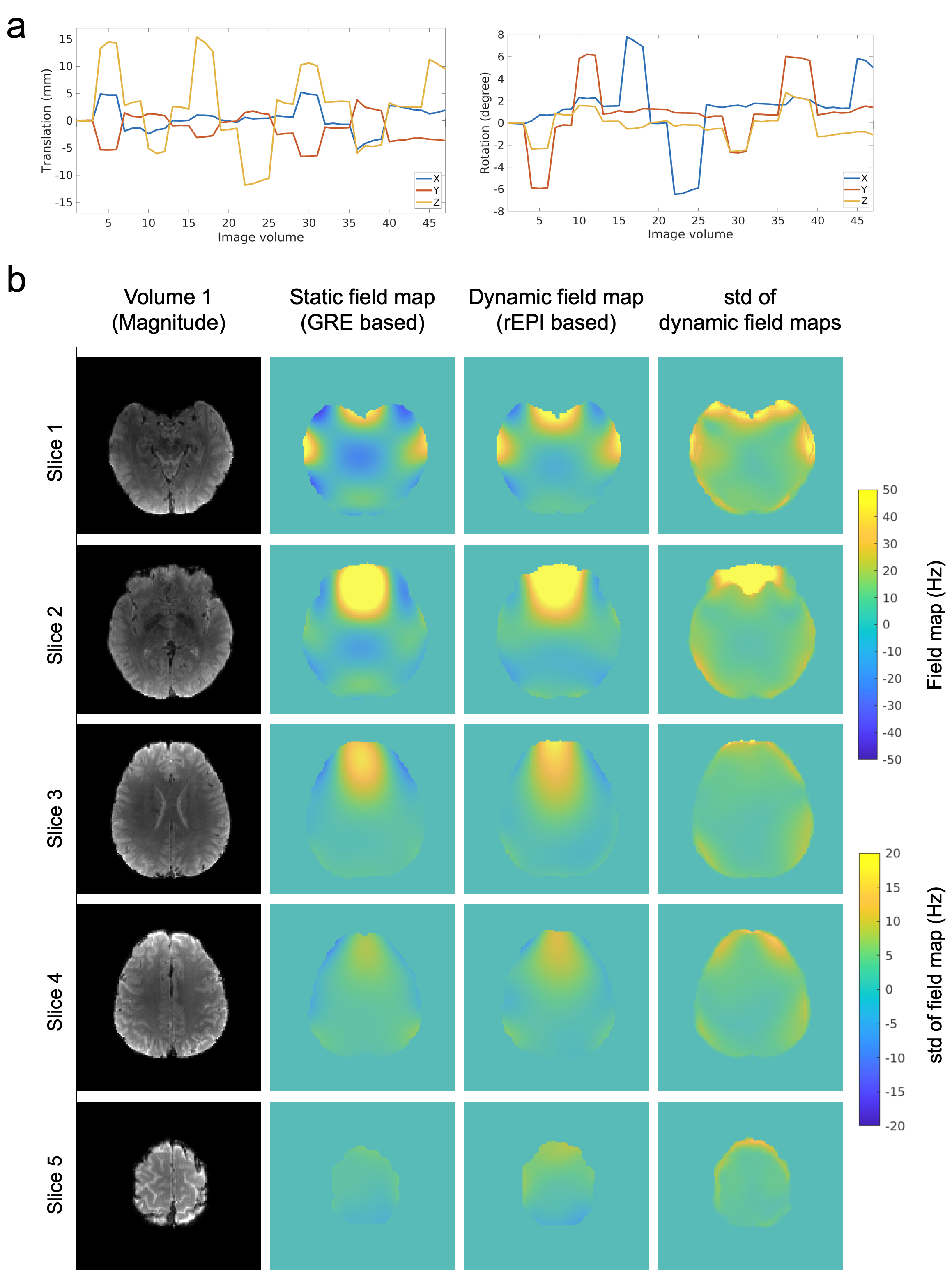
As shown in Fig.3, rEPI generated field maps are highly similar to the field map measured using a GRE sequence, with consistent spatial pattens present in all field maps. Image distortions are effectively corrected using rEPI produced field maps, demonstrating the high accuracy of rEPI based field mapping.In the presence of subject motion (Fig. 4a), B0 field can vary substantially over time, particularly in regions at lower slices and near the frontal area (Fig.4b). These variations can be measured using rEPI due to its capability of dynamic field mapping, which is not possible with GRE based static field mapping.
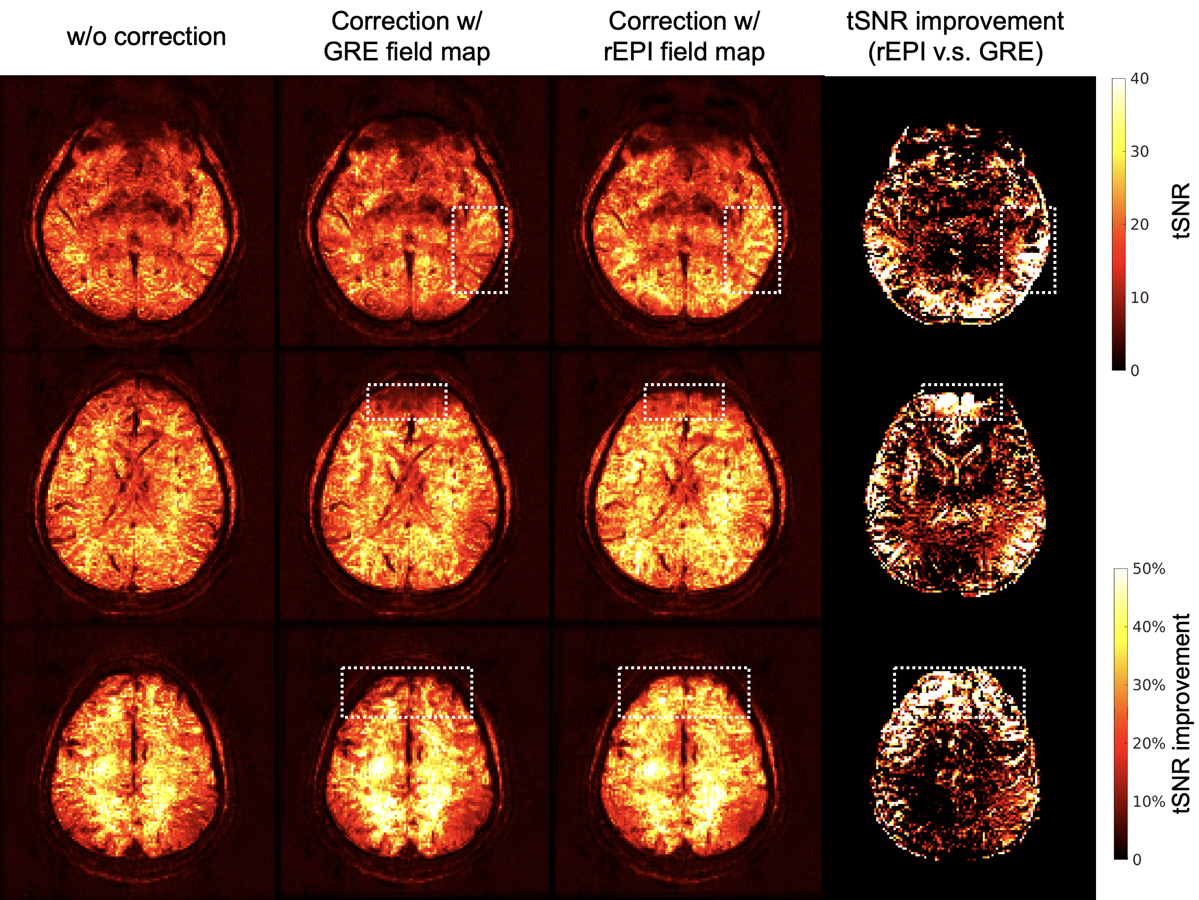
Fig. 5 shows a comparison of temporal SNR (tSNR) for different methods. With dynamic distortion correction, rEPI based corrections provide a higher tSNR than GRE field map based correction (particularly in regions marked with rectangles).
Discussion and Conclusions
In this work, a new method for robust dynamic distortion correction was developed using rEPI acquisition with associated joint multi-echo reconstruction. In vivo results demonstrate high accuracy of the proposed method and its superiority over GRE based static field mapping in the presence of subject motion. Note, rEPI can be calibrated to acquire a different number of echoes, but three-echo rEPI was found to be very robust for field map estimation and thus used here. More evaluations will be performed in the future.Acknowledgements
W.W. is supported by the Royal Academy of Engineering (RF\201819\18\92). The Wellcome Centre for Integrative Neuroimaging is supported by core funding from the Wellcome Trust (203139/Z/16/Z).References
1. Jezzard, P. and Balaban, R.S., 1995. Correction for geometric distortion in echo planar images from B0 field variations. Magnetic resonance in medicine, 34(1), pp.65-73.
2. Visser, E., Poser, B.A., Barth, M. and Zwiers, M.P., 2012. Reference‐free unwarping of EPI data using dynamic off‐resonance correction with multiecho acquisition (DOCMA). Magnetic resonance in medicine, 68(4), pp.1247-1254.
3. Haldar, J.P., 2013. Low-rank modeling of local k-space neighborhoods (LORAKS) for constrained MRI. IEEE transactions on medical imaging, 33(3), pp.668-681.
4. Penny, W.D., Friston, K.J., Ashburner, J.T., Kiebel, S.J. and Nichols, T.E. eds., 2011. Statistical parametric mapping: the analysis of functional brain images. Elsevier.
5. Jenkinson, M., Bannister, P., Brady, J. M. and Smith, S. M. Improved Optimisation for the Robust and Accurate Linear Registration and Motion Correction of Brain Images. NeuroImage, 17(2), 825-841, 2002.
Figures