1156
Diffusion-weighted MRI of the spinal cord near metal implants: A rapid TSE approach with multispectral imaging and reduced field of view1Barrow Neurological Institute, Phoenix, AZ, United States, 2Philips Healthcare, Houston, TX, United States
Synopsis
Keywords: Artifacts, Artifacts, metal, implant, spinal cord, diffusion, multi-spectral imaging
Diffusion-weighted (DW) spinal cord MRI based on single-shot EPI suffers from strong geometric distortion and signal loss artifacts. While strategies have been developed to reduce these artifacts in DW-EPI, their application in spinal cord DWI is challenging when metal implants are present near the spine. A multispectral DW-PROPELLER has been proposed to overcome this challenge; however, this requires long scan times. In this work, we developed a single-shot TSE technique with multispectral imaging and reduced FOV to achieve fast speed / increased SNR for spinal cord DWI near metals. Volunteer and patient results demonstrated reduced artifacts and improved speed/SNR performance.
Introduction
Spinal cord DWI is very challenging due to the anatomy of the cord, susceptibility differences at tissue interfaces, and metallic implants that are commonly present in patients who have undergone surgeries related to cervical myelopathy or traumatic injuries, which limits our ability to objectively monitor post-operative recovery. Spinal cord DWI based on single-shot EPI (ssEPI) suffers from susceptibility-induced distortion and signal loss due to phase errors accumulated during the EPI readout. Several strategies 1 have been developed to mitigate these artifacts such as a) parallel imaging; b) reduced field-of-view (rFOV) imaging; c) multi-shot EPI (msEPI) methods. In addition, advanced correction techniques have also been proposed for distortion-free DW-EPI 2-4. However, even with these improvements, spinal cord DWI is often non-diagnostic in areas near metallic implants due to the large inhomogeneous field they induce. Various techniques for reducing metal-induced artifacts have been proposed for anatomic imaging, such as view angle tilting (VAT) 5 and multi-spectral imaging (MSI) 6,7. Recently, Koch incorporated MSI into DW-PROPELLER to mitigate the artifacts near metals 8 and demonstrated the utility in cervical spine 9; however, the resulting method required long scan times that may limit clinical application and/or our ability to obtain a large number of diffusion directions. In this work, we develop a single-shot TSE-based DWI (DW-ssTSE) with improved efficiency and artifact reduction for spinal cord DWI near metals.Methods
A DW-ssTSE pulse sequence was implemented on a 3.0-T Philips Ingenia scanner (Fig. 1). Unlike EPI, TSE-based sequences are insensitive to off-resonance-induced artifacts (in-plane), as the TSE pulse train refocuses accumulated phase errors 10. SPLICE signal modulation was used to overcome the violation of the non-Carr-Purcell-Meiboom-Gill condition and to maintain a stable echo train 11. Furthermore, rFOV 12,13 imaging was employed to yield a short echo train/TE for a higher SNR. Finally, through-plane distortion was mitigated by incorporating MSI, which selectively excites and refocuses regions along the slice direction with different off-resonance frequencies 14.The performance of the DW-ssTSE technique was tested on two healthy volunteers and evaluated on a cervical myelopathy patient with titanium implants from a C3-C4 anterior cervical discectomy and fusion (ACDF) procedure, whereby an anterior plate was affixed to the spine with four screws. In the healthy volunteers, several staples were padded and attached to the neck (after confirming its safety) to simulate the metal artifacts encountered in patients in a controlled manner.
In all subjects, data were acquired using the 1) proposed DW-ssTSE with and without MSI, 2) standard-of-care DW-msEPI 15, and 3) DW-PROPELLER with/without MSI (in-house implemention comparable Ref. 8 and 16 ). With the prototype of DW-ssTSE-MSI, a single slice was acquired at 4 frequency offsets (-1000, -500, 0, 500 Hz). All scans were closely matched for scan time for comparison, with scan parameters listed in Table 1. Both DW-msEPI and DW-ssTSE used a rFOV = 180x70 mm2 in the sagittal plane; while for DW-PROPELLER a larger FOV = 240x240 mm2 is required to mitigate aliasing due to its non-dedicated foldover direction. One b = 0 and three b = 600 s/mm2 were acquired. Nine signal averages were acquired with DW-ssTSE at high b values to match scan times. To improve the SNR of the diffusion-weighted images, a complex-average strategy was implemented for combining images from multiple signal averages 17,18. The final MSI image was generated from the source images using a root-mean-square method 14.
Results and Discussion
Fig. 2 shows the source images of DW-ssTSE-MSI acquired in a healthy volunteer with metal staples taped on the back of the neck and the final combined image. The source images demonstrate the impact of off-resonance frequency on the target slice. The source image at frequency offset zero represents the results acquired without MSI, illustrating strong signal loss such that a portion of the spinal cord is lost. In the combined image, the signal losses in the spinal cord were well recovered, while some subtle residual artifacts remained due to intravoxel dephasing effects from the strong field inhomogeneities caused by the metal pieces.Fig. 3 compares the performance of DW-msEPI, the proposed DW-ssTSE, and DW-PROPELLER in two healthy volunteers. DW-msEPI exhibited substantial signal loss/distortion of the spinal cord near the metal. DW-ssTSE and DW-PROPELLER without MSI performed better than DW-msEPI, but still suffered from significant artifacts. Both DW-ssTSE-MSI and DW-PROPELLER-MSI successfully recovered signals within the spinal cord. In addition, DW-ssTSE demonstrated higher SNR than DW-PROPELLER.
In the patient (Fig. 4), the susceptibility-induced artifacts in DW-msEPI were reduced in both DW-ssTSE-MSI and DW-PROPELLER-MSI. Moreover, a compression-related lesion visible in the anatomical T2-TSE scan was clearly identified in DWI-ssTSE-MSI, but could not be confidently identified in either a) DW-msEPI due to distortion artifacts and b) DW-PROPELLER-MSI due to lower SNR.
Conclusion
In this work, we developed a novel diffusion-weighted ssTSE technique with rFOV and MSI for imaging the spinal cord near metals. The preliminary results demonstrated its advantages over standard-of-care DW-msEPI, as well as a multi-spectral DW-PROPELLER technique. This may allow for the development of novel diffusion-weighted MRI biomarkers of post-surgical recovery, which are currently unavailable in many individuals with cervical myelopathy and traumatic spinal cord injury due to the impact of metal implants on conventional DWI approaches.Acknowledgements
No acknowledgement found.References
- Holdsworth SJ, O'Halloran R, Setsompop K. The quest for high spatial resolution diffusion-weighted imaging of the human brain in vivo. NMR Biomed. 2019;32:e4056.
- Liao C, Stockmann J, Tian Q, Bilgic B, Arango NS, Manhard MK, Huang SY, Grissom WA, Wald LL, et al. High-fidelity, high-isotropic-resolution diffusion imaging through gSlider acquisition with and T1 corrections and integrated ΔB0/Rx shim array. Magn Reson Med. 2020;83:56-67.
- In M-H, Tan ET, Trzasko JD, Shu Y, Kang D, Yarach U, Tao S, Gray EM, Huston III J, et al. Distortion-free imaging: A double encoding method (DIADEM) combined with multiband imaging for rapid distortion-free high-resolution diffusion imaging on a compact 3T with high-performance gradients. J Magn Reson Imaging. 2020;51:296-310.
- Li S, Wang Y, Hu Z, et al. High-fidelity diffusion tensor imaging of the cervical spinal cord using point-spread-function encoded EPI. Neuroimage 2021;236:118043.
- Cho ZH, Kim DJ, Kim YK. Total inhomogeneity correction including chemical shifts and susceptibility by view angle tilting. Med Phys. 1988;15:7-11.
- Koch KM, Lorbiecki JE, Hinks RS, King KF. A multispectral three-dimensional acquisition technique for imaging near metal implants. Magn Reson Med. 2009;61:381-390.
- Lu W, Pauly KB, Gold GE, Pauly JM, Hargreaves BA. SEMAC: Slice Encoding for Metal Artifact Correction in MRI. Magn Reson Med. 2009;62:66-76.
- Koch KM, Bhave S, Gaddipati A, et al. Multispectral diffusion-weighted imaging near metal implants. Magn Reson Med 2017;79:987-993.
- Koch KM, Bhave S, Kaushik SS, et al. Multispectral diffusion-weighted MRI of the instrumented cervical spinal cord: a preliminary study of 5 cases. European Spine Journal 2020;29:107101077.
- Hennig J, Nauerth A, Friedburg H. RARE imaging: A fast imaging method for clinical MR. Magn Reson Med. 1986;3:823-833.
- Schick F. SPLICE: sub-second diffusion-sensitive MR imaging using a modified fast spin-echo acquisition mode. Magn Reson Med 1997;38:638–644.
- Wilm BJ, Svensson J, Henning A, et al. Reduced field-of-view MRI using outer volume suppression for spinal cord diffusion imaging. Magn Reson Med 2007;57:625-630.
- Symms MR, Wheeler-Kingshott CA, Parker GJM et al. Z Onally-magnified Oblique Multislice (ZOOM) EPI. In Proceedings of the 8th Annual Meeting of ISMRM, Denver, CO, USA, 2000. Abstract 160.13. Wilm BJ, Svensson J, Henning A, et al. Reduced field-of-view MRI using outer volume suppression for spinal cord diffusion imaging. Magn Reson Med 2007;57:625-630.
- Hargreaves BA, Taviani V, Litwiller DV, et al. 2D Multi-spectral imaging for fast MRI near metal. Magn Reson Med 2018;79:968-973.
- Jeong H-K, Gore JC, Anderson AW. High-resolution human diffusion tensor imaging using 2-D navigated multishot SENSE EPI at 7 T. Magn Reson Med. 2013;69:793-802.
- Deng J, Omary RA, Larson AC. Multishot diffusion-weighted SPLICE PROPELLER MRI of the abdomen. Magn Reson Med 2008;59:947-953.
- Walsh DO, Gmitro AF, Marcellin MW. Adaptive reconstruction of phased array MR imagery. Magn Reson Med 2000;43:682-690.
- Kordbacheh H, Seethamraju RT, Weiland E, et al. Image quality and diagnostic accuracy of complex-averaged high b value images in diffusion-weighted MRI of prostate cancer. Abdom Radiol 2019;44:2244-2253.
Figures
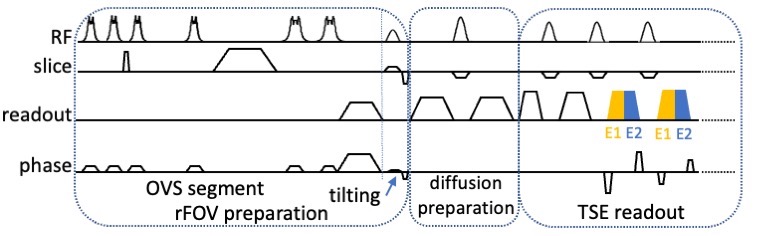
Fig. 1 Diagram of the rFOV DW-ssTSE sequence. An OVS (outer-volume suppression) segment is used, followed by tilting the excitation plane (optional), to reduce the FOV in the phase encoding direction. After the diffusion preparation segment, a single-shot TSE readout with SPLICE signal modulation is played out.
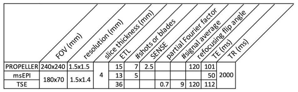
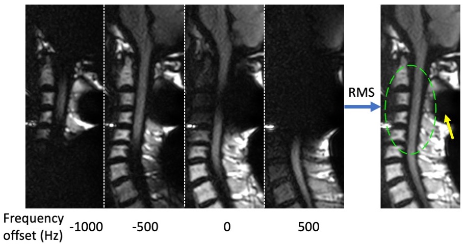
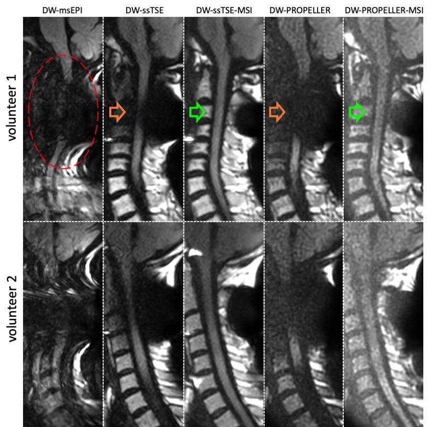
Fig. 3 Comparison of DW-msEPI, DW-ssTSE, DW-ssTSE-MSI, DW-PROPELLLER, and DW-PROPELLER-MSI on two healthy volunteers. Note that the PROPELLER images were cropped from a large FOV. Significant distortion and signal loss are present in DW-msEPI (red oval). These artifacts are mitigated in DW-ssTSE, but part of the spinal cord is still missing (orange arrows). DW-ssTSE-MSI recovers the signal loss and substantially improves the quality of the spinal cord (green arrows). DW-PROPELLER and DW-PROPELLER-MSI also mitigate or recover the signal loss, but exhibit lower SNR than ssTSE.
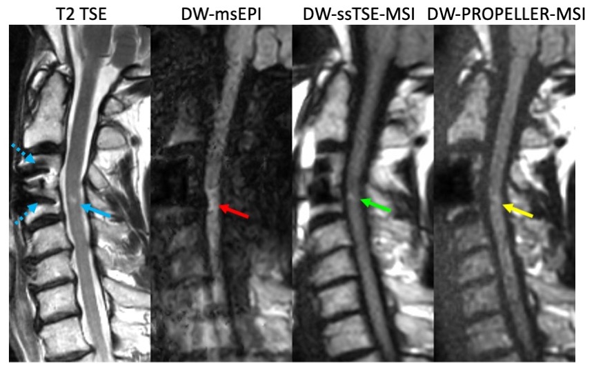
Fig. 4 Results from a patient with C3-C4 ACDF surgery (dotted blue arrows point to the screws). One lesion visible on the T2 TSE image (blue arrow) can not be identified with confidence on the DW-msEPI image due to distortion artifacts. It can be clearly seen on the DW-ssTSE-MSI image (green arrow), while less conspicuous on the DW-PROPELLER-MSI, presumably due to low SNR (yellow arrow).