1153
Optimized Flow Compensation for Wave-CAIPI Post-Contrast 3D-T1 MPRAGE
Min Lang1, Azadeh CD Tabari1, Komal Awan1, Wei Liu2, Clifford Bryan3, Wei-Ching Lo3, Daniel Nicolas Splitthoff4, Stephen Cauley1, Huang Susie1, and Conklin CD John1
1Massachusetts General Hospital, Boston, MA, United States, 2Siemens Shenzhen Magnetic Resonance Ltd., Shenzhen, China, 3Siemens Medical Solutions USA, Boston, MA, United States, 4Siemens Healthcare GmbH, Erlangen, Germany
1Massachusetts General Hospital, Boston, MA, United States, 2Siemens Shenzhen Magnetic Resonance Ltd., Shenzhen, China, 3Siemens Medical Solutions USA, Boston, MA, United States, 4Siemens Healthcare GmbH, Erlangen, Germany
Synopsis
Keywords: Artifacts, Data Acquisition, MR Value, Clinical Application, Neuro, Artifacts, Flow, Data Acquisition
Flow-related artifacts have been consistently observed in highly accelerated Wave-CAIPI post-contrast 3D-T1 MPRAGE and have an atypical appearance. Such artifacts introduce a diagnostic conundrum as they can mimic enhancing lesions and may require callback for repeat imaging, posing a critical barrier to wider clinical adoption of this technique. To address this, we developed an optimized flow-mitigated Wave-CAIPI post-contrast 3D T1 MPRAGE acquisition, tested it in a novel flow phantom, and deployed it in 17 patients undergoing contrast-enhanced brain MRI. Flow-mitigation was successful at reducing flow-related artifacts in most cases without sacrificing SNR, gray-white matter contrast, or enhancing lesion conspicuity.Introduction
Fast MRI acquisition techniques are increasingly being adopted clinically to meet the growing demand for medical imaging [1]. MR acceleration techniques are continuously being refined to balance image quality, artifact, and scan time. Wave-CAIPI is a relatively new parallel imaging-based technique developed in the past decade that combines 2D-CAIPI and bunch phase encoding approach to maximize 3D coil sensitivity and achieve controlled aliasing in all three spatial directions [2], resulting in high acceleration factors with negligible g-factor penalty. Wave-CAIPI has been validated for clinical 3D volumetric T1-weighted magnetization-prepared rapid gradient echo (MPRAGE) [3,4], fluid-attenuated inversion recovery (FLAIR) [5], and susceptibility weighted imaging [6], showing equivalent visualization of pathology and overall diagnostic quality compared to their conventional counterparts.Flow artifacts are caused by pulsatile laminar flow that produces a complex multilayered band from flow-related dephasing and can propagate in the phase encoding direction. These flow-related artifacts are well known in MRI and may have anomalous appearances, depending on the k-space sampling pattern. Flow-related artifacts have been observed in Wave-CAIPI post-contrast 3D-T1 MPRAGE and have an atypical appearance, manifesting as smearing of T1 hyperintense signal in the brainstem, subcortical nuclei and other areas of the brain parenchyma [3]. Such artifacts introduce a key diagnostic conundrum for the interpreting radiologist as they can mimic enhancing lesions and may require callback for repeat imaging with conventional non-accelerated MR sequences, posing a critical barrier to wider clinical adoption of this technique.The goal of this study was to characterize the source of flow-related artifact in Wave-CAIPI post-contrast 3D T1 MPRAGE on a novel flow phantom and develop an optimized flow-mitigated Wave-CAIPI post-contrast 3D T1 MPRAGE acquisition. The optimized protocol was then deployed in a clinical setting, and the resulting image quality and diagnostic performance were evaluated against Wave-CAIPI post-contrast 3D T1 MPRAGE performed without flow mitigation.Methods
A flow phantom was constructed consisting of a water bath containing tubing with pineapple juice. Flow was controlled in the tubing by a peristaltic pump and used to simulate vascular flow (~200 mL/min) in the cerebral arteries (Fig. 1). T1-weighted MPRAGE images of the flow phantom were acquired using Wave-CAIPI. Flow compensation techniques were incorporated into the Wave-CAIPI protocol to determine the optimal parameters for mitigating flow-related artifacts. Flow compensation strategies included the addition of flow compensation gradients as well as the use of a radial reordering of the k-space data acquisition [3]. The readout bandwidth of the flow-compensated protocol was increased to compensate for increased echo time (TE) and to preserve tissue contrast.The optimized flow-mitigation Wave-CAIPI T1-weighted MPRAGE protocol was then deployed in 17 consecutive patients undergoing contrast-enhanced brain MRI at an outpatient imaging center in October 2022 (Fig. 1A). The brain MRIs were performed on a 3T MRI scanner (MAGNETOM Vida, Siemens Healthcare, Erlangen, Germany) using a 20-channel head coil. The MRI protocols included the conventional Wave-CAIPI post-contrast 3D T1 MPRAGE sequence and an optimized flow-mitigated version of Wave-CAIPI post-contrast 3D T1 MPRAGE (Fig. 1B) [4].Two neuroradiologists (with 12 and 10 years of experience), blinded to sequence type, independently reviewed and assessed the clinical imaging data sets for presence of artifacts, signal-to-noise ratio (SNR), gray-white matter contrast, enhancing lesion contrast, and image blurring on a 3-point Likert scale (image A preferred to image B, no significant difference, or image B preferred to image A). Statistical analysis was performed using the Wilcoxon rank sum test.Results
In the flow phantom experiments, the combination of flow compensation gradients and radial reordering k-space acquisition provided the greatest level of flow artifact reduction (Fig. 2). These findings led to the development of the optimized flow-mitigation protocol. In clinical evaluation, the optimized flow-mitigation protocol reduced flow-related artifacts in 15 of 17 (88%) cases and 16 of 17 (94%) cases for raters 1 and 2, respectively. SNR, gray-white matter contrast, and enhancing lesion contrast were rated to be equivalent for the standard Wave-CAIPI MPRAGE image and the flow-mitigated images in all patients by both raters. The optimized flow-mitigation protocol was the preferred sequence for reduced flow-related artifacts by both raters (P<0.001). Figure 3 shows a representative case with an area of potential enhancement on conventional T1-weighted post-contrast Wave-CAIPI MPRAGE images that mimics the appearance of an enhancing lesion in the pons, likely arising from flow in the basilar artery/venous plexus. No abnormal enhancement was seen in the pons on the flow-mitigated images, confirming the finding was artifactual. Figure 4 shows another case with a small focus of apparent enhancement in the inferior frontal lobe on the conventional T1-weighted post-contrast Wave-CAIPI MPRAGE images. No enhancement was noted in the inferior frontal lobe on the flow-mitigated images, suggesting that the area of apparent enhancement was likely flow-related artifact.Conclusion
This study evaluated the efficacy of optimized flow-mitigated Wave-CAIPI-MPRAGE for reduction of flow-related artifacts in post-contrast T1-weighted imaging. Flow-mitigation was successful at reducing flow-related artifacts in most cases without sacrificing SNR, gray-white matter contrast, or enhancing lesion conspicuity. As fast MRI becomes increasingly adopted in clinical practice, this study highlights the need to minimize the presence of unexpected artifacts and reduction in image quality as potential compromises to achieving short scan times.Acknowledgements
This work was supported by the National Institutes of Health (grant number P41EB030006) and a research grant from Siemens Healthineers.References
- Lang M, Cartmell S, Tabari A, Briggs D, Pianykh O, Kirsch J, Cauley S, Lo WC, Risacher S, Filho AG, Succi MD, Rapalino O, Schaefer P, Conklin J, Huang SY. Evaluation of the Aggregated Time Savings in Adopting Fast Brain MRI Techniques for Outpatient Brain MRI. Acad Radiol. 2021 Oct 8;S1076-6332(21)00319-6. doi: 10.1016/j.acra.2021.07.011.
- Bilgic B, Gagoski BA, Cauley SF, Fan AP, Polimeni JR, Grant PE, Wald LL, Setsompop K. Wave-CAIPI for highly accelerated 3D imaging. Magn Reson Med. 2015 Jun;73(6):2152-62. doi: 10.1002/mrm.25347.
- Longo MGF, Conklin JC, Cauley SF, Setsompop K, Tian Q, Polak D, Polackal M, Splitthoff D, Liu W, Gonzalez RG, Schaefer PW, Kirsch JE, Rapalino O, Huang SY. Evaluation of ultrafast Wave-CAIPI Magnetization Prepared-Rapid Gradient-Echo (MPRAGE) for visual grading and automated measurement of brain tissue volume. American Journal of Neuroradiology. 2020 Aug;41(8):1388-1396. doi: 10.3174/ajnr.A6703.
- Goncalves Filho ALM, Awan K, Conklin J, Ngamsombat C, Cauley SF, Setsompop K, Liu W, Splithoff DN, Lo WC, Kirsch JE, Schaefer PW, Rapalino O, Huang SY. Validation of a Highly Accelerated Post-Contrast Wave-Controlled Aliasing in Parallel Imaging (CAIPI) 3D-T1 MPRAGE Compared to Standard 3D-T1 MPRAGE for Detection of Intracranial Enhancing Lesions on 3T MRI. 2022 European Radiology (in press).
- Ngamsombat C, Filho ALMG, Longo MGF, Cauley SF, Setsompop K, Kirsch JE, Tian Q, Fan Q, Polak D, Liu W, Lo WC, Gonzalez RG, Schaefer PW, Rapalino O, Conklin J, Huang SY. Evaluation of ultrafast Wave-CAIPI 3D FLAIR in the visualization and volumetric estimation of cerebral white matter lesions. American Journal of Neuroradiology. 2021 Jul 08. doi: 10.3174/ajnr.A7191.
- Conklin J, Longo MGF, Cauley SF, Setsompop K, Kirsch JE, Gonzalez RG, Schaefer PW, Rapalino O, Huang SY. Validation of highly-accelerated Wave-CAIPI susceptibility-weighted imaging (SWI) compared to conventional SWI and T2*-weighted gradient-echo for routine clinical brain MRI at 3T. American Journal of Neuroradiology. 2019 Dec;40(12):2073-2080.
Figures
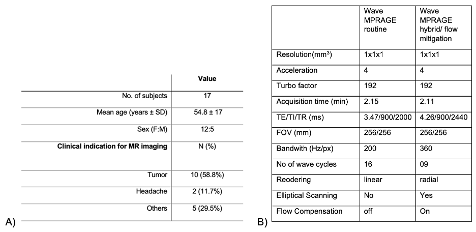
Fig 1: A) Demographic details of the patient population. B) Acquisition parameters for the conventional and flow-mitigated Wave-CAIPI T1 MPRAGE protocols.
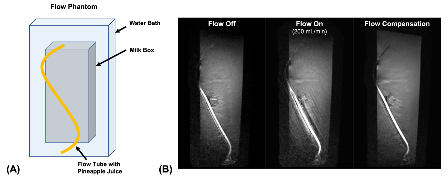
Fig 2: A) Phantom flow experimentation apparatus. Pineapple juice was used as the contrast agent to mimic contrast enhancement in the phantom test. B) Representative images demonstrating artifact simulation by turning on flow within the flow tube (200 ml/min) and reduction of this artifact by turning on flow compensation.
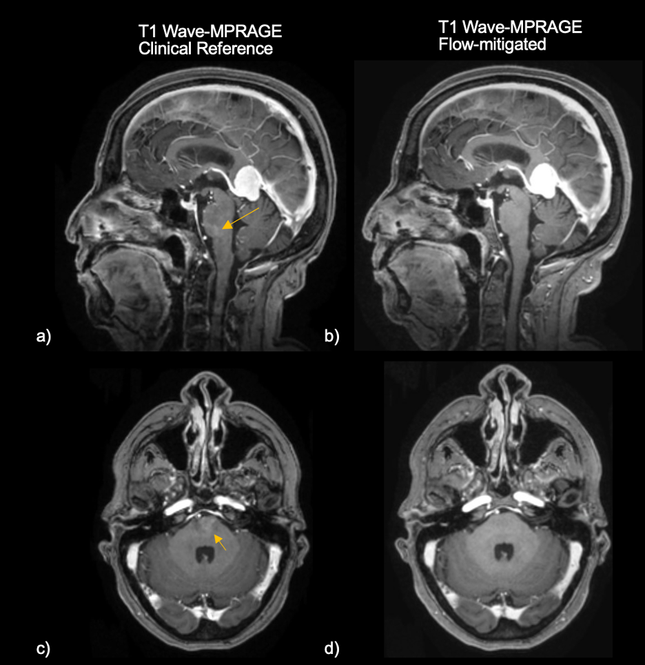
Fig 3: Representative images of a 64-year-old female with a homogeneously enhancing mass in the pineal region and abutting the falx. A & C) Conventional T1-weighted Wave-MPRAGE images in sagittal and axial planes demonstrating an enhancing pseudo-lesion in the inferior pons, likely arising from flow in the basilar artery/venous plexus. B & D) Flow-mitigated T1-weighted Wave-MPRAGE images demonstrate no abnormal enhancement in the pons, confirming that the enhancement seen on the conventional Wave-MPRAGE images was artifactual.
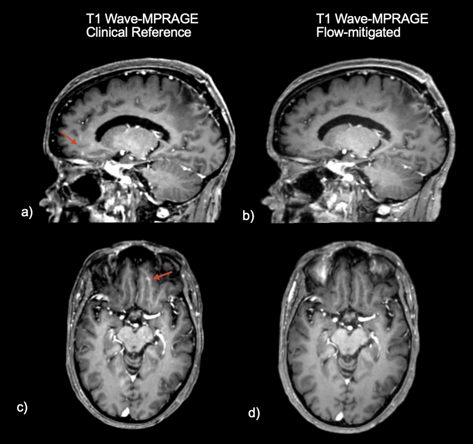
Fig 4: Representative images of a 67-year-old female status post left parietal craniotomy for tumor resection with small focus of apparent enhancement in the inferior frontal lobe. A) Sagittal and C) axial views of conventional T1-weighted post-contrast Wave-MPRAGE imaging. No enhancement was noted in the inferior frontal lobe on corresponding B) sagittal and D) axial planes of the flow-mitigated Wave-MPRAGE images, suggesting that the area of apparent enhancement was consistent with flow-related artifact.
DOI: https://doi.org/10.58530/2023/1153