1152
Automatic coil selection to suppress motion artifacts in exercise real-time cine imaging
Chong Chen1, Yingmin Liu2, Yu Ding3, Mathew Tong4, Yuchi Han4, and Rizwan Ahmad5
1Biomedical Engineering, The Ohio State Univerity, Columbus, OH, United States, 2Davis Heart and Lung Research Institute, The Ohio Sate University, Columbus, OH, United States, 3Davis Heart and Lung Research Institute, The Ohio State University, Columbus, OH, United States, 4Internal Medicine, The Ohio State University, Colubmus, OH, United States, 5Biomedical Engineering, The Ohio Sate University, Columbus, OH, United States
1Biomedical Engineering, The Ohio State Univerity, Columbus, OH, United States, 2Davis Heart and Lung Research Institute, The Ohio Sate University, Columbus, OH, United States, 3Davis Heart and Lung Research Institute, The Ohio State University, Columbus, OH, United States, 4Internal Medicine, The Ohio State University, Colubmus, OH, United States, 5Biomedical Engineering, The Ohio Sate University, Columbus, OH, United States
Synopsis
Keywords: Artifacts, Artifacts
We propose a novel method to automatically identify and discard coils that strongly contribute to image artifacts. This is achieved by projecting coil images to the space spanned by the ESPIRiT coil sensitivity maps. The proposed method is evaluated using the real-time cine data collected from twelve volunteers during exercise. The artifacts in the reconstructed real-time cine images are suppressed significantly with the proposed coil selection method.Background
In exercise stress CMR, compressive sensing (CS) based real-time cine (RT-Cine) imaging has been used to assess cardiac function with acceptable spatial and temporal resolution [1]. However, significant movement of body coils during exercise can degrade image quality due to the temporal varying coil sensitively maps. The artifacts caused by the coil movement are also visible in the temporally averaged coil images. To suppress the motion artifacts, the k-space from certain coils can be excluded by visual inspection of the coil images. However, visual inspection is not practical for routine use. In this abstract, we propose a method to automatically identify coils that introduce a high level of artifacts to the reconstructed image. Using the exercise real-time cine data collected from 12 volunteers, we demonstrate that the motion artifacts in the reconstructed real-time cine images are suppressed significantly with the proposed coil selection algorithm.Method
Twelve healthy volunteers were scanned under free-breathing conditions using a prototype bSSFP RT-Cine sequence on a 3T scanner (Vida, Siemens Healthcare, Erlangen, Germany). The volunteers were instructed to exercise on a supine cycle ergometer (Lode BV, Netherlands) with resistances 20 W, 40 W, and 60 W respectively. 14-slice short-axis (SAX) stack covering the whole heart and a two-chamber (2CH)) slice were acquired at each exercise stage using an 18-channel body array coil combined with a 12-channel posterior spine array coil, resulting in 30 channels. The other imaging parameters are: TE/TR 1.1/2.55—1.29/2.9 ms, flip angle 29-44 degrees, spatial resolution 1.82x1.82—2.27x2.27 mm2, temporal resolution 35.7-50.2 ms, acquisition time 3-6 s/slice and acceleration rate 7-8 with GRO [2] sampling pattern.Typically, the RT-Cine data collected at 40 W or 60 W have severe motion artifacts and were used to evaluate the performance of the proposed method. We first averaged the k-space of all the frames to generate the time-averaged coil image $$$\{x_i\}_{i=1}^N$$$. Then, the coil images were projected to the the space spanned by the ESPIRiT [3] coil sensitivity maps $$$\{S_i\}_{i=1}^N$$$: $$\tilde{x}_i = \left( \sum_{n=1}^N x_n S_n^* \right) S_i $$ where $$$\{\tilde{x}_i\}_{i=1}^N$$$ are the projected images and $$$ N $$$ is the number of coils. Fig. 1 shows the images from two physical coils, one (coil-4) with significant motion artifacts and the other (coil-11) without. The difference between $$$x_i$$$ and $$$\tilde{x}_i$$$ can be mainly attributed to motion artifacts. We propose to use the residual signal $$$ \| \tilde{x}_i - x_i \|_2 $$$ to identify the coils that strongly contribute to image artifacts.
The five physical coils with the highest values of $$$ \| \tilde{x}_i - x_i \|_2 $$$ were automatically discarded before the reconstruction. The data from the remaining coils were compressed to 12 virtual coils and reconstructed using a parameter-free SENSE-based CS method SCoRe [4]. For comparison, the data without coil selection were also reconstructed using the same method. The RT-Cine images reconstructed using both methods from each volunteer were visually scored by two cardiologists in terms of the level of image artifact in the cardiac region. The images were scored using a five-point scale: 1--Unusable, 2--Severe artifacts obscuring useful information, 3--Moderate artifacts with some loss of information but still diagnostic, 4--Minor artifacts with minimal loss of information, 5--No artifacts.
Results and Discussion
Fig. 2 shows the time-averaged images and the residual signal which cannot be characterized using one set of ESPIRiT coil sensitivity maps from a representative dataset. As highlighted by the red circle and boxes, the proposed method successfully identifies the coils with a high level of motion artifacts. Fig. 3 demonstrates the representative RT-Cine images of 2CH and SAX views, where Fig. 3a and 3c show images reconstructed using all the physical coils, and Fig. 3b and 3d show images after automatic coil selection. Even though there was some signal loss due to the coil rejection, especially on the chest wall, the motion artifacts were reduced significantly as highlighted by the red arrows. The temporal profiles across the heart are also displayed, demonstrating that the artifacts inside the blood pool and the myocardium are both suppressed. Results from visual scoring of image quality by two cardiologists are listed in Table 1. The image quality reconstructed with coil selection (4.4±0.7) was better than that without coil selection (3.6±0.8).Conclusion
By projecting the time-averaged images to the space spanned by the ESPIRiT coil sensitivity maps, we propose a method to automatically identify the coils with a high level of artifacts. We demonstrate that the proposed method can reduce motion artifacts in the reconstructed RT-Cine images.Acknowledgements
This work was funded by NIH project R01HL151697References
[1] C. Chen et al. SCMR 2019 Abstract #550349
[2] M. Joshi Et al. arXiv:2206.03630
[3] M. Uecker et al. Magnetic Resonance in Medicine 71 (3), 990–1001 (2014).
[4] R. Ahmad et al. IEEE Transactions on Computational Imaging 1 (4), 220-235 (2015)
Figures
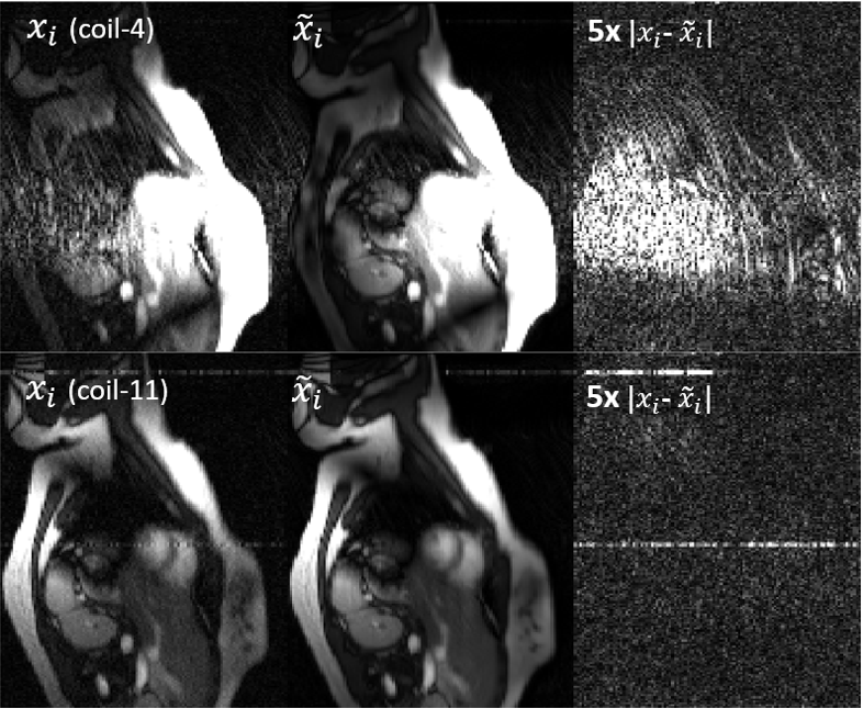
Figure 1. Time-averaged coil images,$$$ x_i $$$, and their
projection, $$$ \tilde{x}_i$$$ , on the space
spanned by the ESPIRiT coil sensitivity maps and their difference, $$$| \tilde{x}_i - x_i|$$$. The images from two
physical coils, one (coil-4) with significant motion artifacts and the other without
(coil-11), are shown here. The horizonal lines in coil-11 are the RF
interference from the in-scanner exercise device (supine cycle ergometer).
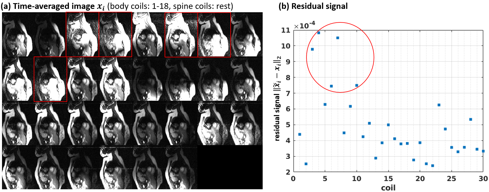
Figure 2. Time-averaged images of all physical coils (a)
and the residual signal which cannot be characterized using one set of ESPIRiT
coil sensitivity maps (b). The red boxes and circle highlight the five physical
coils we discarded before reconstruction.
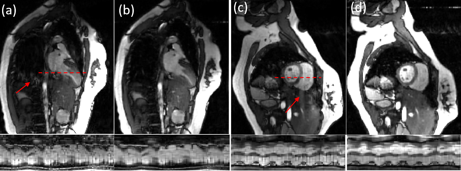
Figure 3. Representative RT-Cine images of 2CH (a,b) and
SAX (c,d) views. (a,c) are the images
reconstructed using all the physical coils and (b,d) are the ones after
automatically discarding five physical coils with high level of motion
artifacts. The temporal profile is along the red dashed line and the red arrows
highlight the motion artifacts.
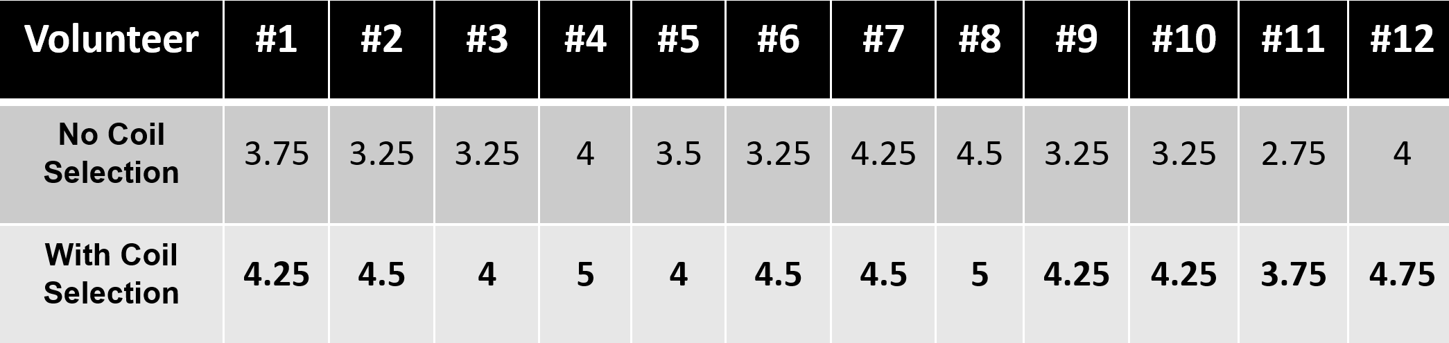
Table 1. Scores in terms of the image quality in the heart region. 1: Unusable, 2: Severe artifacts
obscuring useful information, 3: Moderate artifacts with some loss of
information but still diagnostic, 4: Minor artifacts with minimal loss of
information, 5: No artifacts. The values presented are the average of the
scores across two slices and two readers.
DOI: https://doi.org/10.58530/2023/1152