1130
Tractwise perfusion-microstructure relationships in the aging white matter based on the Lifespan Human Connectome Project - Aging1Rotman Research Institute, Baycrest, Toronto, ON, Canada, 2Medical Biophysics, University of Toronto, Toronto, ON, Canada, 3Biomedical Engineering, University of Toronto, Toronto, ON, Canada
Synopsis
Keywords: White Matter, Perfusion
This study examined baseline and age-related differences in white matter perfusion and microstructural integrity across ten tracts of interest using 535 adult subjects of the Human Connectome Project in Aging. Whole-brain and tractwise relationships were identified between age, cerebral blood flow, mean diffusivity, and fractional anisotropy. Additionally, regional differences in age-related perfusion and microstructural trajectories were identified, representing one of the first direct examinations of regional variation in white matter perfusion in aging.Introduction
Increasing understanding of the importance of neurovascular health in maintaining tissue integrity has led to the examination of tissue structure-perfusion associations in aging5,6. The current literature mainly addresses cortical perfusion and its associations with cortical morphology and declining WM integrity2. Although white-matter (WM) microstructure as an earlier indicator of tissue degeneration should associate more directly with WM perfusion than morphology, the structural-perfusion relationship in the aging WM has not been examined, largely due to challenges in WM perfusion quantification3,6,8,10. This study seeks to examine tractwise age-related variations in the WM perfusion, as well as the relationships between tractwise WM integrity and perfusion. We hypothesize that lower baseline perfusion would be associated with lower microstructural integrity. Secondarily, we hypothesize that regional trajectories of age-related decline in WM microstructure would correspond with those in WM perfusion.Methods
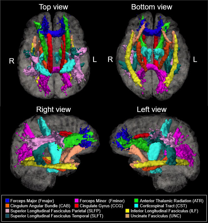
535 healthy adult subjects (300 female, aged 36-100) were drawn from the Human Connectome Project in Aging (HCP-A) dataset (OMB Control# 0925-0667)4. All subjects were in generally good health and without pathological cognitive impairment (i.e. stroke, clinical dementia). The study accessed whole-brain T1-weighted structural MRI, diffusion-weighted MRI (dMRI), and multi-delay pseudo-continuous arterial spin labeling (pCASL) measures collected using four matched Siemens Prisma 3T MRI scanners, with a (1.5mm)3 voxel resolution, MB=4, with 93 directions at b=1500s/mm2. CBF was quantified using the general kinetic model that accounts for arterial-transit delay (FSL oxford_ASL). dMRI data were corrected for eddy-current and susceptibility-related distortions via EDDY, and FA and MD maps were derived using Dipy’s DKI tool, which provides kurtosis-corrected DTI metrics (given the high b-value used). Eighteen major WM tracts were reconstructed from the dMRI data using the Tracts Constrained by Underlying Anatomy (TRACULA) tool in Freesurfer version 7.2.07,11. FSL’s fslmaths function was then used to produce per-subject masks of each WM tract in local space9. The eighteen reconstructed tracts were combined bilaterally to produce ten tracts of interest for analysis (Figure 1). All analyses in this study were conducted in R (version 4.1.1). Relationships between whole-brain and tract-wise measures of mean WM CBF, MD, and FA were assessed using multivariate regression to determine relationships between CBF and MD/FA. Additionally, baseline perfusion/microstructural values extracted from the youngest sub-group, and used for computing fractional variations in CBF, MD and FA.Results
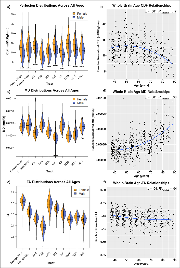
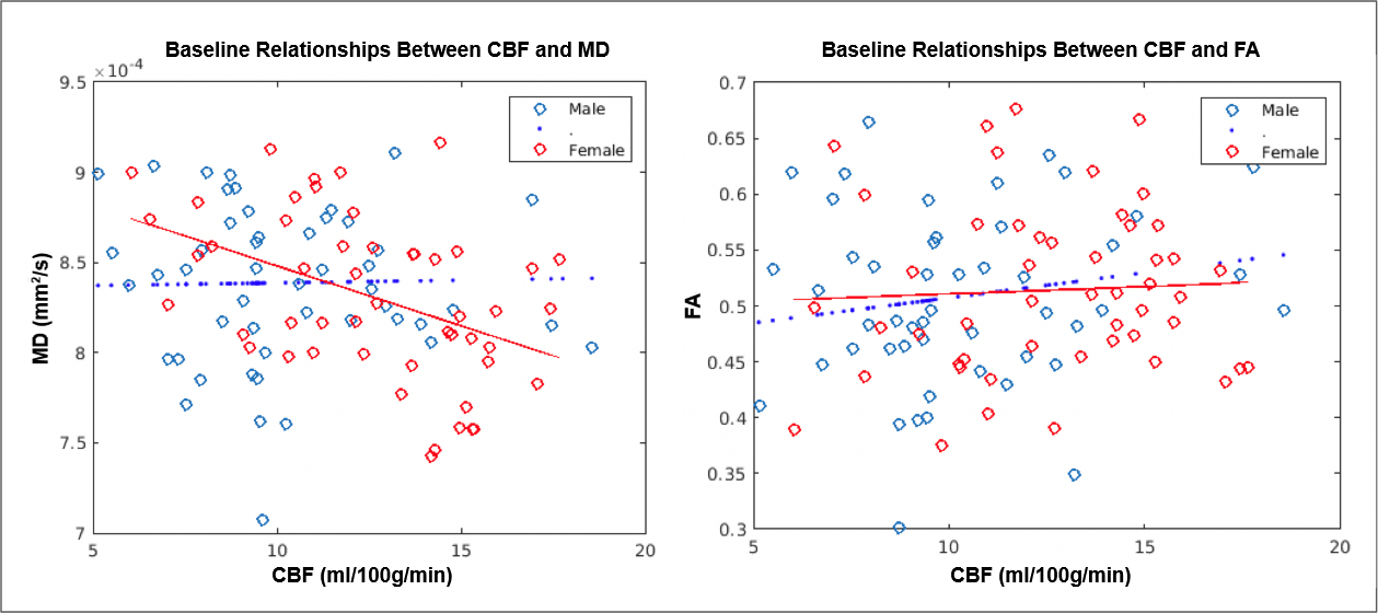
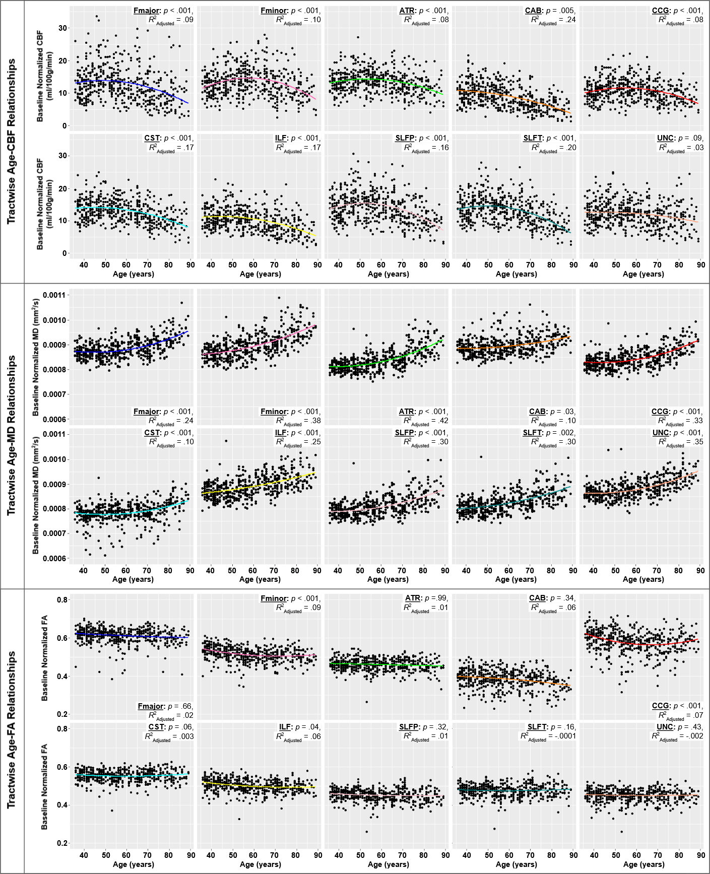
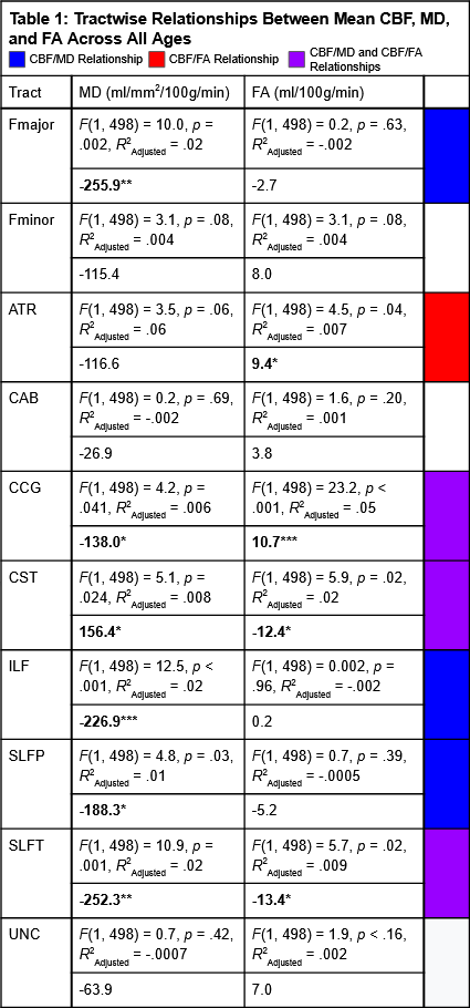
Across ages, we observed significant sex differences in both WM CBF and WM microstructure (Figure 2a, c, e). We also found a quadratic relationship to provide a better fit than a linear model to the age-associations in CBF MD and FA, when averaged across all WM tracts (Figure 2b, d, f). Across ages, we found a significant negative relationship between whole-WM perfusion and MD (F(1,498) = -2.62, p = .009, R2Adjusted = .01), but not between CBF and FA. In the youngest sub-group only, we found a significant negative baseline relationship between CBF and MD across tracts, only in the females; again, no significant baseline relationship between CBF and FA was found. Tractwise comparisons found significant relationships between CBF and MD in the Fmajor, CCG, CST, ILF, SLFP, SLFT (see Figure 1 for definitions of the acronyms). Tractwise comparisons of CBF and FA identified significant relationships in the ATR, CCG, CST, and SLFT (Table 1). Following normalization of perfusion and microstructure values by the young-baseline, significant quadratic relationships between age and perfusion were identified in all tracts, with the exception of the UNC. Significant quadratic relationships between age and MD were identified in all tracts of interest, and between age and FA in the Fminor, CCG, and ILF (Figure 3).Discussion
In addition to the well-established MD and FA associations with age, we also found significant and consistent CBF associations with WM microstructural integrity. Specifically, CBF declines with advancing age in the vast majority of WM tracts, mirroring the age-associated increases in tractwise MD (Figure 4). CBF appears to demonstrate a peak in middle-age, with the peak point varying by tract. However, FA was not uniformly associated with age across tracts, indicating variability in patterns or rates of WM decline. Moreover, these trajectories of perfusion and microstructural declines vary considerably by tract, with some demonstrating distinct quadratic patterns, and others indicating more linear declines. Additionally, while previous literature suggested a negative association between CBF and FA in healthy WM1, we found no such association in our youngest participants across WM tracts (Figure 3), in part agreeing with our first hypothesis. We acknowledge that the CBF-FA relationship may in fact be regionally specific. Furthermore, addressing our second hypothesis, CBF and MD are negatively associated in the majority of tracts, while CBF and FA are positively associated in 2 of the 10 tracts (Table 1). However, there are exceptions to these directions, such as CST, which was associated positively with MD and negatively with FA, and the SLFT, which was associated negatively with both MD and FA. Further quantitative comparison of trajectories of declines are expected to clarify the interactions between WM perfusion and microstructural integrity in aging. Lastly, while this is a novel demonstration of regional differences in WM perfusion, our data also provides arterial-transit delay estimates, which should be incorporated into future analyses.Acknowledgements
This study was supported through grant funding by the Canadian Institutes of Health Research (CIHR)
Data used in this study was provided by the Human Connectome Project in Aging (HCP-A)4
References
1. Aslan, S., Huang, H., Uh, J., Mishra, V., Xiao, G., van Osch, M. J. P., & Lu, H. (2011). White matter cerebral blood flow is inversely correlated with structural and functional connectivity in the human brain. NeuroImage, 56(3), 1145–1153
2. Baker, L. M., Laidlaw, D. H., Conturo, T. E., Hogan, J., Zhao, Y., Luo, X., Correia, S., Cabeen, R., Lane, E. M., Heaps, J. M., Bolzenius, J., Salminen, L. E., Akbudak, E., McMichael, A. R., Usher, C., Behrman, A., & Paul, R. H. (2014). White matter changes with age utilizing quantitative diffusion MRI. Neurology, 83(3), 247–252. https://doi.org/10.1212/WNL.0000000000000597
3. Bernbaum, M., Menon, B. K., Fick, G., Smith, E. E., Goyal, M., Frayne, R., & Coutts, S. B. (2015). Reduced blood flow in normal white matter predicts development of leukoaraiosis. Journal of cerebral blood flow and metabolism : official journal of the International Society of Cerebral Blood Flow and Metabolism, 35(10), 1610–1615. https://doi.org/10.1038/jcbfm.2015.92
4. Bookheimer, S.Y., Salat, D.H., Terpstra, M., Ances, B.M., Barch, D.M., Buckner, R.L., Burgess, G.C., Curtiss, S.W., Diaz-Santos, M., Elam, J.S., Fischl, B., Greve, D.N., Hagy, H.A., Harms, M.P., Hatch, O.M., Hedden, T., Hodge, C., Japardi, K.C., Kuhn, T.P., Ly, T.K., Smith, S.M., Somerville, L.H., Uğurbil, K., van der Kouwe, A., Van Essen, D., Woods, R.P., Yacoub, E., 2019. The Lifespan Human Connectome Project in Aging: An overview. Neuroimage 185, 335–348.
5. Chen, J. J., Rosas, H. D., & Salat, D. H. (2011). Age-associated reductions in cerebral blood flow are independent from regional atrophy. NeuroImage, 55(2), 468–478.
6. Chen, J.J., Rosas, H.D., Salat, D.H., (2013). The relationship between cortical blood flow and sub-cortical white-matter health across the adult age span. PLoS One 8, e56733.
7. Maffei, C., Lee, C., Planich, M., Ramprasad, M., Ravi, N., Trainor, D., Urban, Z., Kim, M., Jones, R., Henin, A., Hofmann, S., Pizzagalli, D., Auerbach, R., Gabrieli, J., Whitfield-Gabrieli, S., Greve, D., Haber, N., Yendiki, A. Using diffusion MRI data acquired with ultra-high gradients to improve tractography in routine-quality data. bioRxiv 2021.06.28.450265; doi: https://doi.org/10.1101/2021.06.28.450265
8. O'Sullivan M, Lythgoe DJ, Pereira AC, Summers PE, Jarosz JM, Williams SC, Markus HS. Patterns of cerebral blood flow reduction in patients with ischemic leukoaraiosis. Neurology. 2002 Aug 13;59(3):321-6. doi: 10.1212/wnl.59.3.321. PMID: 12177363.
9. Smith, S.M., Jenkinson, M., Woolrich, M.W., Beckmann, C.F., Behrens, T.E.J., Johansen-Berg, H., Bannister, P.R., De Luca, M., Drobnjak, I., Flitney, D.E., Niazy, R., Saunders, J., Vickers, J., Zhang, Y., De Stefano, N., Brady, J.M., and Matthews, P.M. Advances in functional and structural MR image analysis and implementation as FSL. NeuroImage, 23(S1):208-19, 2004
10. van Dalen, J. W., Mutsaerts, H., Nederveen, A. J., Vrenken, H., Steenwijk, M. D., Caan, M., Majoie, C., van Gool, W. A., & Richard, E. (2016). White Matter Hyperintensity Volume and Cerebral Perfusion in Older Individuals with Hypertension Using Arterial Spin-Labeling. AJNR. American journal of neuroradiology, 37(10), 1824–1830. https://doi.org/10.3174/ajnr.A4828
11. Yendiki, A., Panneck, P., Srinivasan, P., Stevens, A., Zöllei, L., Augustinack, J., Wang, R., Salat, D., Ehrlich, S., Behrens, T., Jbabdi, S., Gollub, R., & Fischl, B. (2011). Automated probabilistic reconstruction of white-matter pathways in health and disease using an atlas of the underlying anatomy. Frontiers in neuroinformatics, 5, 23. https://doi.org/10.3389/fninf.2011.00023
Figures