1129
7T MR DTI and Tractography of a 1-Week Postmortem Fixed Human Brainstem-Cerebellum: Novel Imaging Methodology Producing Extraordinary Images1Department of Neurosurgery, Hacettepe University, Ankara, Turkey, 2Department of Neurosurgery, Barrow Neurological Institute, Phoenix, AZ, United States, 3Barrow Neurological Institute, Phoenix, AZ, United States
Synopsis
Keywords: White Matter, Diffusion Tensor Imaging, 7T MRI, Tractography, Connectivity, Postmortem
Diffusion-tensor imaging (DTI) is routinely used to estimate the pattern, orientation, and directionality of white matter tracts in human living subjects. However, diffusion imaging of ex-vivo brain specimens is complex due to the diffusivity properties of fixed brain tissue. We developed a novel methodology for tractography of a 1-week postmortem fixed human brainstem and cerebellum specimen utilizing a deterministic fiber tracking algorithm on a 7T MR DTI diffusion scheme registered to an MNI space through landmark-based affine registration. The results showed highly detailed tractography images and agreement between left and right crossing and non-crossing fibers.Introduction
Diffusion-tensor imaging (DTI) is an extension of diffusion-weighted MR imaging (DWI) that, when applied to the brain, can estimate the pattern, orientation, and directionality of white matter tracts (i.e., 3D maps of white matter connectivity). Although classically used on in-vivo tissue (animal models or human subjects), DTI can be expanded to ex vivo (i.e., postmortem/cadaveric) tissue. However, diffusion imaging of ex-vivo specimens requires high-gradient magnetic strengths and longer scanning times due to reduced proton density, T1, T2, and overall diffusivity of fixed tissue [1]. On the other hand, diffusion imaging of the ex-vivo human brainstem is superior to in-vivo due to the small size and complexity of the anatomic area, suffering from low spatial resolution, magnetic susceptibility artifacts, and echo-planar imaging-induced eddy current distortions [2]. Therefore, ex-vivo human brainstem and cerebellum are optimal for studying white matter tractography because the small anatomic area can fit in between the coils of a high magnetic MRI and be scanned for long periods, enabling high spatial resolution for reconstructing small and complex tracts. High-resolution tractography has not been produced using standard postmortem cadaveric brain tissue until now.Methods
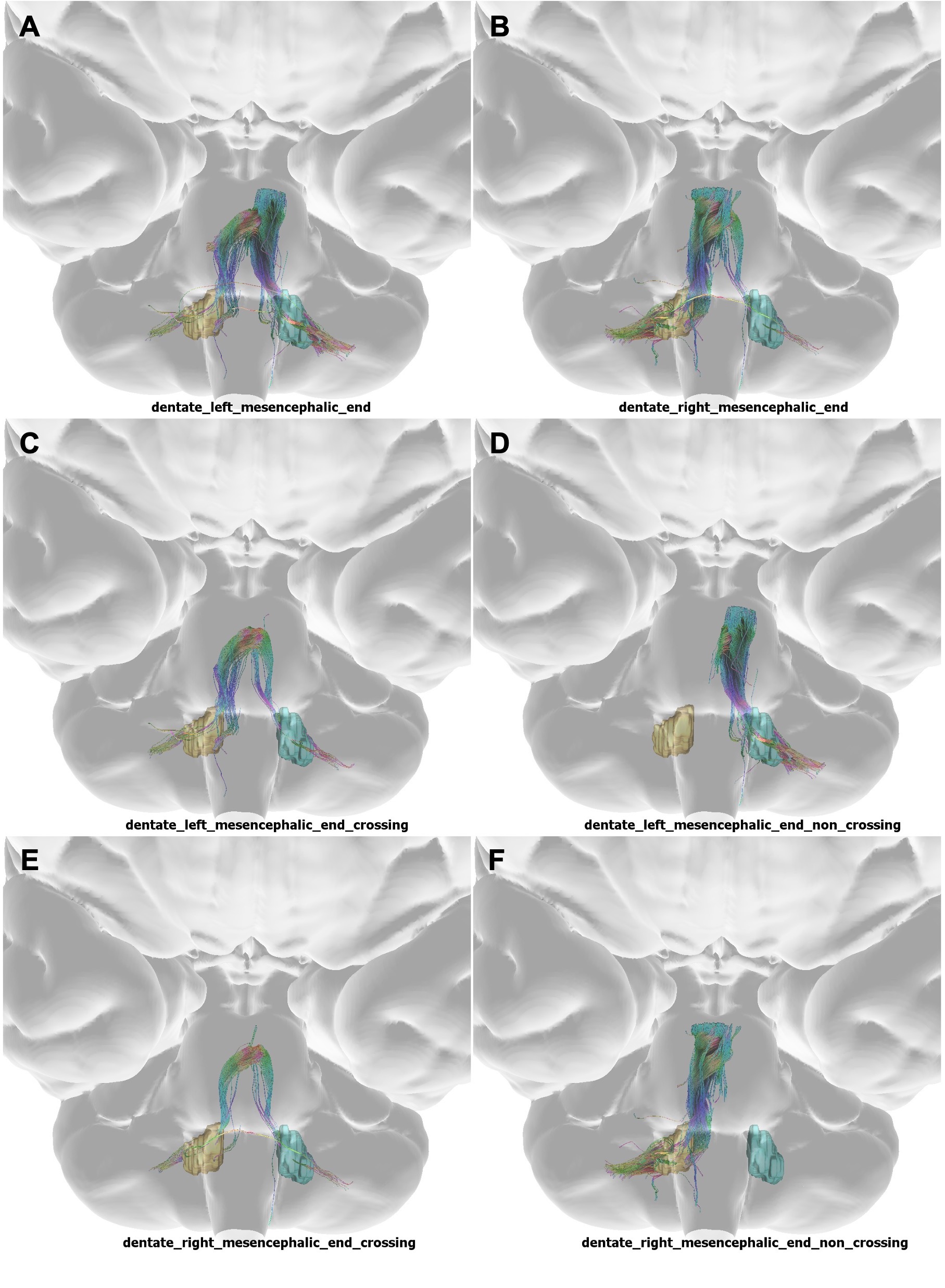
One formalin-fixed whole brainstem-cerebellum specimen (85 y/o M) was imaged. Time from death to fixation was 7 days (168 hours). Before imaging, the specimen was re-hydrated in normal saline solution for 7d and later degassed for 30 minutes in an inert, oxidatively stable solution with continuous vibration. 7T MRI was used for anatomic (acquisition time: 3h 23min) and DTI (acquisition time: 48h 48min) imaging. The parameters used for the anatomic scan were: TR/TE=4000/9 ms, 4 averages, and 78.125 x 78.125 x 500 mm3 resolution. The parameters for the diffusion scan were: TR/TE= 2500/27 ms, b=3000 s/mm2, 60 directions, ∂/∆= 7/14 ms, and 555 x 555 x 500 mm3 resolution. Diffusion maps were generated with DIPY and fractional anisotropy (FA) maps were generated using MATLAB. B0 images were registered to MNI space with a landmark-based affine registration, and the diffusion images were resampled using the same transformation matrix in 3D Slicer.Whole brainstem tractography was performed and visualized using DSI studio. A DTI methodology was employed, and 60 diffusion sampling directions were acquired. The b- value was 3105 s/mm2. The in-plane resolution and slice thickness were 0.55 and 0.5 mm, respectively. The b-table was checked by an automatic quality control routine to ensure accuracy. A deterministic algorithm [3] was used for identification and tracking of fibers. A seeding region was placed at the surface of the whole specimen. The angular threshold was 60 degrees. The step size was randomly selected from 0.5 voxels to 1.5 voxels. Tracks with lengths shorter than 50 mm or longer than 200 mm were discarded. A total of 106 seeds were placed. Fibers connecting the mesencephalic end of our specimen to the bilateral dentate nuclei were isolated and analyzed.
Results
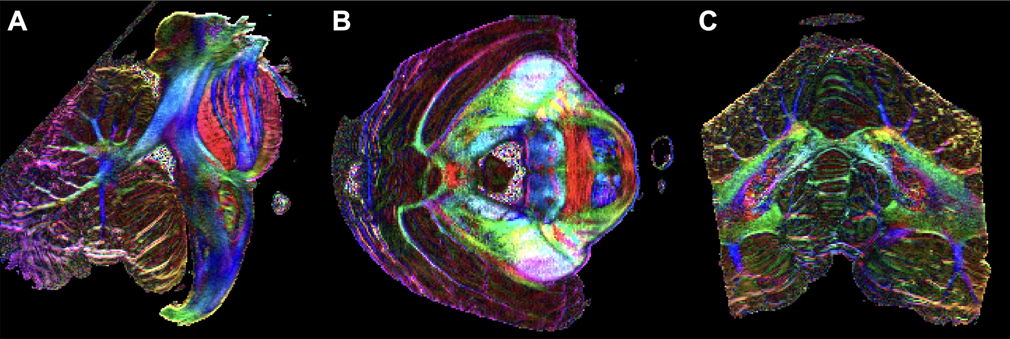
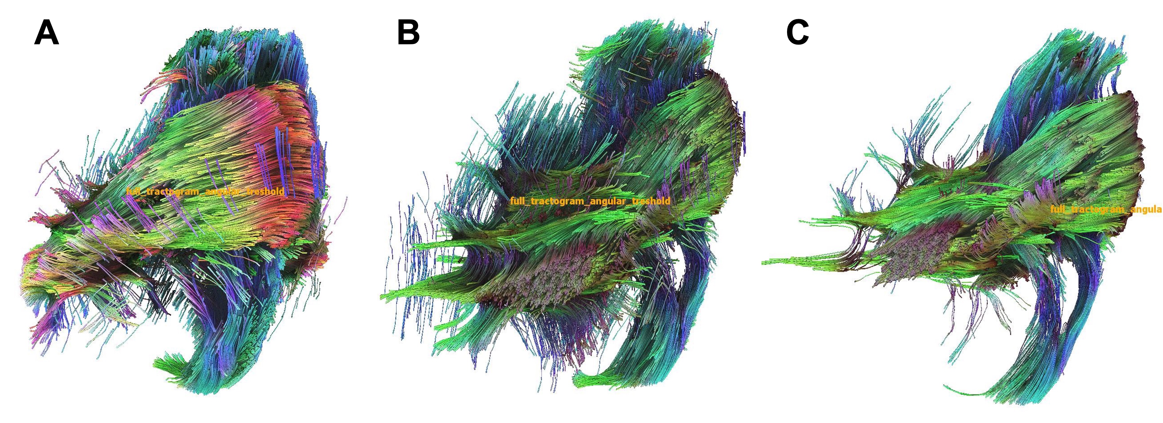
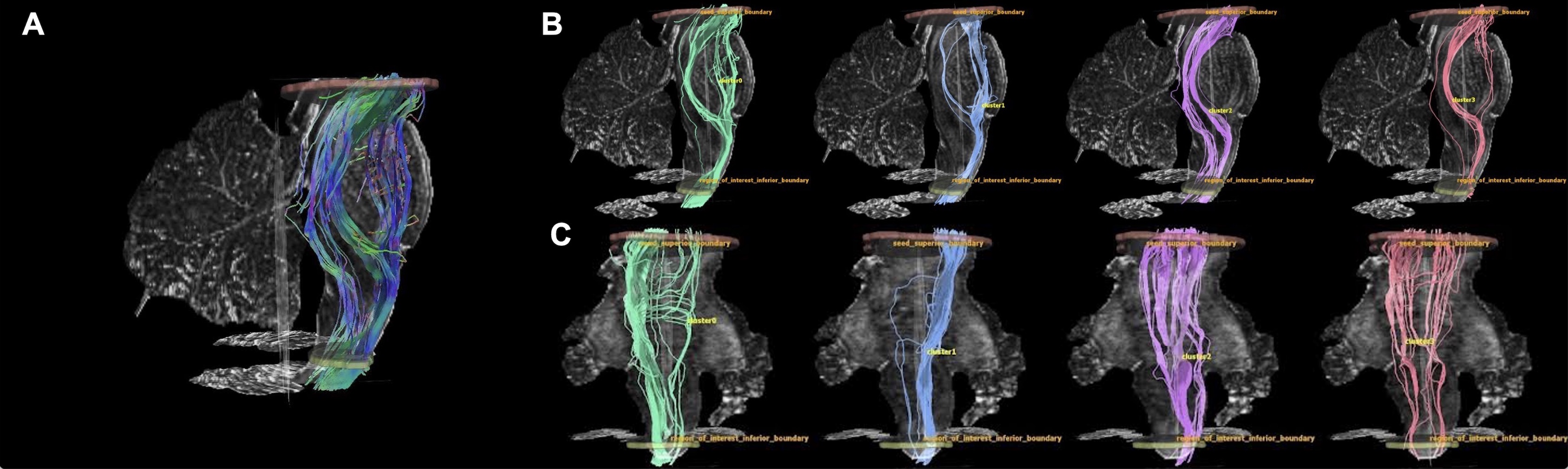
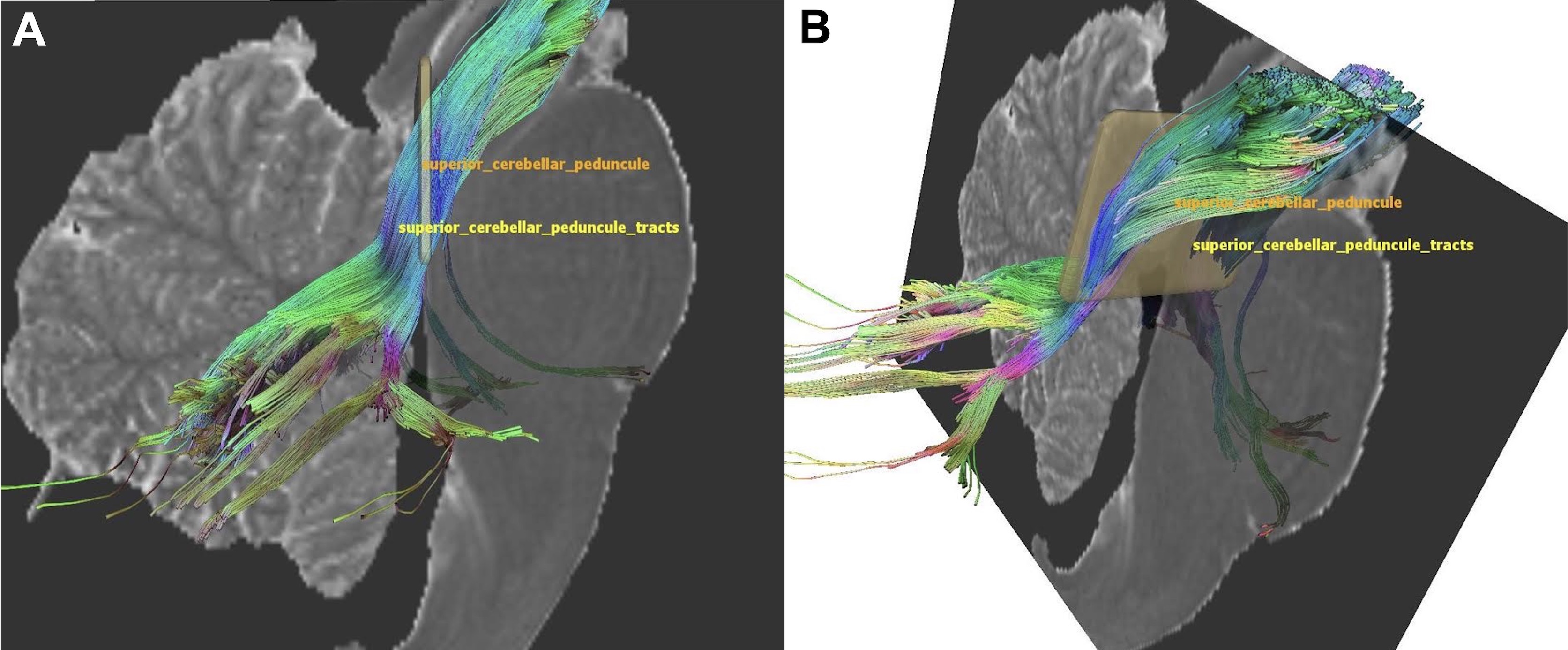
Clear FA mapping (Figure 1) and tractography (Figure 2) were successfully created from a 7-day postmortem brainstem-cerebellum specimen. A total of 21,641 streamline tracts were generated. White matter fibers traveling from mesencephalon to medulla, horizontally traversing the pons, fasciculi of superior/middle/inferior cerebellar peduncles, and decussation fibers are visible (Figure 3 and 4). K-means clustering of whole specimen surface seeding tractography shows anatomically accurate fiber distribution with clear left-right and superior-inferior preference clusters generated. Regarding mesencephalic-dentate connectivity, a total of 5186 streamlines (left: 2155, right: 3031) were bilaterally generated, seen in Figure 5. The mean length of all streamlines was 69.4 mm. The mean length of the crossing and non-crossing fibers were 90.9 and 64.1 mm, respectively. The diameter, tract number (number of generated streamlines), and radius were consistent between right and left crossing and non-crossing fibers. Tract numbers of left and right non-crossing fibers were 1795 and 2793, respectively, while the left and right crossing fibers were 309 and 238, respectively.Discussion
Until now, high-resolution tractography has not been reliably and clearly produced using standard postmortem cadaveric brain tissue. Previous studies that conducted diffusion imaging on ex-vivo human brains had specimens of 65 years of age with death to fixation times between 48 and 24 hours [4,5]. Our specimen died at 85 years, and the time to fixation was 1-week (168 hours). Both times to fixation and donor age are known to affect the volume of brain structures. Regardless of the specimen age and time to fixation, we produced remarkable results with agreement between left and right and crossing and non-crossing connectivity. Our results indicate that standard cadaveric specimens can be reliably used for diffusion imaging and white matter connectivity research. Unlike living specimens, cadaveric tissue can fit between the coils of a high-field MRI, and diffusion resolution can be maximized in deep, complex areas.Conclusion
Highly detailed and precise white matter tractography was created from a standard postmortem specimen, even when fixed 1-week after death. This methodology and results are ground-breaking for diffusion imaging and tractography research because standard cadaveric specimens obtained from donor organizations can be reliably used. Diffusion resolution of small, complex brain areas can be maximized in cadaveric specimens.Acknowledgements
No acknowledgement found.References
1. Roebroeck A, Miller KL, Aggarwal M. Ex vivo diffusion MRI of the human brain: Technical challenges and recent advances. NMR Biomed. 2019;32(4):e3941.
2. Le Bihan D, Poupon C, Amadon A, et al. Artifacts and pitfalls in diffusion MRI. Journal of Magnetic Resonance Imaging. 2006;24(3):478-488.
3. Yeh F-C, Verstynen TD, Wang Y, et al. Deterministic Diffusion Fiber Tracking Improved by Quantitative Anisotropy. PLOS ONE. 2013;8(11):e80713.
4. McNab JA, Jbabdi S, Deoni SC, et al. High resolution diffusion-weighted imaging in fixed human brain using diffusion-weighted steady state free precession. Neuroimage. 2009;46(3):775-785.
5. Calabrese E, Hickey P, Hulette C, et al. Postmortem diffusion MRI of the human brainstem and thalamus for deep brain stimulator electrode localization. Hum Brain Mapp. 2015;36(8):3167-3178.
Figures