1108
Locally low-rank denoising in transform domains.1Radiology, University of Minnesota, Minneapolis, MN, United States, 2Veterinary Clinical Sciences, University of Minnesota, Minneapolis, MN, United States, 3Electrical and Computer Engineering, University of Minnesota, Minneapolis, MN, United States
Synopsis
Keywords: Quantitative Imaging, Data Processing
The concept of transform processing domain with locally low rank denoising is proposed as T-NORDIC and demonstrated for MSK and brain applications. The improvements on quantitative maps may be leveraged for faster acquisitions by relaxing the number of averages needed to obtain sufficient SNR for high resolution acquisitions and for application of low rank denoising to common clinical acquisitions.Introduction
Low-rank techniques are frequently used to extract image features and suppress image noise1. Low-rank techniques are either applied globally to the whole image series or in a local manner to image patches2. The latter technique, referred to as locally low-rank (LLR) regularization, has been widely used for MR image series3. The LLR technique relies on either a larger number (tens) of images in the series or identification of many regions across images with similar features for noise suppression1. Current LLR techniques for post reconstruction improvements4-8 utilize an approach where a local patch with 43 – 113 voxels is extracted from each time-point. The similarity over tens of time-points is used to separate signal from noise. We propose a new method where the use of LLR is applicable for as few as 4 volumes, extending the use of LLR for many clinical protocols.Conventional locally low-rank reconstruction (LLR) extracts voxels in a patch of size NxNxN, from M volumes. Analysis is done and from an SVD on the N3xM Casorati matrix, followed by soft/hard thresholding of singular values to suppress noise components. In this work, we propose the use of LLR in unitary transform domains, such as Fourier or wavelet domains, to improve the underlying low-rankness of the patches, preserve high-frequency content, while maintaining the independently identically distributed (i.i.d.) nature of noise across coordinates in the transformed representation. Results on MSK and brain data show that the proposed method substantially improves the quality of quantitative maps.
Methods
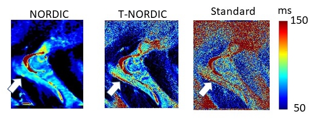
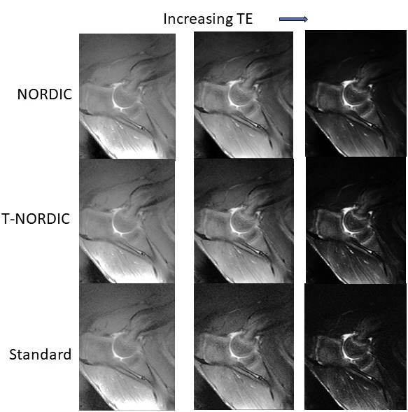
Transform-domain LLR denoising: Each image in the images series are transformed using a unitary transformation, which are FFT and Haar wavelet transforms in this study. Then local Casorati matrices are generated from these transform domain image series, similar to standard LLR processing. Finally, hard singular value thresholding is applied with a heuristic threshold, referred to as T-LLR; or using a threshold based on a non-asymptotic random matrix characterization of the thermal noise (as introduced in NORDIC6), referred to as T-NORDIC.Denoising experiments: T-NORDIC was first compared with NORDIC in parametric mapping with limited series/volumes in an in-vivo porcine application. Distal humerus (elbow joint) was imaged with a T2 mapping multi-slice multi-echo acquisition at 3T using an 8 channel coil to detect naturally-occuring osteochondritis dissecans (OCD) precursor lesions in the epiphyseal cartilage. T-NORDIC was applied in FFT domain on each channel independently, which were combined with a SENSE-1 reconstruction9.
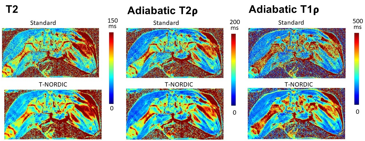
Subsequently, T-NORDIC was evaluated in high-resolution 3T quantitative MRI experiments, including quantitative T2 mapping of the femoral trochlea (knee joint) to detect surgically-induced OCD precursor lesions (18-channel coil); and a magnetization-prepared 3D SPACE sequence with T2, adiabatic T1𝞀, and adiabatic T2𝞀 contrast of proximal femora (hip joint) for detecting surgically-induced ischemia to the femoral head (8-channel coil). T-NORDIC was applied with patch-size 53x8 for the former, and 43 x 4 for the latter, for each channel independently and then combined with SENSE-1.
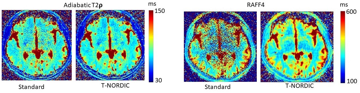
Finally, a human volunteer was scanned on a 3T Siemens scanner with a 64 channel head-coil using a multi-slice 2D GRE sequence to obtain T1𝞀, T2𝞀 and Relaxation Along a Fictitious Field in the rotating frame of rank n = 4 (RAFF4)10 contrasts. For each voxel the signal was fitted as described in11. T-NORDIC was applied with 43 x 4 on the SENSE-1 combined GRAPPA reconstruction.
Results
Figures 1 and 2 show a comparison of T-NORDIC and NORDIC, both applied individually to single-channel fully-sampled acquisitions and then SENSE1 combined. Results show T-NORDIC lads to sharper maps and images compared to NORDIC.Figure 3 depicts T-NORDIC applied to an undersampled acquisition before reconstruction with GRAPPA. The maps show that T-NORDIC leads to high-quality quantitative maps from as few as 4 images in the series.
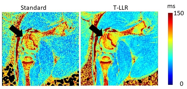
Figure 4 shows T-LLR using conventional (GRE) quantitative maps from vendor reconstructed data . The maps show that T-LLR leads to high-quality quantitative maps from as few as 8 images in the series.
Figure 5 depicts the use of T-NORDIC after using SENSE1 combined images utilizing series with 4 volumes. T-NORDIC visibly improves parametric mapping from 4 volumes at high-resolution.
Discussion/Conclusion
The combination of unitary transform processing with LLR denoising leverages low-rank properties of patches in transform domains while preserving noise distributions. For FFT, image edge information across different contrast weightings is consistent across the image series, and can be captured with few singular values/vectors. For short series/volumes/replicas the use of T-LLR reduces the dominance of an average image in the LLR, and the associated blurring effects, typically observed with image-domain LLR processing. Such improvements may be leveraged for faster acquisitions by reducing the number of averages needed to obtain sufficient SNR for high-resolution acquisitions.For quantitative mapping with few volumes, both T-LLR and T-NORDIC have been evaluated in different ways, using channel-by-channel processing, using SENSE1 combined images, and when applied to undersampled acquisitions before reconstruction with GRAPPA, with series of length 4-12 volumes. The use of T-LLR is flexible, and it can be applied at different points in the reconstruction pipeline.
The use of T-NORDIC improves the clarity of quantitative maps for in-vivo applications. It enables the use of higher resolution mapping in feasible acquisition times and is compatible with previously acquired data.
Acknowledgements
Grant support: NIH, Grant numbers: R56AR078315, R56AR078209, P41EB027061, P30 NS076408, R01 HL153146, U01 EB025144, NSF CAREER CCF-1651825References
Kostadin Dabov, Alessandro Foi, Vladimir Katkovnik, Karen Egiazarian, Image denoising with block-matching and 3D filtering. Proceedings Volume 6064, Image Processing: Algorithms and Systems, Neural Networks, and Machine Learning; 606414 (2006) https://doi.org/10.1117/12.643267
Joshua Trzasko, Armando Manduca Local Versus Global Low-Rank Promotion in Dynamic MRI Series Reconstruction, ISMRM, 2011, page 4371
Mathews Jacob, Merry P. Mani, Jong Chul Ye. Structured Low-Rank Algorithms: Theory, Magnetic Resonance Applications, and Links to Machine Learning. IEEE Signal Process Mag. 2020 Jan; 37(1): 54–68. doi: 10.1109/msp.2019.2950432
Manjón, J. V., Coupé, P., Concha, L., Buades, A., Collins, D. L., and Robles, M. (2013). Diffusion weighted image denoising using overcomplete local PCA. PLoS One 8:e73021. doi: 10.1371/journal.pone.0073021
Veraart, J., Novikov, D. S., Christiaens, D., Ades-aron, B., Sijbers, J., and Fieremans, E. (2016). Denoising of diffusion MRI using random matrix theory. NeuroImage 142, 394–406. doi: 10.1016/j.neuroimage.2016.08.016
Meyer et al. "Locally low-rank denoising of complex-valued EPI reconstructions preceding task fMRI analysis." ISMRM 2020, 3877.
Vizioli, L., Moeller, S., Dowdle, L. et al. Lowering the thermal noise barrier in functional brain mapping with magnetic resonance imaging. Nat Commun 12, 5181 (2021). https://doi.org/10.1038/s41467-021-25431-8
Pierre-Louis Bazin , Anneke Alkemade, Wietske van der Zwaag, Matthan Caan, Martijn Mulder and Birte U. Forstmann, Front. Neurosci., 09 October 2019, Sec. Brain Imaging Methods, Volume 13, Article 1066, https://doi.org/10.3389/fnins.2019.01066
S. N. Sotiropoulos,S. Moeller,S. Jbabdi,J. Xu,J. L. Andersson,E. J. Auerbach,E. Yacoub,D. Feinberg,K. Setsompop,L. L. Wald,T. E. J. Behrens,K. Ugurbil,C. Lenglet. Effects of image reconstruction on fiber orientation mapping from multichannel diffusion MRI: Reducing the noise floor using SENSE. Magn Reson Med. 2013, Volume70, Issue 6 Pages 1682-1689, https://doi.org/10.1002/mrm.24623
Liimatainen T, Hakkarainen H, Mangia S, Huttunen JM, Storino C, Idiyatullin D, Sorce D, Garwood M, Michaeli S. MRI contrasts in high rank rotating frames. Magn Reson Med. 2015 Jan;73(1):254-62.
Pavel Filip, Alena Svatkova, Adam F Carpenter, Lynn E Eberly, Igor Nestrasil, Mikko J Nissi, Shalom Michaeli, Silvia Mangia, Rotating frame MRI relaxations as markers of diffuse white matter abnormalities in multiple sclerosis,Neuroimage Clinical, Volume 26, 2020, 102234, DOI: 10.1016/j.nicl.2020.102234
Figures