1087
Motion artifact reduction in self-gated CMR 4D flow imaging under exercise stress
Syed Murtaza Arshad1, Chong Chen2, Yingmin Liu3, Preethi Chandrasekaran3, Christopher Crabtree4, Ning Jin5, and Rizwan Ahmad2
1Department of Electrical & Computer Engineering, The Ohio State University, Columbus, OH, United States, 2Department of Biomedical Engineering, The Ohio State University, Columbus, OH, United States, 3Davis Heart and Lung Research Institute, The Ohio State University, Columbus, OH, United States, 4Department of Human Sciences, The Ohio State University, Columbus, OH, United States, 5Cardiovascular MR R&D, Siemens Medical Solutions Inc, Columbus, OH, United States
1Department of Electrical & Computer Engineering, The Ohio State University, Columbus, OH, United States, 2Department of Biomedical Engineering, The Ohio State University, Columbus, OH, United States, 3Davis Heart and Lung Research Institute, The Ohio State University, Columbus, OH, United States, 4Department of Human Sciences, The Ohio State University, Columbus, OH, United States, 5Cardiovascular MR R&D, Siemens Medical Solutions Inc, Columbus, OH, United States
Synopsis
Keywords: Flow, Image Reconstruction, 4D Flow
Free-breathing self-gated CMR 4D flow imaging using traditional Compressed Sensing (CS) methods invariably contains motion artifacts due to the inaccuracy of self-gating signal. Self-gating signal degrades even further in the case of exercise stress imaging due to excessive movement of the subject. We propose Compressive recovery with Outlier Rejection (CORe) to reduce the motion artifacts. Using data from a 2D digital phantom and 4D flow data under rest and stress conditions, we demonstrate that CORe is effective in suppressing motion artifacts while maintaining agreement with 2D-PC based flow quantification.
Introduction
In free-breathing Cardiac Magnetic Resonance (CMR) 4D flow imaging, the respiratory motion is compensated either by prospective navigator gating or by retrospective self-gating. Typically, these respiratory compensation methods do not suppress respiratory motion completely, leading to image artifacts. In the case of exercise stress imaging, the quality of the self-gating signal degrades even further due to irregular and exaggerated breathing patterns, and contamination from exercise-induced torso movement. In this work, we integrate an outlier rejection scheme into the image reconstruction process. This technique minimizes motion artifacts by suppressing contributions from k-space samples that have been erroneously assigned to the end-expiratory bin.Methods
Typically used Compressed Sensing (CS) methods for accelerated 4D flow imaging employ an L2 norm for data fidelity and an L1 norm for regularization, i.e.,$$$\boldsymbol{\hat{x}} = \operatorname*{arg\,min}_x\left\{\frac{1}{2}\left\|\boldsymbol{Ax}-\boldsymbol{y}\right\|_2^2+\lambda_1 \left\| \boldsymbol{Wx}\right\|_1 \right\},$$$ (Eq. 1)
where $$$\boldsymbol{\hat{x}}$$$ is the recovered MR image, $$$\boldsymbol{x}$$$ represents the true image that we intend to reconstruct, $$$\boldsymbol{y}$$$ is the measured k-space data which has been retrospectively sorted into a motion resolved respiratory bin by self-gating, $$$\boldsymbol{A}$$$ is the sensing matrix, $$$\boldsymbol{W}$$$ refers to undecimated wavelet transform, and $$$\lambda_1>0$$$ is the regularization parameter.
In self-gated 4D flow imaging, data are collected continuously for several minutes. Then, the collected data are sorted into various respiratory bins using self-gating. Due to imperfections in the extracted self-gating signal, bin assignment for a small fraction of readouts is invariably incorrect. The quality of the images reconstructed in each bin with traditional CS (Eq. 1) can degrade substantially in presence of these outliers. We implement a motion robust extension of CS1 which reduces the motion artifacts in the reconstructed image by suppressing the outliers in measured data. This model, termed Compressive recovery with Outlier Rejection (CORe), entails solving the following optimization problem:
$$$\boldsymbol{\hat{x}} = \operatorname*{arg\,min}_{x,v}\left\{\frac{1}{2}\left\|\boldsymbol{Ax}-(\boldsymbol{y}-\boldsymbol{v})\right\|_2^2+\lambda_1 \left\| \boldsymbol{Wx}\right\|_1+\lambda_2 \left\| \boldsymbol{v}\right\|_1 \right\},$$$ (Eq. 2)
where $$$\boldsymbol{v}$$$ represents the outliers in data and parameter $$$\lambda_2>0$$$ is a Lagrange multiplier. The term $$$\lambda_2 \left\| \boldsymbol{v}\right\|_1$$$ ensures sparsity of the outliers in the measurement domain. This model in Eq. 2 assumes that data contains zero-mean Gaussian noise and sparse outliers, and reconstructs the image by (partially) eliminating the contribution of $$$\boldsymbol{v}$$$.
We compare both models, CS and CORe, for the reconstruction of a 2D image originating from a bimodal dynamic phantom. The dynamic phantom is simulated to transition between the expiratory and inspiratory phases (Figure 1). To simulate contamination with outliers, approximately 10% of the k-space data is randomly sampled from the inspiratory phase and the rest of the k-space is randomly filled with data from the expiratory phase. This contaminated k-space is used to reconstruct the expiratory phase image. The simulation is repeated 20 times, each with a random realization of the outlier locations in k-space. The acceleration rate is fixed at 3.5.
To validate the improvement offered by CORe in 4D flow image reconstruction, two free-running, free-breathing, and fully self-gated 4D flow datasets were collected for a fixed acquisition time of 5 minutes using a research sequence with cartesian sampling2. The datasets were acquired from a healthy volunteer at rest and exercise stress (at 20 W load) using a clinical 3T scanner (MAGNETOM Vida, Siemens Healthcare, Erlangen, Germany) and a cycle ergometer (MR Ergometer Pedal, Lode, The Netherlands). To compare blood flow quantification using CORe and CS, real-time 2D phase-contrast MRI (2D-PC) was collected as a reference. Aortic net flow quantification over a cardiac cycle was performed using 2D-PC, CORe, and CS.
Results
In the dynamic phantom simulation, reconstruction of the expiratory phase using CS and CORe for 20 realizations yielded average PSNR values (in dB) of 24±2.6 and 29.7±1.8, respectively. The improvement in recovered image quality is also evident in the example shown in Figure 1. Representative 4D flow magnitude and phase images from a single slice in axial view at systole are shown in Figure 2 for CS and CORe. The comparison demonstrates that the suggested model is more effective in suppressing motion artifacts, with the reduction in artifacts being more evident under exercise. Net aortic flow values measured using CORe and CS are comparable with 2D-PC reference as shown in Table 1, with CS showing marginal overestimation (4.5 ml) and CORe showing marginal underestimation (2.5 ml). While flow quantifications from CS and CORe are comparable, the latter is more effective in suppressing motion artifacts as evident in Figures 2 and 3.Discussion
The parameters $$$\lambda_1$$$ and $$$\lambda_2$$$ used in CORe are chosen empirically. The values of $$$\lambda_1$$$ and $$$\lambda_2$$$ depend on the level of Gaussian noise; for a higher noise level, the values of $$$\lambda_1$$$ and $$$\lambda_2$$$ should be larger. The parameter $$$\lambda_2$$$ additionally depends on the percentage of outliers in data. A higher value of $$$\lambda_2$$$ makes $$$\boldsymbol{v}$$$ sparser which implies less suppression of outliers. The computation time for both CS and CORe reconstruction algorithms is comparable.Conclusion
The proposed method, CORe, integrates outlier rejection into the reconstruction framework. Data from a digital phantom and 4D flow exercise stress imaging demonstrates that CORe is more effective in suppressing motion artifacts than traditional CS techniques.Acknowledgements
This work was funded by NIH project R01HL151697.References
- Dong B, Ji H, Li J, et al. Wavelet frame based blind image inpainting. Applied and Computational Harmonic Analysis. 2012;32(2):268-279
- Pruitt A, Rich A, Liu Y, et al. Fully self-gated whole-heart 4D flow imaging from a 5-minute scan. Magn Reson Med. 2021;85(3):1222-1236
Figures
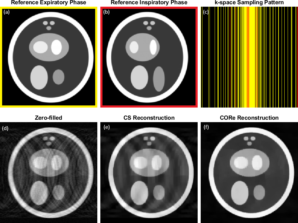
Figure 1: A realization from the dynamic phantom study. (a) Fully sampled reference frame from the expiratory phase of the bimodal dynamic phantom, (b) Fully sampled reference frame from the inspiratory phase of the phantom, (c) k-space sampling pattern, black represents encodings not sampled, yellow and red represent encodings sampled from expiratory and inspiratory phases, respectively, (d) zero-filled image obtained by inverse Fourier transform of the contaminated k-space, (e) expiratory phase image recovered using CS, and (f) expiratory phase image recovered using CORe.
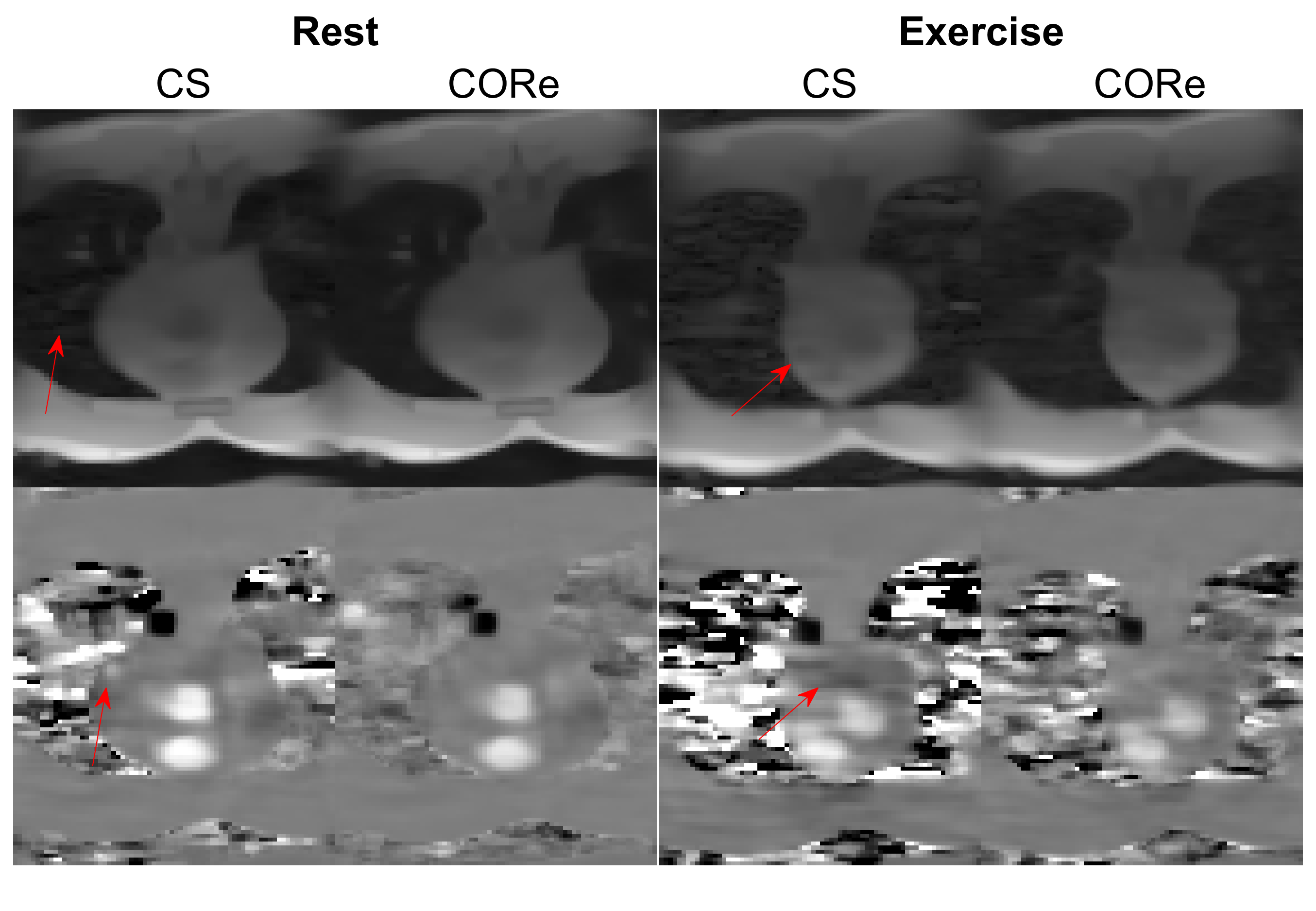
Figure 2: Visual comparison of magnitude and a velocity component of
4D flow images reconstructed using CS and CORe. A single axial slice is shown
at systole (peak flow). The red arrows highlight some of the visible artifacts
in CS images that have been suppressed by CORe. The VENC values for rest and stress were 150 cm/s and 250 cm/s, respectively.
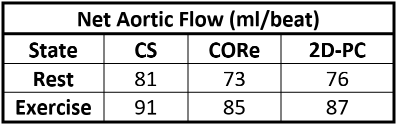
Table 1: Net aortic flow measured from reconstructed 4D flow images using CS, CORe, and real-time 2D phase-contrast (2D-PC), at rest and exercise.
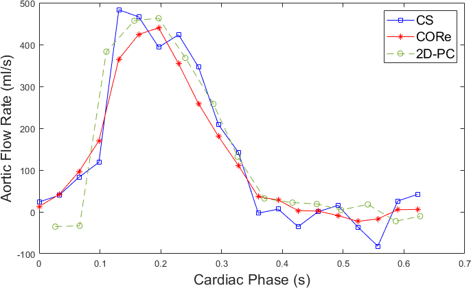
Figure 3: Comparison of CS and CORe aortic flow rate profiles measured from the reconstructed 4D flow images with 2D-PC reference under exercise stress. The motion-induced oscillations are visible in CS but not in CORe.
DOI: https://doi.org/10.58530/2023/1087