1082
Improved Simultaneous Intracranial Vessel Wall Imaging and dynamic MRA Using Low-rank Reconstruction1Department of Bioengineering, University of Washington, Seattle, WA, United States, 2Institute of Science and Technology for Brain-Inspired Intelligence, Fudan University, Shanghai, China, 3Department of Electrical Engineering, University of Washington, Seattle, WA, United States, 4Department of Radiology, University of Washington, Seattle, WA, United States, 5Department of Surgery, University of Washington, Seattle, WA, United States, 6Department of Radiology and Imaging Sciences, University of Utah, Salt Lake City, UT, United States
Synopsis
Keywords: Vessel Wall, Blood vessels
Acquiring multi-contrast images of intracranial vessels from one single MRI sequence is beneficial for comprehensive neurovascular disease diagnosis. Vessel wall (VW) images and dynamic MRA (dMRA) can be obtained from a time-efficient multi-contrast sequence named iSNAP. However, the previously adopted k-space sharing reconstruction method generated high-quality dMRA but low-quality VW images. In this study, we aim to develop a novel low-rank-based image reconstruction method to reconstruct VW and dMRA from highly undersampled 4D MRI. High quality VW images with sharper wall delineation are obtained, while hemodynamic information from dMRA is preserved.Introduction:
Acquiring multi-contrast images of intracranial vessels from one single MRI sequence is beneficial for comprehensive neurovascular disease diagnosis. Previously, a highly time-efficient multi-contrast sequence named iSNAP was proposed1. It utilizes 3D golden angle radial acquisition and can simultaneously obtain intracranial vessel wall (VW) images and whole brain dynamic MRA (dMRA). Reconstruction of the VW images from iSNAP provides high spatial resolution and good blood suppression for delineation of the vessel wall in the proximal large intracranial arteries (ICA, BA, VA, MCA-M1, PCA-P1, ACA-A1), while the dMRA provides luminal hemodynamic information for both proximal and distal arteries ( MCA-M2/3, PCA-P2/3, ACA-A2/3). However, the previously adopted reconstruction method for iSNAP, i.e. k-space weighted image contrast (KWIC), generated high-quality dMRA but low-quality VW images2. In this study, we aim to develop a novel low-rank-based image reconstruction method targeted for highly undersampled 4D multi-contrast MRI. The proposed low-rank reconstruction can improve the lumen and wall delineation for VWI and preserve hemodynamic information from dMRA at the same time. With low-rank-based reconstruction, the 4D multi-contrast MRI, i.e., iSNAP, can simultaneously provide high-quality dMRA and VW images that can better characterize the cerebral vasculature.Methods:
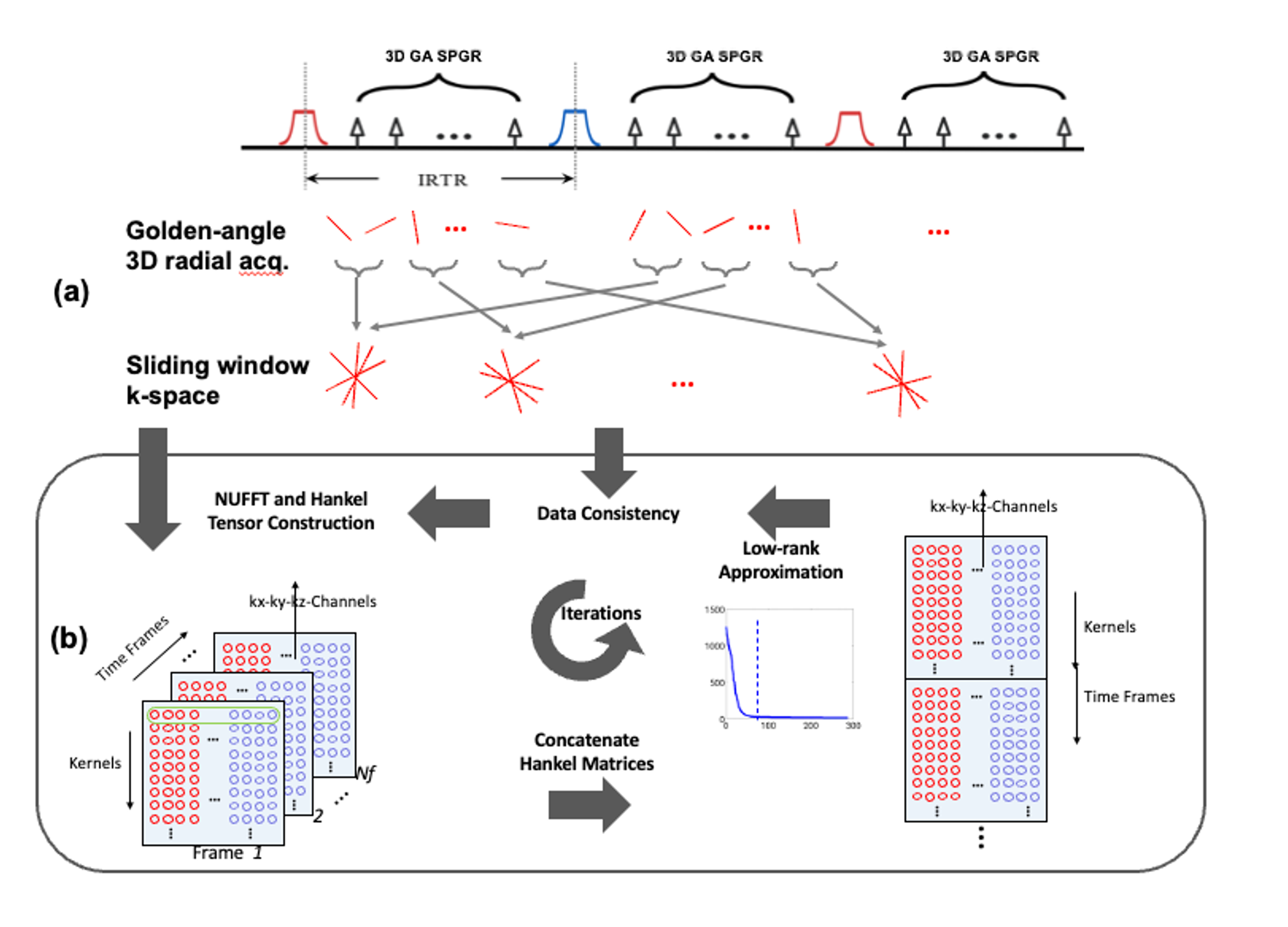
Data Acquisition: The sequence scheme and inversion manipulation of iSNAP are shown in Figure 1 (a), and the details can be found in the original paper1. In vivo experiments were performed on a Philips Ingenia 3T MR scanner (Philips Healthcare, Best, The Netherlands) using a 32-channel head coil. Four subjects with atherosclerotic cerebrovascular disease (male, age 68, 73, 76, and 80 years) were recruited.Image Reconstruction: We propose a novel low-rank joint reconstruction for highly undersampled 4D MRI data. The sharable structural information across time frames was utilized to facilitate the reconstruction. A 3D Hankel tensor was constructed for each frame and concatenated for low-rank approximation3,4,5, with a kernel size of $$$3\times3\times3$$$, targeted rank of 50, and iteration number of 50(Figure 1(b)). A narrower band (4 rings) was used to reduce the contrast mixture. As a reference, KWIC reconstruction was performed using 17 rings and 4 rings. After images of all TIs (temporal resolution=150ms) were jointly reconstructed, dMRA images were obtained by subtraction between acquisitions with small and large inversion, and VW images were obtained by choosing large-slab inversion images at a TI near the blood nulling point.
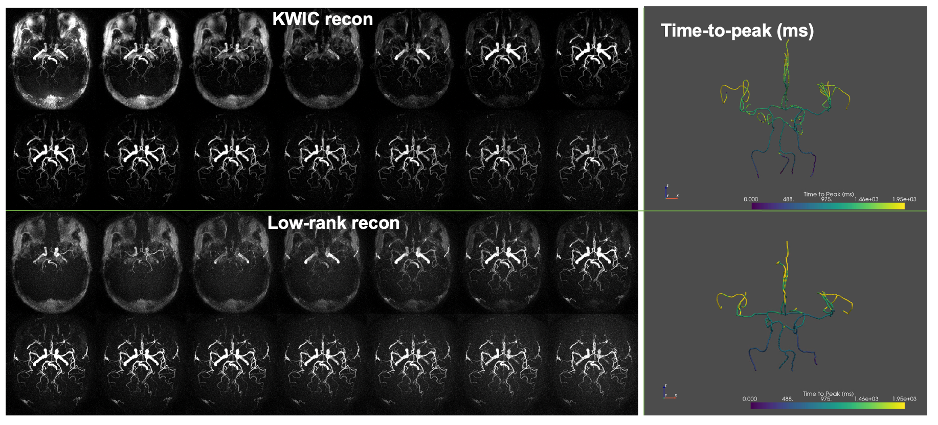
Image Processing: Reconstructed images were interpolated to 0.5mm isotropic. Intracranial arterial centerlines were traced and labeled from the maximum intensity projection of dMRA images using AICafe6,7. Then, multiplanar reformatting (MPR) view images of the proximal arteries were extracted. Time-to-peak value (TTP) was calculated based on the multi-frame data along arterial centerlines, including both proximal and distal arteries, to evaluate the hemodynamic information.
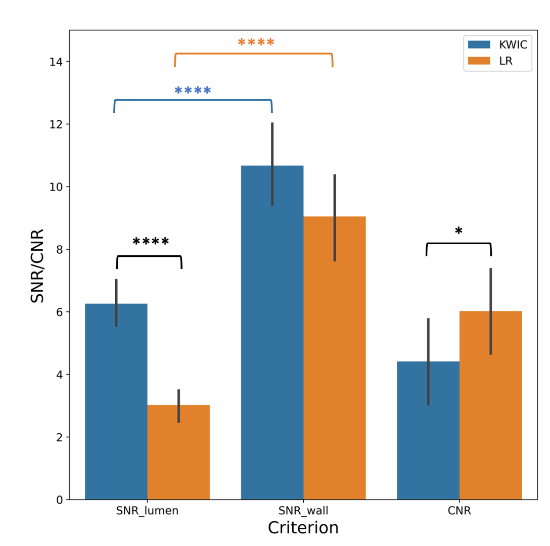
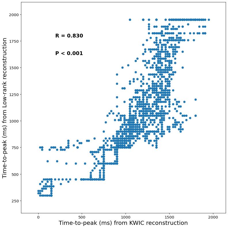
Evaluation: For VW images, quantitative signal-to-noise ratio (SNR) and contrast-to-noise ratio (CNR) were evaluated at the middle slices of ICA, BA, VA, M1, A1, and P1 segments on both sides based on the cross-sectional images. 42 segments from 4 subjects were selected for VW image quality evaluation (2 segments were missing due to occlusion). Lumen signal ($$$S_l$$$) was defined as the mean intensity within an ROI manually drawn including the whole lumen, while wall signal ($$$S_w$$$) was defined as the mean signal intensity within an ROI covering the vessel wall. Noise ($$$\sigma_n$$$) was defined as the standard deviation within an ROI manually drawn in the white matter adjacent to the intracranial segment instead of the background air8. Lumen and wall SNR can be calculated as $$$SNR_l = S_l / \sigma_n$$$ and $$$SNR_w = S_w / \sigma_n$$$, while CNR can be calculated as $$$CNR_w = SNR_w - SNR_l$$$. For dMRA, the TTP value was defined as the time when signal intensity reaches the peak. Linear regression was used to compare the time-to-peak values between the two reconstruction methods along the 3D vascular tree.
Results:
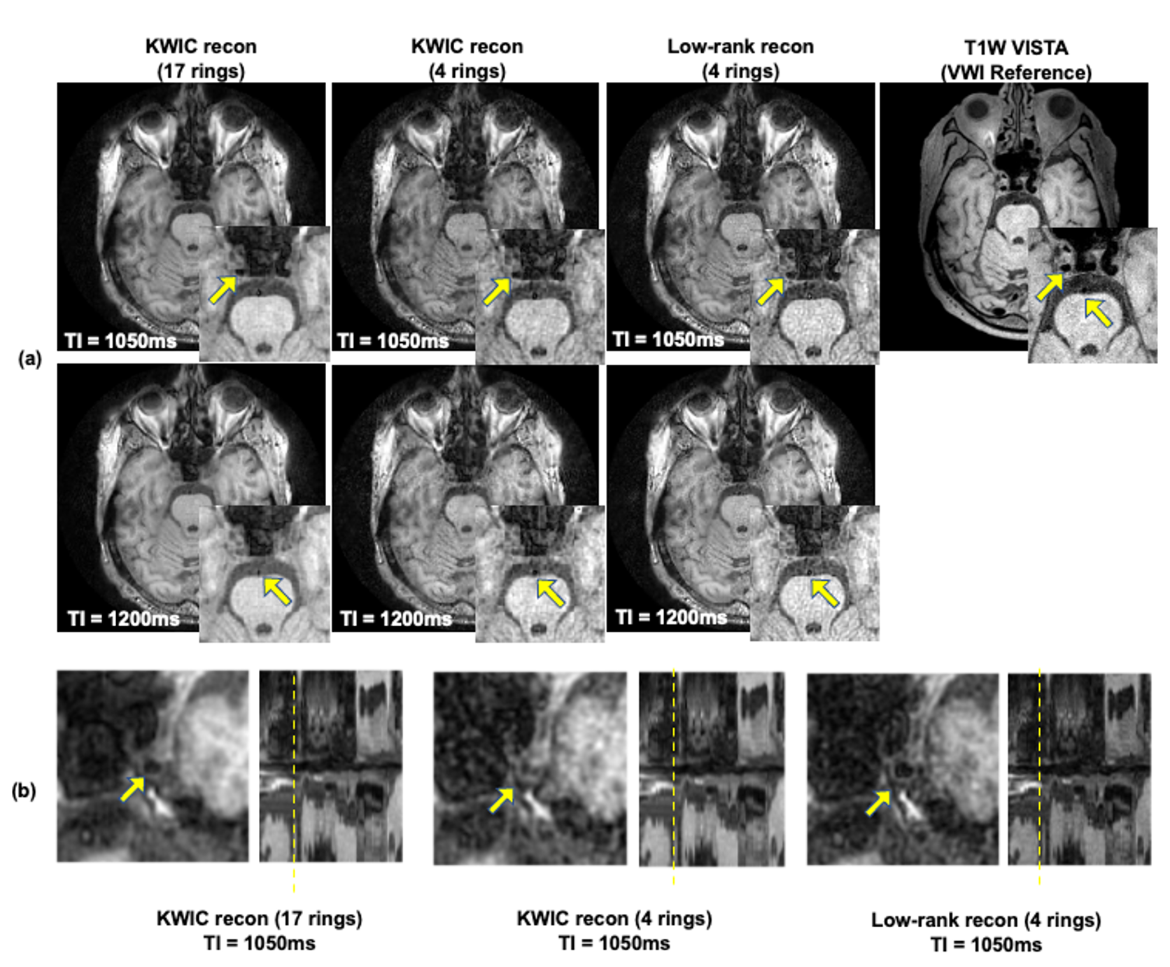
Compared with the original KWIC reconstruction using 17 rings, the proposed low-rank reconstruction improved the image quality by enhancing the sharpness (Figure 2 (a)), so the vessel wall can be better visualized, for example, in cross-sectional view (Figure 2 (b)). In addition, low-rank reconstruction reduced aliasing artifacts compared with KWIC reconstruction using 4 rings. Better avoidance of blood contamination can also be found in our proposed method compared to KWIC, as can be seen in the MPR-view images in Figure 2 (b). The SNR and CNR bar charts for the two reconstruction methods demonstrate that although there is no significant difference between the vessel wall SNRs from KWIC and low-rank reconstruction methods, better suppression of blood signal contamination from adjacent frames improves the CNR between lumen and vessel wall on low-rank reconstruction.The dMRA images reconstructed by KWIC and low-rank appeared similar, though the low-rank method slightly improves the visualization of distal arteries (Figure 3). The 3D TTP maps were also shown in Figure 3. There is a good agreement of TTP values between the two reconstruction methods, as shown in Figure 4.Conclusion:
We proposed a low-rank reconstruction method for 4D multi-contrast MRI (iSNAP) that can improve the image quality of VW images and preserve the hemodynamic information. The improved image quality is beneficial to comprehensively evaluate the cerebral vasculature including stenosis, vessel wall characterization, and blood flow.Acknowledgements
No acknowledgement found.References
1. Chen Z., et al. A novel sequence for simultaneous measurement of whole‐brain static and dynamic MRA, intracranial vessel wall image, and T1‐weighted structural brain MRI, Magn. Res. Med. 85(1), 316-325 (2020).
2. Song, Hee Kwon, and Lawrence Dougherty. "Dynamic MRI with projection reconstruction and KWIC processing for simultaneous high spatial and temporal resolution." Magnetic Resonance in Medicine: An Official Journal of the International Society for Magnetic Resonance in Medicine 52.4 (2004): 815-824.
3. Ma, Xiaodong, et al. "Accelerating Navigator-free Multi-shot Spiral DTI via Joint Calibrationless Reconstruction with Low-Rank Tensor Completion."
4. P. J. Shin, P. E. Larson, M. A. Ohliger, M. Elad, J. M. Pauly, D. B. Vigneron, et al., "Calibrationless parallel imaging reconstruction based on structured low-rank matrix completion," Magn Reson Med, vol. 72, pp. 959-70, Oct 2014
5. Yilong Liu, Jun Cao, Mengye Lyu, and Ed X. Wu, Calibrationless Parallel Imaging Reconstruction Using Hankel Tensor Completion (HTC), ISMRM 2017, #0445
6. Chen, Li, et al. "Deep Open Snake Tracker for Vessel Tracing." International Conference on Medical Image Computing and Computer-Assisted Intervention. Springer, Cham, 2021.
7. Chen, Li, et al. "Development of a quantitative intracranial vascular features extraction tool on 3 D MRA using semiautomated open‐curve active contour vessel tracing." Magnetic resonance in medicine 79.6 (2018): 3229-3238.
8. Zhou, Zechen, et al. "Evaluation of 3D multi-contrast joint intra-and extracranial vessel wall cardiovascular magnetic resonance." Journal of Cardiovascular Magnetic Resonance 17.1 (2015): 1-11.
Figures