1070
Automatic 3D Segmentation of perforating arteries from ultra-high resolution 7T Compressed Sensing MRA images.1MR Collaboration, Siemens Healthineers Ltd., Beijing, China, 2Tiantan Neuroimaging Center of Excellence, Beijing, China, 3Beijing Tiantan Hospital, Capital Medical University, China National Clinical Research Centre for Neurological Diseases, Beijing, China, 4Beijing Tiantan Hospital, Capital Medical University, Neurology Department, Beijing, China
Synopsis
Keywords: Vessels, Segmentation, perforating arteries segmentation
The evaluation of perforating arteries is important for the diagnosis of small vessel disease. It’s challenging to image perforating arteries because of their small caliber size, which requires an extremely high resolution. Moreover, to determine pathological changes, the vessel trees need to be segmented and quantified. In this study, an automatic 3D Segmentation method was introduced and perforating arteries around the Circle of Willis were segmented and quantified. The number of stems of perforators was counted and compared based on segmentation results and MIPs images. The results revealed that the segmentation method robustly achieved the segmentation of perforators.Introduction
Analysis of small vessels is essential for the diagnosis of different cerebral serious diseases. For example, vascular dementia or ischemic stroke can be caused by abnormalities of small cerebral vessels [1]. To determine pathological changes, the vessel trees need to be segmented and quantified. With the introduced 7T MRI, high-resolution 3D TOF images can be acquired non-invasively. These images contain considerably more thin vessels compared to 1.5T or 3T MRA images [2, 3]. Many studies have attempted to segment small cerebral vessels, but most of them have focused on larger perforating arteries (PAs) such as the lenticulostriate artery (LSA) [4][5]. To our knowledge, no studies have attempted to segment other smaller perforating arteries, limited by the resolution achievable by conventional TOF. It was recently demonstrated that the novel compressed sensing (CS) technique was implemented in TOF MRA at 7T, which enables the achievement of ultra-high-resolution imaging (200um iso) while preserving the image quality [6] [7]. This greatly improves the visualization of small intracranial vessels, especially perforating arteries. Our other submitted abstract described the feasibility of visualizing all PAs around the Circle of Willis using a 200-um CS TOF. In this study, we consider the automatic segmentation of PAs in 3D 7T MRA images. An automatic post-processing method was designed and adjusted to produce PAs vascular volume.Methods
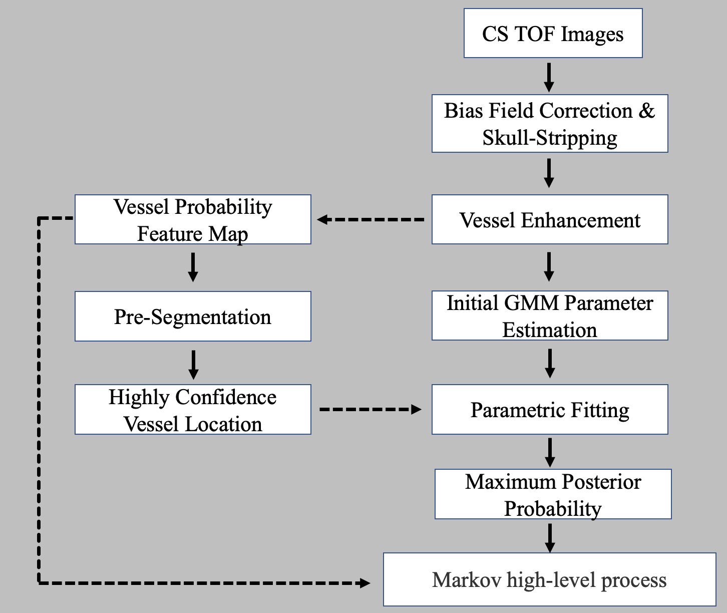
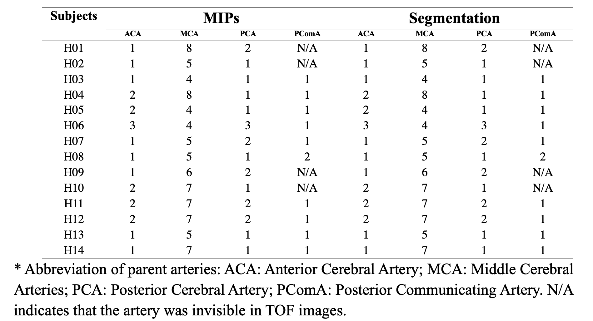
All measurements were performed on a 7T MR system (MAGNETOM Terra, Siemens Healthcare, Erlangen, Germany) using a 32- channel Rx/8Tx head-coil (Nova Medical, Wilmington, Massachusetts, USA). Seven healthy volunteers were included in this initial study. A prototype CS TOF MRA was used to achieve 0.2 mm isotropic LSA imaging. The imaging parameters were consistent with our previous studies [6] [7]. The segmented perforators are divided into several groups according to their parent artery, the anterior cerebral artery (ACA) perforating branches, the middle cerebral arteries (MCA) perforating branches, the posterior cerebral artery (PCA) perforating branches, and the posterior communicating artery (PComA) perforating branches.The segmentation method was based on the adaptive algorithm [8]and modified the finite mixture models into Gaussian distributions mixture models, and a knowledge-based expectation-maximization algorithm is explored to obtain the Gaussian model parameters. To improve the identification of small vessels, and rich in vascular structure, a probability feature map is captured according to the estimated vascular distribution weight in Gaussian mixture models and then is embedded into the Markov high-level process [9]. The algorithms are implemented by using MATLAB 2021b, and the schematic diagram of the segmentation algorithm is shown in Figure 1. The perforators stem number was counted based on these segmentation results and MIPs images. Wilcoxon signed-rank test was used to compare the numbers of stems between the two results.Results
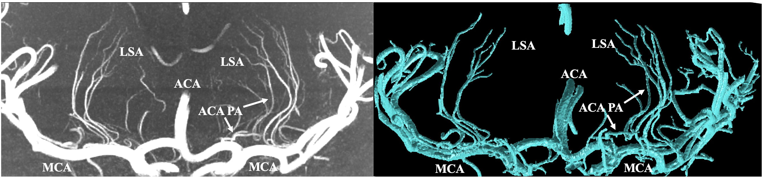
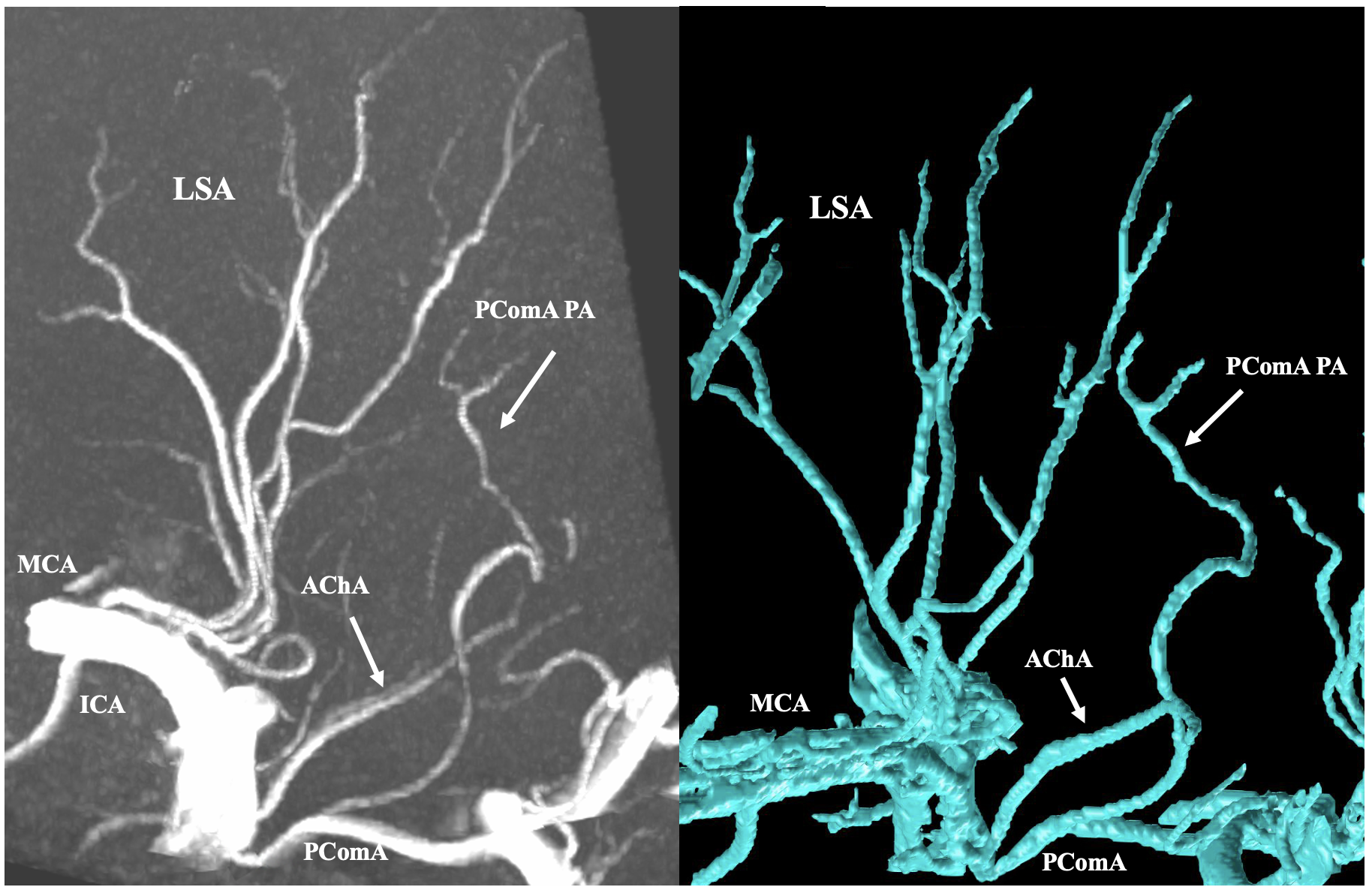
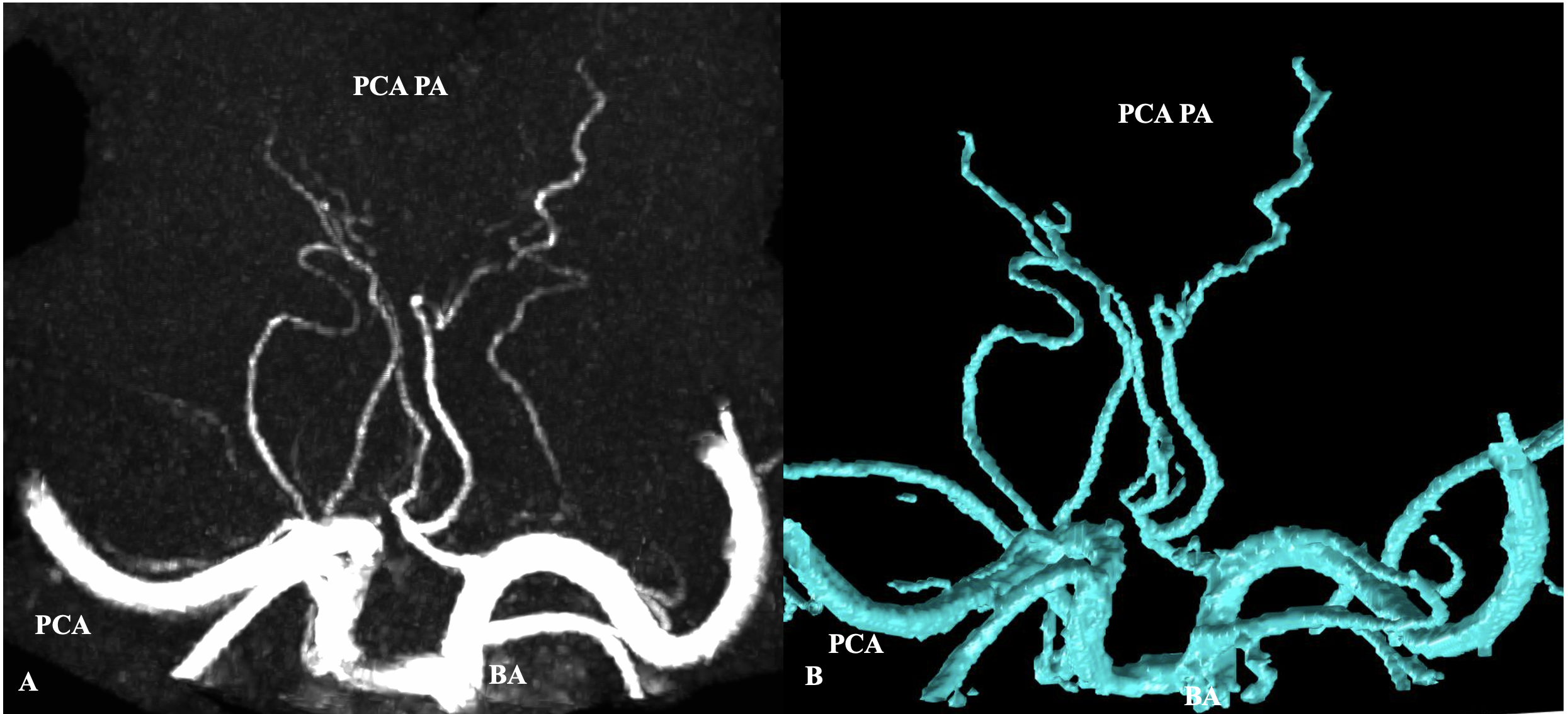
The number of PAs stems were shown in Table 1. The number of perforators based on segmentation results was consistent with that of MIP images. The representative ACA perforator was Heubner’s artery, as shown in Fig. 2. Heubner’s artery most often courses along the A1 segment in a reverse direction, usually in front of the MCA perforators. The MCA perforators, known as the lenticulostriate arteries (Fig. 2), always originate from the M1 segment of the middle cerebral artery, either as singular vessels or by their own common trunks. The PComA, which interconnects the PCA and ICA, most often possesses only one perforating branch (Figs. 3) known as the premamillary artery. The representative perforating artery from PCA is shown in figure 4.Discussion
This is the first time that cerebral PAs segmentation around the circle of Willis has been demonstrated. Previous studies mainly focused on LSAs Segmentation, which was larger PAs and had a relatively faster blood flow [4]. Segmenting other smaller PAs is a challenging task since many PAs are relatively thin and long, and are often located close to each other. In addition, other typical challenges of 7T MRA data include high noise levels, highly curved vessels, low image contrast, and tubular artifacts. This work overcomes this challenge in two ways. Firstly, segmentation was based on ultra-high resolution (200um) CS TOF images, which enable smaller voxel size. A higher spatial resolution reduces the partial volume effect, so the smaller perforator delineation is significantly improved in the high-resolution TOF images. Additionally, CS reconstruction enables better noise reduction and improved contrast between arteries and tissues, which further improves the visibility of small PAs. Second, the segmentation method was based on the MAP-MRF framework and has 3 contributions, i.e., data preprocessing, knowledge-based EM estimation of GMM parameters, and Markov high-level model with novel neighborhood constraint energy function, which make the method more effective, better segmentation accuracy and the sensibility to small-sized vessels.In our future research, more subjects would be enrolled to verify the performance of segmentation. And patients with cerebral vascular diseases should be evaluated. The performance of the proposed method applied to conventional TOF could be also explored.Conclusion
Benefiting from the ultra-high resolution of CS TOF images at 7T, an automatic post-processing method was designed and the perforators around Circle of Willis were successfully segmented. This method may be a promising tool for analyzing cerebral small vessel disease.Acknowledgements
No acknowledgement found.References
1. Román GC, Erkinjuntti T, Wallin A, et al (2002) Subcortical ischaemic vascular dementia. The Lancet Neurology 1:426–436. https://doi.org/10.1016/S1474-4422(02)00190-4
2. Morze C von, Xu D, Purcell DD, et al (2007) Intracranial time-of-flight MR angiography at 7T with comparison to 3T. Journal of Magnetic Resonance Imaging 26:900–904. https://doi.org/10.1002/jmri.21097
3. Cho Z-H, Kang C-K, Han J-Y, et al (2008) Observation of the Lenticulostriate Arteries in the Human Brain In Vivo Using 7.0T MR Angiography. Stroke 39:1604–1606. https://doi.org/10.1161/STROKEAHA.107.508002
4. Vogels V, Dammers R, van Bilsen M, Volovici V (2021) Deep Cerebral Perforators: Anatomical Distribution and Clinical Symptoms: An Overview. Stroke 52:. https://doi.org/10.1161/STROKEAHA.120.034096
5. Liao W, Rohr K, Kang C-K, et al (2016) Automatic 3D Segmentation and Quantification of Lenticulostriate Arteries from High-Resolution 7 Tesla MRA Images. IEEE Trans on Image Process 25:400–413. https://doi.org/10.1109/TIP.2015.2499085
6. Zhe Zhang, Qingle Kong, Jing jing, et al. 0.20 mm isotropic intracranial perforating arteries imaging using compressed sensing TOF-MRA at 7T. In Proceedings of the 31st Annual Meeting of ISMRM, London, England, UK, 2022. Abstract 4142
7. Zhe Zhang, Qingle Kong, Yingkui Zhang, et al. Improved Characterization of Lenticulostriate Arteries using Compressed Sensing Time-of-Flight at 7T. European radiology. Under Review.
8. Wilson DL, Noble JA (1999) An adaptive segmentation algorithm for time-of-flight MRA data. IEEE Trans Med Imaging 18:938–945. https://doi.org/10.1109/42.811277
9. Hassouna MS, Farag AA, Hushek S, Moriarty T (2006) Cerebrovascular segmentation from TOF using stochastic models. Medical Image Analysis 10:2–18. https://doi.org/10.1016/j.media.2004.11.009
Figures