1065
Inflatable RF Coil with Liquid Metal for MR Imaging1Arizona State University, Tempe, AZ, United States, 2Yonsei University, Gangwon-do, Korea, Republic of
Synopsis
Keywords: Non-Array RF Coils, Antennas & Waveguides, New Devices, Liquid metal RF coil
In this work, we present a novel inflatable radio-frequency receive coil for Magnetic Resonance Imaging at 7T. A small sample was imaged with and without inflation, and the overall signal-to-noise ratio of the sample was improved by 12.7% in dB with inflation. This coil design will offer many opportunities in the field of endorectal imaging, imaging for irregular sample shapes, and reducing motion artifacts. Inflation also changes the tuning and matching conditions, which compensate for loading effects by precisely adjusting the air volume inside the cavity.Introduction
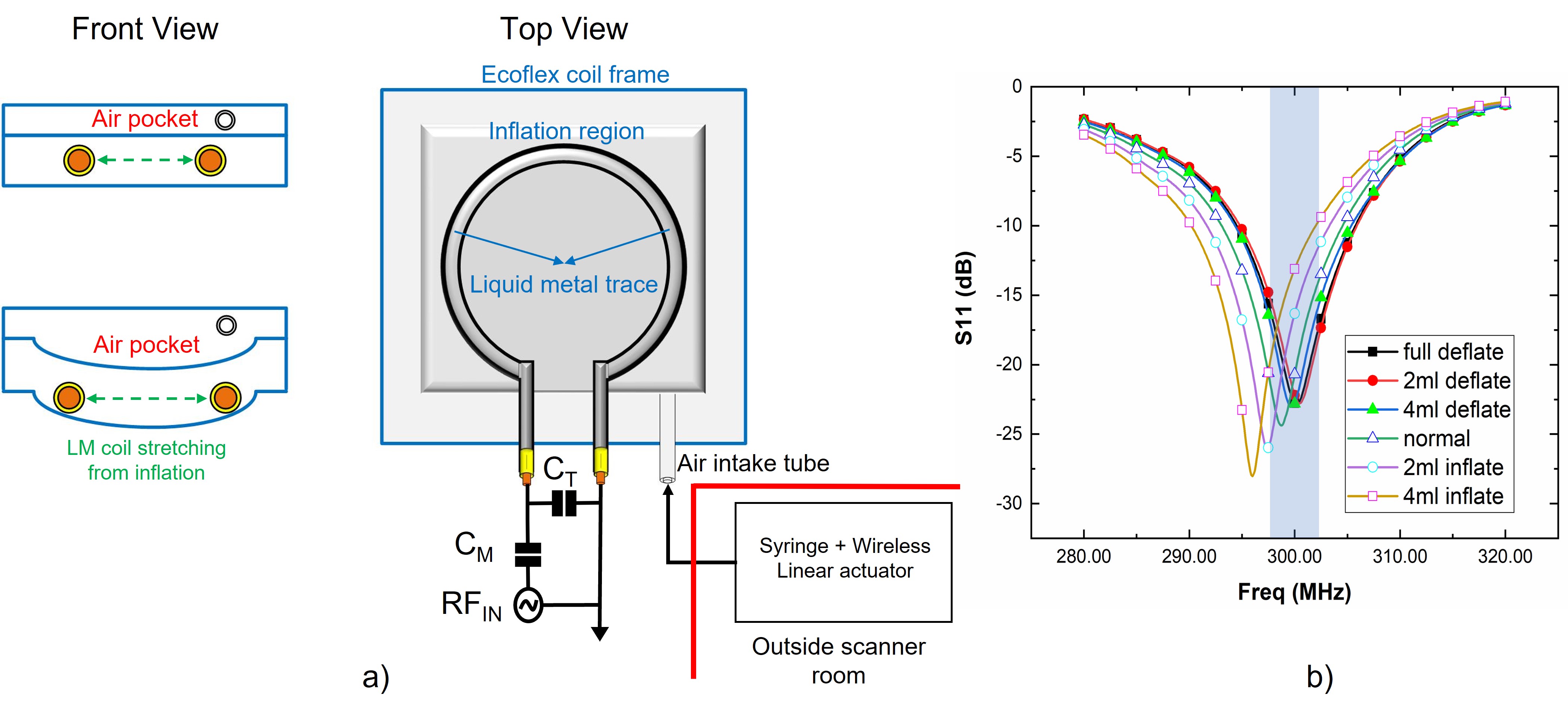
Liquid metal antennas have been actively developed for stretchable and flexible sensors with discrete or continuous tuning control.1 Flexible and stretchable liquid metal (LM) based radio frequency (RF) coils are shown to improve the signal-to-noise ratio (SNR) of the image at high field strengths.2-4 In this work, we introduce an inflatable RF coil with the ability to physically reconfigure and move closer to sample by pushing air into a small cavity. This allows the user to place the RF coil close to the sample structure which improves peripheral SNR and reduces the motion artifact by restricting movement. The induced magnetic field (B1) in a loop coil is defined as B1=(µ0 IR2)/〖2(R2+d2 )〗(3⁄2) by the Biot-Savart Law, where, R is the radius of the coil, I is the magnitude of current flow and d is the distance between a coil and a subject. As the B1 field intensity is proportional to and , to the distance and radius of the coil respectively, an inflatable coil can have superior performance in MR signal induction and reception in comparison with all other coil types including flexible and conformal RF coils as the coil is moves much closer to the region-of-interest by inflation.Method
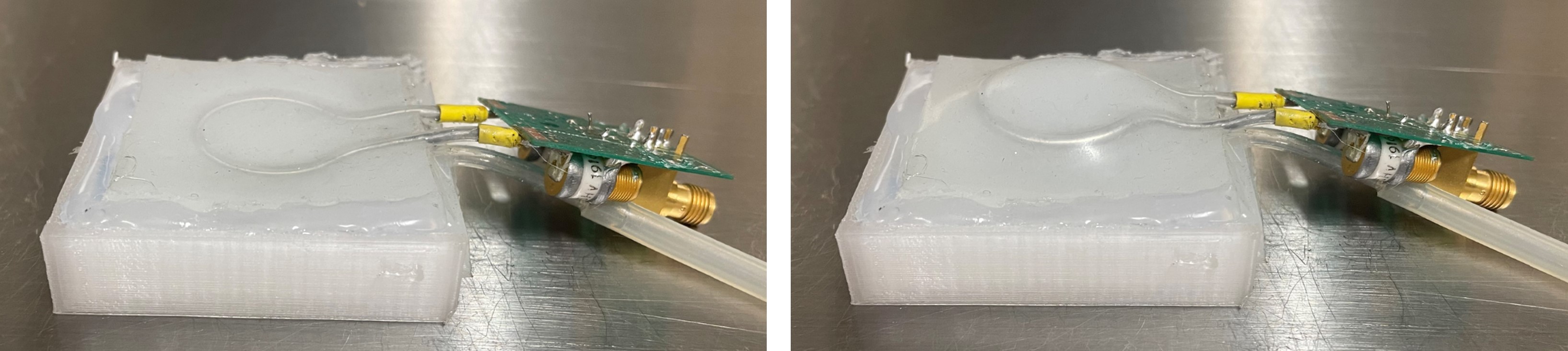
Inflatable RF coil was designed in two steps i) Elastic silicone polymer (Ecoflex Series, Smooth-On, USA) is used to facilitate a flexible and stretchable material for inflation and ii) LM loop coil element was designed to bend and stretch along with the inflating base structure. A 3D printed mold (30 mm x 30 mm x 2 mm) was designed to make elastic substrate layers. Two layers are stacked to create an air cavity with an intake tube and a third layer containing the LM coil is placed on top of the air cavity to move the RF coil as shown in Fig.1a. An L-matching network with two variable trimmer capacitors is connected to the coil element for a 50 Ω match. This stack is placed in a 3D print coil frame to provide one-directional inflation i.e., towards the sample as shown in Fig.1b. An air intake tube is located between the bottom two layers which is connected to a syringe. This syringe is connected to a wirelessly controlled linear actuator to create a push-pull motion to inflate and deflate the cavity. The coil is placed 3 mm away from the sample and at full inflation can raise up to 4.5 mm from the base. Elastomers such as Ecoflex provide high elasticity, therefore enabling 100% shape retention of the LM coil after inflation.Results and Discussion
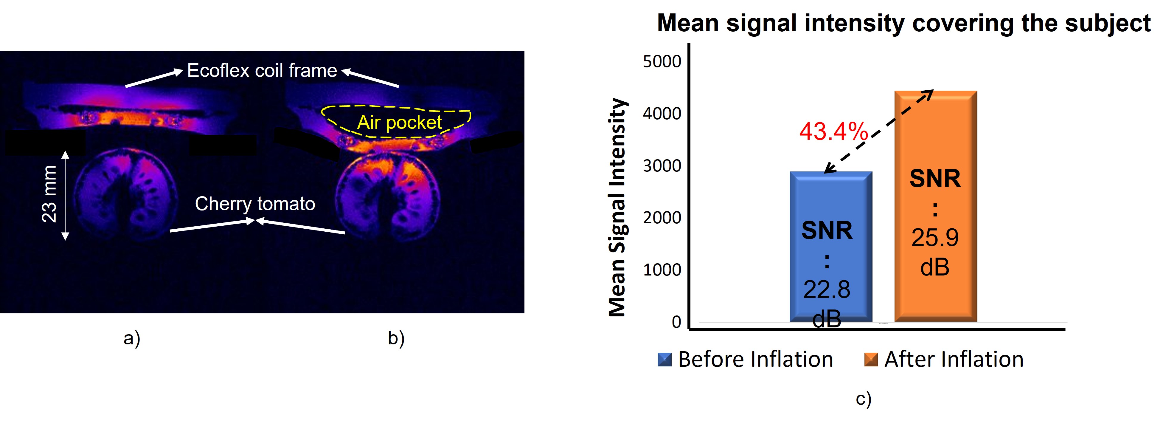
A two-port VNA (FieldFox N9923A, Keysight Technologies, USA) was used to gather the S-parameters of the inflatable coil with a saline phantom for various inflation ranges. Fig.2a shows the bench test setup along with front and top views of the inflatable LM coil frame. Fig.2b shows the impact of inflation on the coil resonance based on the volume of air inside the coil cavity. The impedance mismatch caused by inflating the coil and increasing its dimension was compensated with two variable capacitors located on the L-matching network. All the MR imaging studies were conducted at Barrow Neurological Institute - Arizona State University (BNI-ASU), Center for Preclinical Imaging, using a 7T small-animal, 30-cm horizontal-bore magnet and BioSpec Avance III spectrometer (Bruker, Billerica, MA) with a 116-mm high-power gradient set (600 mT/m). Fast-Low-Angle-Shot (FLASH) Sequence with a repetition time (TR) of 350 ms, echo time (TE) of 5.4 ms, and a flip angle (α) of 20 degrees was used for MR imaging experiments. A field of view (FOV) of 40 x 40 mm and 256 x 256 matrix leading to an in-plane resolution of 156 x 156 μm, in addition 5 slices were acquired along the sample with a slice thickness of 1 mm. A Bruker Linear Birdcage coil was used as the transmit coil. The peak power used for the FLASH sequence was 700 W. A rat bed of 72 mm diameter was used to hold the receive coil housing. Cherry tomato was then placed inside the receive coil housing and fed into the scanner. Fig.3a and 3b show the MR images before and after inflation respectively. All the parameters of the imaging sequence were kept same including the reciever gain to validate the performace of the inflateble coil. Same slices (5/5) were of the MR images were selected to calculate the SNR and mean signal intensity as shown in Fig.3c. 12.7% (from 22.8 dB to 25.9 dB) in SNR and 43.4% (from xxx to xxx) in signal intensity are observed with inflation. This novel design will offer opportunities in many imaging fields and further studies are being carried out to carefully analyze the benefits and drawback of the coil in multiple scenarios.Conclusion
In this work, we have designed, fabricated, and tested a novel inflatable RF coil with liquid metal and elastic silicone polymer. By inflating, the coil closer to the sample, we have demonstrated the novel design method for physically reconfigurable RF coils at a preclinical 7T.Acknowledgements
This work was supported by the National Institute of Biomedical Imaging And Bioengineering of the National Institutes of Health under Award Number R00EB020058.References
1. Motovilova, Elizaveta, and Shao Ying Huang. 2020. "A Review on Reconfigurable Liquid Dielectric Antennas" Materials 13, no. 8: 1863. https://doi.org/10.3390/ma13081863
2. Motovilova, E., Tan, E.T., Taracila, V. et al. Stretchable self-tuning MRI receive coils based on liquid metal technology (LiquiTune). Sci Rep 11, 16228 (2021). https://doi.org/10.1038/s41598-021-95335-6
3. Martin JF, Hajek P, Baker L, Gylys-Morin V, Fitzmorris-Glass R, Mattrey RR. Inflatable surface coil for MR imaging of the prostate. Radiology. 1988 Apr;167(1):268-70. https://doi.org/10.1148/radiology.167.1.3347731 PMID: 3347731
4. Noworolski, S.M., Crane, J.C., Vigneron, D.B. and Kurhanewicz, J. (2008), A clinical comparison of rigid and inflatable endorectal-coil probes for MRI and 3D MR spectroscopic imaging (MRSI) of the prostate. J. Magn. Reson. Imaging, 27: 1077-1082. https://doi.org/10.1002/jmri.21331
Figures