1055
The FACE: Flexible Array for Cervical & Extraspinal 3T MR Imaging1Hospital for Special Surgery, New York, NY, United States, 2Tesla Dynamic Coils, Zaltbommel, Netherlands, 3GE HealthCare, Aurora, OH, United States
Synopsis
Keywords: RF Arrays & Systems, RF Arrays & Systems
Conventional cervical coils lack the flexibility to closely conform to the inherently curved neck region, particularly at its head/shoulder junctions. High SNR imaging of the c-spine and extraspinal soft tissues (including small peripheral nerves) relies on close proximity of receive elements to these targeted structures. This study evaluates the performance of a novel, conformal, 23-channel Flexible Array for Cervical & Extraspinal (FACE) 3T MR Imaging with increased flexibility to enhance image quality and SNR compared to conventional coils. SNR measurements on phantoms and high resolution 2D and 3D in vivo c-spine and peripheral nerve neck imaging were performed at 3T.INTRODUCTION
Imaging of the cervical spine (c-spine) and neck region, including extra-spinal nerves, requires high signal-to-noise ratio (SNR) to acquire high spatial resolution. SNR and parallel imaging factors are increased by close proximity of receiver radiofrequency (RF) coil elements to the target structure and increasing the number of receive elements, respectively1,2. However, designing a cervical coil with coverage of the anterolateral neck region and its junction with both the shoulder and head is challenging due to the inherent concavities of this anatomical region and range of body habiti across the general population3,4. Conventional c-spine coils utilize rigid designs that lack flexibility to conform to variable neck shapes/sizes. Newer, flexible phased-array coil designs are now commercially available but provide suboptimal coverage and SNR for the neck, as they are not specifically designed for the cervical region.We designed and implemented a novel, 23-channel, Flexible Array for Cervical & Extraspinal (FACE) coil for imaging at 3-Tesla (3T), with a default shape conformal to the average adult face, neck and upper shoulder regions with flexibility in three dimensions to accommodate varying body habiti. In this preliminary evaluation in phantoms and human volunteers, we hypothesized that the performance of the FACE coil would be superior to commercially available, 21-channel head-and-neck unit (HNU), and 16-channel surface coils (16CH).
METHODS
An initial comparison of body habiti and c-spine/neck imaging requirements determined that 20-24 elements arranged in 3 linear rows with diameters between 5-8 cm were needed to achieve a biconical, conformal 2D array-shape (Fig. 1A). Following computer aided design of the coil layout (Fig. 1B) for targeted coil coverage from the orbital level to the clavicles, a 23-channel receive array was implemented (Tesla Dynamic Coils, Zaltbommel, The Netherlands), consisting of high impedance coil elements5 embedded in a flexible mask and a rigid posterior head-rest housing (Fig. 1C).The posterior housing for the 23-channel FACE array was designed to accommodate average body dimensions6 (neck circumferences between 35.5-cm and 48.8cm). The circumferential shape was designed with software (Rhinoceros®, Robert McNeel & Associates) based on non-uniform rational B-splines model for the representation of curves and freeform surfaces. The housing was 3D-printed with thermoplastic polyurethane material. The interfacing circuitry, pre-amps and cable traps were placed within the posterior housing, including the connector for interfacing to the system and coil ID detection for evaluation at 3-Tesla with resonance at 127.7 ± 0.5Mhz.
Safety testing and imaging were performed at 3T (Premier, GE HealthCare). SNR was measured in a 11-cm diameter, cylindrical silicon oil phantom at isocenter and 4.2 cm off-isocenter, and compared to a 21-channel HNU including the anterior piece (GE HealthCare) using a 3D gradient echo sequence. Five volunteers were recruited to perform comparisons of the FACE coil under an IRB-approved protocol for evaluating prototype RF coils. Imaging was repeated with identical 2D and 3D sequence parameters, optimized for the c-spine and peripheral nerves, using the FACE coil, the HNU, and a 16CH coil (Variety Multipurpose Coil, Noras MRI Coils).
RESULTS
The FACE coil provided higher SNR than the HNU throughout the silicon oil phantom (Fig. 2A-B), except directly anteriorly. Mean SNR with the FACE coil was 4% higher than the HNU in the isocenter of the phantom and 141-164% higher at left/right/posterior positions off-isocenter (Fig. 2C).In vivo imaging with the FACE coil provided comparable image quality in a routine, sagittal T2-weighted c-spine exam and higher SNR posteriorly, but slightly lower signal in the direct anterior neck region compared to the HNU (Fig. 3A-B). High-resolution (0.6x0.6x0.8mm), 3D MR neurography with the FACE coil provided overall higher image quality, particularly in the peripheral neck regions, showing sharper delineation of the extraspinal nerve roots and the spinal accessory nerve in comparison to the HNU (Fig. 4A-F). 2D MR neurography with the FACE coil demonstrated superior circumferential signal around the neck and superior conspicuity of the right facial nerve as compared against the 16CH positioned unilaterally (Fig. 5A-D). Volunteers reported no discomfort, including sensation of ‘overheating’, with any of the coils.
DISCUSSION
The FACE coil accommodated the biconical shape of the head/neck and upper shoulder region and provided extended flexibility compared to standard coils. Preliminary, anecdotal evaluation with this FACE coil showed comparable image quality in the c-spine, but superior image quality for neck MR neurography compared to the HNU. SNR of the bilateral, peripheral neck regions was increased when compared to a set of flexible surface coils. Nerve conspicuity was markedly increased in 2D and 3D MR neurography. The reasons for SNR improvements in the FACE coil are attributed to the increased number of smaller coil elements at closer proximity to the targeted anatomy, and possibly as well from improved parallel imaging performance.Future work will include analysis of differences in a large patient cohort between the FACE coil, the HNU and 16CH coils. Compared to the HNU, the lack of an anterior piece covering the face might improve patient tolerance with the FACE coil, but additional elements may be needed over the anterior/lower neck region to enhance SNR in this region.
CONCLUSION
Early, preliminary evaluation of a conformal 23-channel c-spine/neck array demonstrate comparable or superior image quality for c-spine and neck imaging.Acknowledgements
The authors thank Yan Wen and Julie Poujol from GE Healthcare for technical support.References
1. May MW, Hansen SJD, Mahmutovic M, et al. A patient‐friendly 16‐channel transmit/64‐channel receive coil array for combined head–neck MRI at 7 Tesla. Magnet Reson Med. 2022;88(3):1419-1433. doi:10.1002/mrm.29288
2. Hayes CE, Tsuruda JS, Mathis CM, Maravilla KR, Kliot M, Filler AG. Brachial plexus: MR imaging with a dedicated phased array of surface coils. Radiology. 1997;203(1):286-289. doi:10.1148/radiology.203.1.9122409
3. Davidson EJ, Tan ET, Pedrick EG, Sneag DB. Brachial Plexus Magnetic Resonance Neurography: Technical Challenges and Solutions. Invest Radiol. 2022;Publish Ahead of Print. doi:10.1097/rli.0000000000000906
4. Beck MJ, Parker DL, Bolster BD, et al. Interchangeable neck shape–specific coils for a clinically realizable anterior neck phased array system. Magnet Reson Med. 2017;78(6):2460-2468. doi:10.1002/mrm.26632
5. Zhang B, Sodickson DK, Cloos MA. A high-impedance detector-array glove for magnetic resonance imaging of the hand. Nat Biomed Eng. 2018;2(8):570-577. doi:10.1038/s41551-018-0233-y
6. Molenbroek, J.F.M. (Johan) (2018): DINED - anthropometric database. 4TU.ResearchData. Collection. https://doi.org/10.4121/uuid:199467d8-5c40-4a1f-a2f2-f2040db26270
Figures

Fig 1. The initial design (A) included a biconical 2D array-shape with 3 linear rows (in the superior-inferior (z) direction) of varying coil diameters. The neck array design (B) with 23 coil elements (red) overlapped neighboring elements for coil decoupling, and had matching boards attached to each element. The final prototype FACE coil (C) consisted of the flexible mask coil and a posterior head-rest that housed preamplifiers and interfacing components.
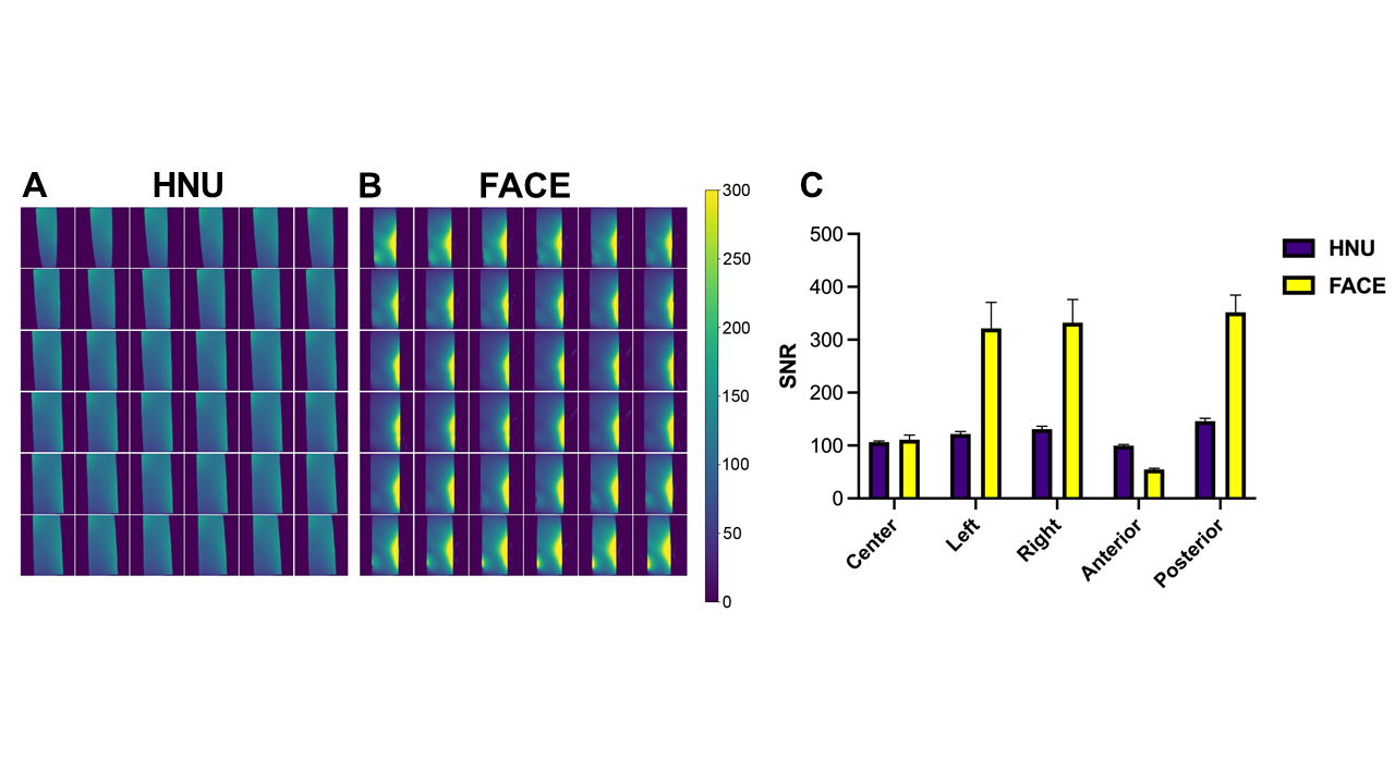
Fig 2. Sagittal images of silicon oil phantom SNR maps using the 21-channel head/neck unit (HNU, A) compared to the prototype conformal 23-channel c-spine/neck coil (FACE, B). Quantitative SNR comparisons (mean ± standard deviation, C) in the phantom isocenter and 4.2 cm left, right, anterior, and posterior off-isocenter.
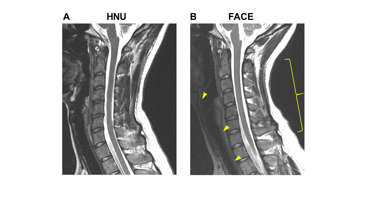
Fig 3. Sagittal 2D, T2-weighted fast spin echo, 22cm FOV, 3mm-thick c-spine obtained with the FACE coil (B) demonstrated similar image quality of the c-spine with improved SNR posteriorly (bracket, B) and less signal of the anterior neck region (arrowheads, B) compared to the HNU with anterior piece (A).
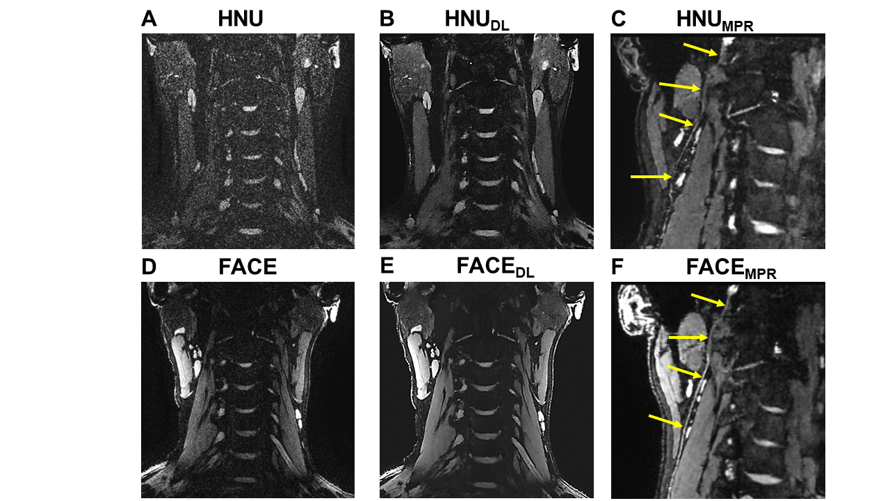
Fig 4. Coronal 3D double echo steady state (DESS) of the neck (TR/TE=15/5-10ms, voxel size=0.6x0.6x0.8mm) without (A,D) and with 3D deep learning (DL) reconstruction (B,E). The prototype FACE coil (D,E) provides overall higher SNR, especially of the peripheral neck region, in comparison to the HNU with anterior piece (A,B). Curved multiplanar reconstruction (MPR) of the same 3D DESS images (C,F) demonstrate sharper delineation of the right spinal accessory nerve (arrows) with FACE (F) vs. HNU (C).
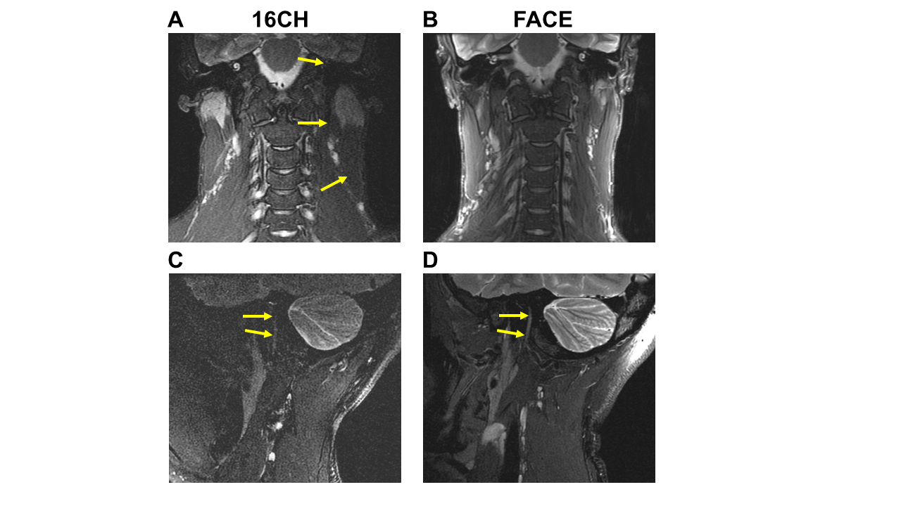
Fig 5. Coronal 2D, T2-weighted Dixon water image with 20cm FOV, 3mm-thick (A,B) demonstrates improved SNR of the central and bilateral peripheral neck region with the FACE coil (B) compared to 16CH (A) positioned on the right side; note deficient SNR on the left side for the 16CH unit where coil elements are lacking (arrows, A). Oblique-sagittal fat-suppressed T2-weighted, 16cm FOV, 1mm-thick (C,D) reveal superior SNR and conspicuity of the right facial nerve (arrows) with the FACE coil (D) compared to the 16CH (C).