1054
First in vivo results with a modular system of flexible coil arrays for 3 T MRI (ModFlex)1High Field MR Center, Center for Medical Physics and Biomedical Engineering, Medical University of Vienna, Vienna, Austria
Synopsis
Keywords: RF Arrays & Systems, RF Arrays & Systems
Form-fitting radiofrequency coil arrays have the potential to enhance the signal-to-noise ratio, enable faster imaging and improve patient comfort in MRI. We developed a flexible modular coil array system for 3T MRI (ModFlex) with 16 receive channels. In neck, spine, ankle and hip imaging, we measured an SNR gain for 4 out of 6 anatomical target regions and similar SNR for 2 out of 6 as compared to commercial reference coils. The coil’s versatility is beneficial for different use cases with varying subject sizes.Introduction
In 3T MRI, receive-only radiofrequency (RF) coil arrays optimized for a dedicated application are often used in combination with the whole-body coil for RF transmission. Form-fitting coil design maximizes the electromagnetic coupling to the sample, and thus, the achievable signal-to-noise ratio (SNR). In addition to conformal design, the overall array geometry has to be adapted to cover the desired field-of-view (FOV). Furthermore, the ideal coil element size for SNR-optimal operation has to be chosen with respect to noise contributions depending on the target penetration depth1. The aim of this work was to propose a versatile coil array that can be tailored to the application and has the potential to enhance SNR, to enable faster imaging and to improve comfort as compared to bulky rigid coils: a modular system of lightweight flexible 4-channel coaxial RF coil arrays2 (“ModFlex”). Here, we present a 16-channel setup together with first in vivo MR measurements for different use cases, and benchmark the multi-purpose ModFlex system against dedicated commercial coils.Methods
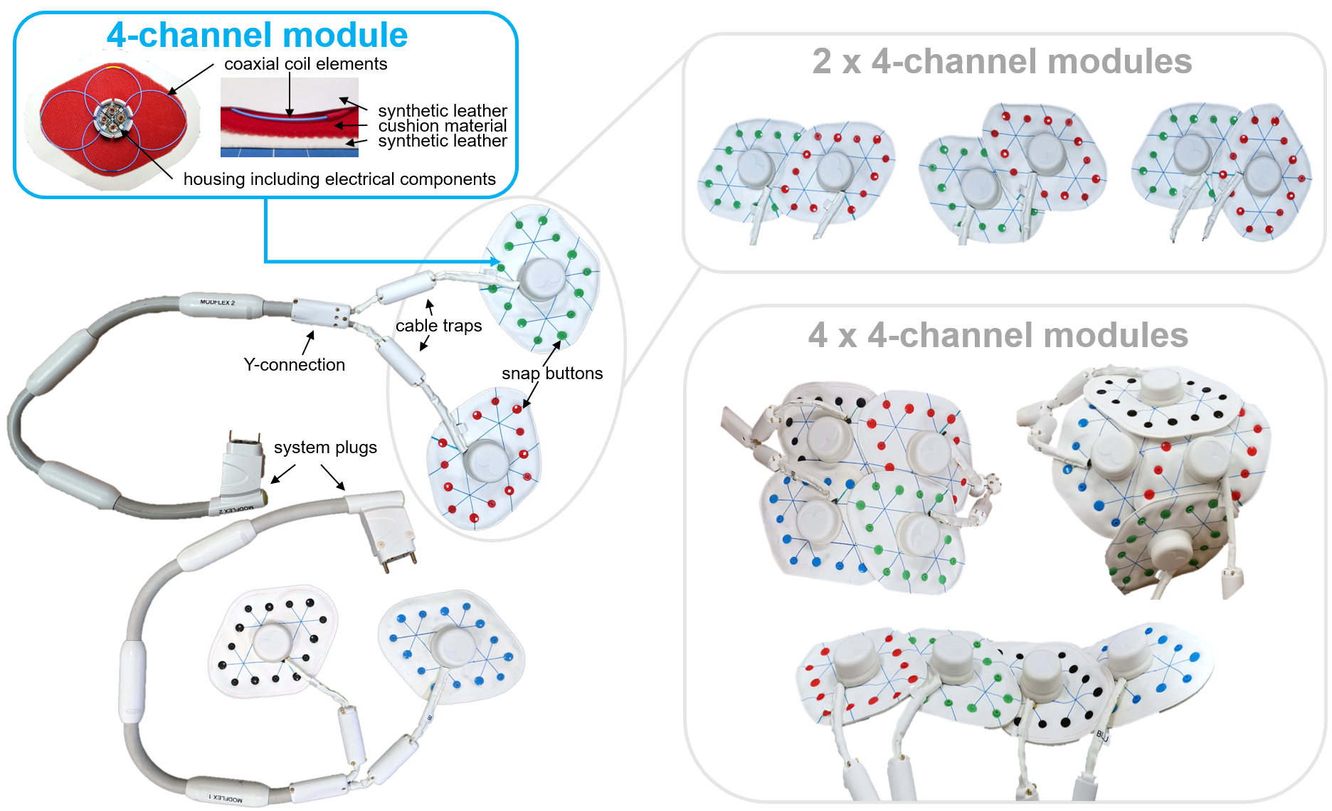
Coil featuresThe 8-channel ModFlex shown in Fig.1 consists of two flexible coil modules with four single-gap coaxial coils3,4 (8cm diameter) each. Each module is equipped with snap buttons that allow a stable mechanical connection between modules and ensure correct coil overlap to minimize inter-element coupling. Technical details, bench measurements and phantom MR data were presented in previous work5. Possible use cases with 1-2 ModFlex coils (4-16 receive channels) include e.g., the neck, breast, spine, abdomen, hip or extremities. Each 4-element building block can be individually turned on or off via the scanner’s console.
In vivo MR measurements
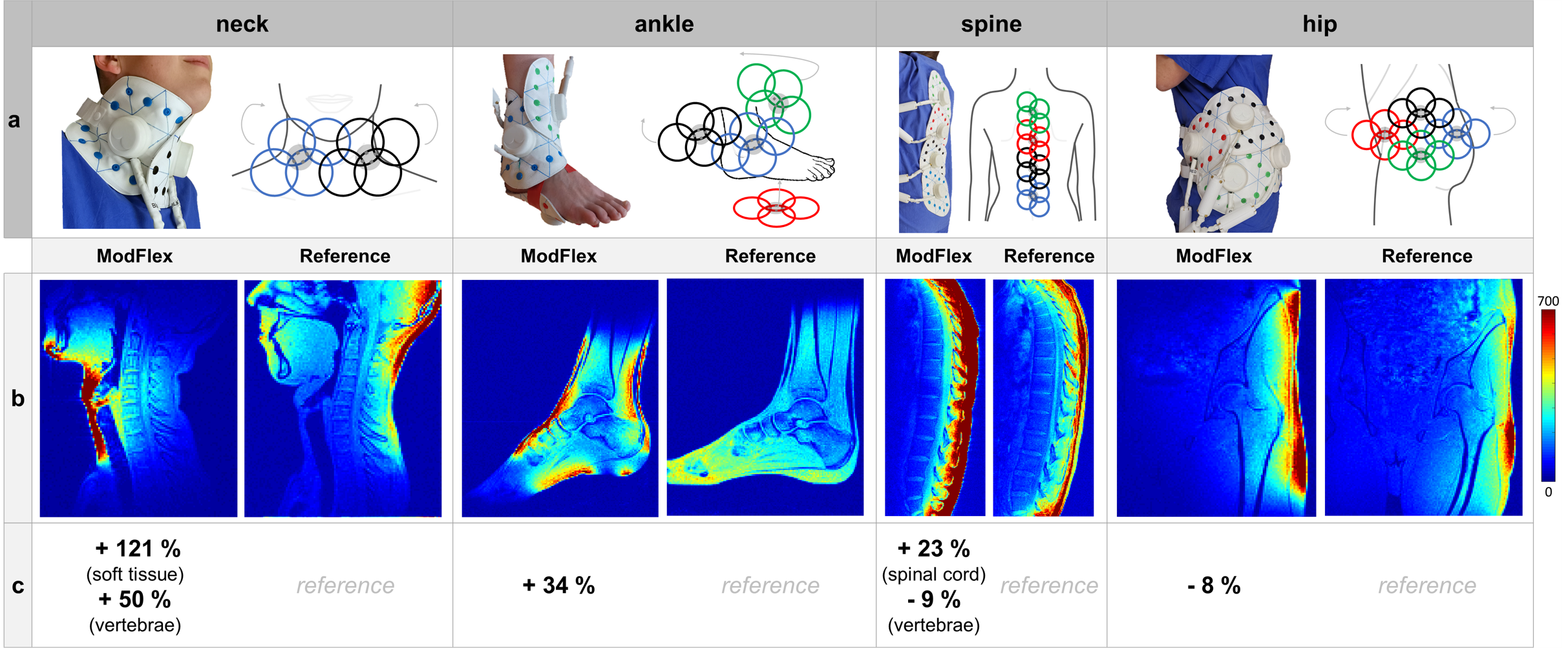
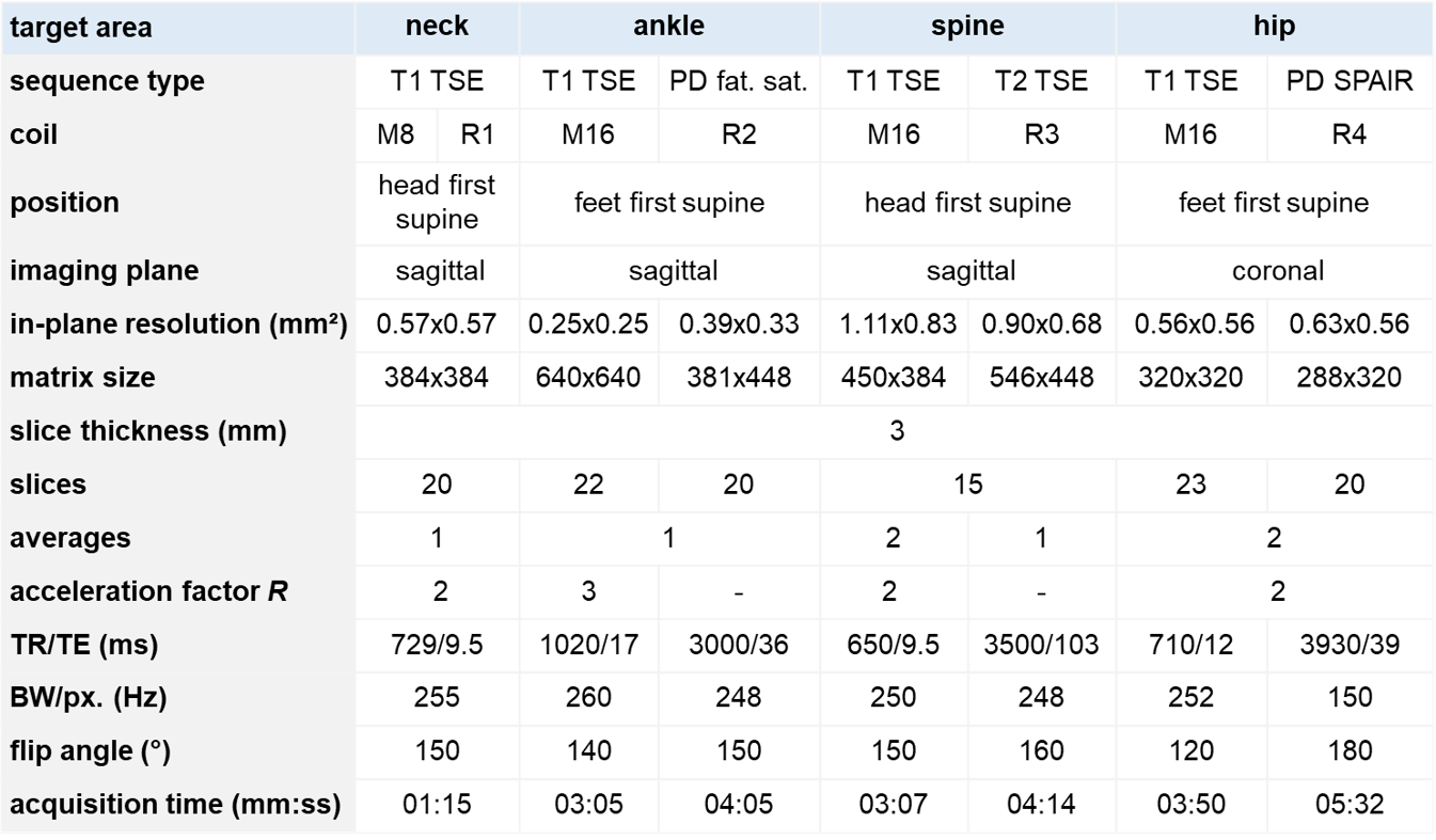
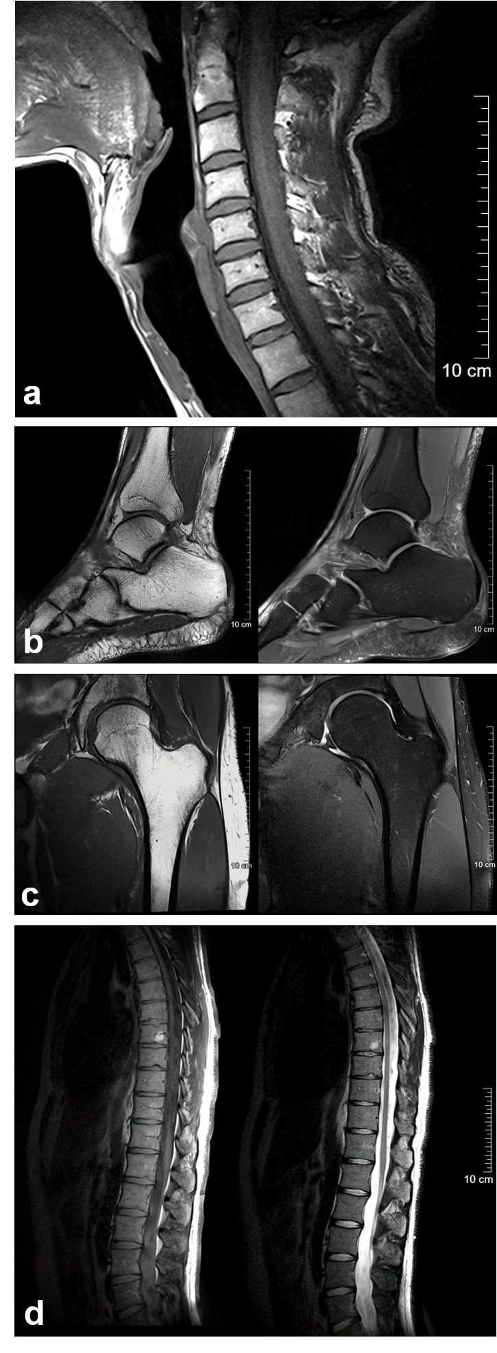
The study was approved by the Ethical Committee of the Medical University of Vienna (nr. 2137/2021) and informed written consent was obtained from all four volunteers.In vivo MR experiments were performed on a 3T MR scanner (Prisma Fit, Siemens Healthineers, Erlangen, Germany). The ModFlex coil performance was evaluated compared to dedicated reference coils for four different anatomical areas (one per volunteer):
- neck: 8-channel ModFlex vs. rigid head/neck coil (38 out of 64 coil channels activated),
- ankle: 16-channel ModFlex vs. rigid 16-channel foot/ankle coil,
- spine: 16-channel ModFlex vs. rigid 32-channel spine coil,
- hip: 16-channel ModFlex vs. semi-flexible multi-purpose 18-channel coil placed around the left hip.
Results
The SNR maps in Fig.3 and the calculated relative SNR differences demonstrate that the mean SNR in the target ROIs with ModFlex is comparable to or higher than with the respective reference coils:- neck: +120 % SNR in soft tissue from tongue to collarbone, +50 % SNR in the cervical spine
- ankle: +34 % SNR between lower tibia and metatarsal bones
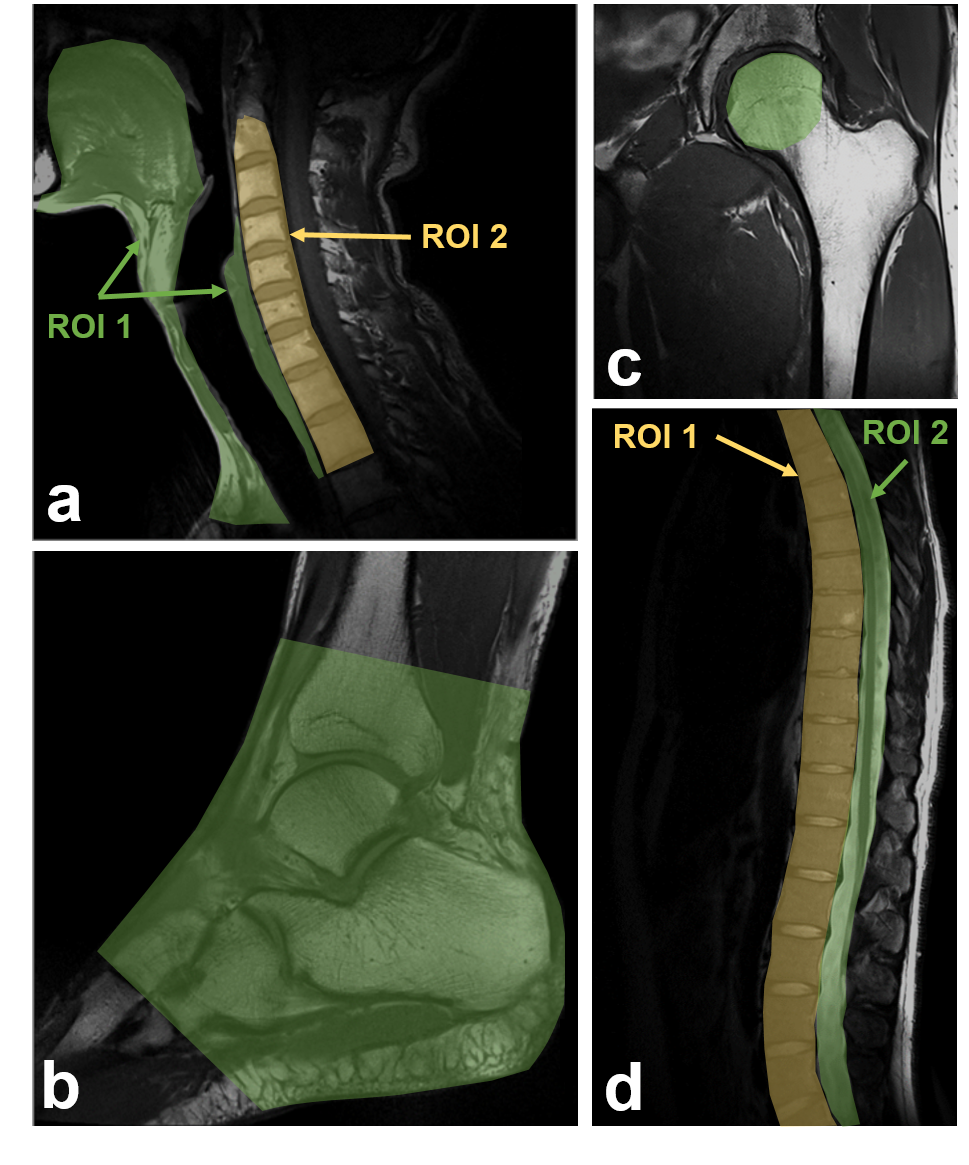
- spine: -9 % SNR in the spine (Th3-S1), +23 % SNR in the spinal cord (along Th3-S1)
- hip: -8 % SNR in the the femoral head
Discussion
High local SNR close to the coil conductors, typical for surface coil arrays, can be observed in Fig.3 and Fig.4. We show that this is especially beneficial e.g. for ankle, spinal cord and neck imaging. For hip and spine imaging, it can be demonstrated that the 8cm coil elements achieve less SNR in larger penetration depth than the reference coils (with larger elements). This could possibly be compensated by combining more than one 16-channel ModFlex wrapped around the area of interest or by fabricating a similar flexible coil with larger individual elements. Nevertheless, the ModFlex coil’s SNR in these ROIs stays in a range comparable to commercial coils and is deemed to be sufficient for a large range of subjects. The coil robustness in terms of mechanical stability and electrical performance for different use cases was not compromised by the modularity of the coil design approach.Conclusion
We developed a light-weight and flexible modular coil array concept (“ModFlex”) with a broad range of possible applications. The SNR gain compared to reference coils and its ability of form-fitting make ModFlex a versatile tool for different use cases and subject sizes. In clinical practice, ModFlex could help make the coil setup more convenient and improve patient comfort. Current work is focused on parallel imaging evaluation, hardware optimization, and the inclusion of on-coil motion sensors7,8. In related work, the coil concept was exploited for the design of a wearable breast MR coil (“BraCoil”, companion abstract).Acknowledgements
This work was funded by the joint Austrian/French grant “BRACOIL“ (Austrian Science Fund FWF Nr. I-3618/Agence Nationale de Recherche ANR-17-CE19-0022).References
1. Kumar, A., Edelstein, W. A. & Bottomley, P. A. Noise figure limits for circular loop MR coils. Magn. Reson. Med. 61, 1201–1209 (2009).
2. Laistler, E., Obermann, M., Nohava, L. & Roat, S. Coil module for magnetic resonance imaging applications. Patent. Application number EP21020242.0 - 1126 (2021).
3. Zhang, B., Sodickson, D. K. & Cloos, M. A. A high-impedance detector-array glove for magnetic resonance imaging of the hand. Nat Biomed Eng 2, 570–577 (2018).
4. Nohava, L. et al. Flexible Multi-Turn Multi-Gap Coaxial RF Coils: Design Concept and Implementation for Magnetic Resonance Imaging at 3 and 7 Tesla. IEEE Trans. Med. Imaging 40, 1267–1278 (2021).
5. Nohava, L. et al. ModFlex – a modular system of flexible receive-only coil arrays for 3 T MRI. in Proc. Intl. Soc. Mag. Reson. Med. 30 (2022) 191.
6. Robson, P. M. et al. Comprehensive quantification of signal-to-noise ratio and g-factor for image-based and k-space-based parallel imaging reconstructions. Magnetic Resonance in Medicine 60, 895–907 (2008).
7. Chen, B. et al. Design and Validation of a Novel MR-Compatible Sensor for Respiratory Motion Modeling and Correction. IEEE Trans. Biomed. Eng. 64, 123–133 (2017).
8. Pancoast, L., Brantner, D., Wiggins, R., Walczyk, J. & Brown, R. Wireless Body Sensor Data Acquisition Platform for Motion Tracking. in Proc. Intl. Soc. Mag. Reson. Med. 29 (2021) 566.
Figures