1020
Towards contact-free motion sensing technique at low-field MRI using beat pilot tone1School of Biomedical Engineering, Shanghai Jiao Tong University, Shanghai, China, 2Department of Electrical Engineering and Computer Sciences, University of California, Berkeley, Berkeley, CA, United States
Synopsis
Keywords: Motion Correction, Low-Field MRI, Pilot Tone
To improve the image quality in low field, the conventional method is to repeat scanning many times, and finally increase the SNR by averaging multiple imaging data. Unfortunately, long scan time can make images highly susceptible to motion artifacts. A recent contact-free motion detection technology Beat Pilot Tone (BPT) improves the sensitivity compared with Pilot Tone (PT) and is not limited by Larmor frequency. We introduce BPT in a 0.25T low-field MRI system, and successfully reduce the motion artifacts while improving SNR by binning the continuously acquired data into different motion states in image domain and k-space via BPT signal.Introduction
Motion sensing technologies are important in MRI to reduce the motion artifacts caused by unavoidable physiological movements such as cardiac and respiratory motion. At low-field MRI, motion detection is significant because it may suffer more motion artifacts besides physiological signals for repeated and longer scanning[1]. This is a compromise to improve its low SNR. Recently a contact-free motion sensor Beat Pilot Tone (BPT) is proposed[2]. It overcomes Larmor frequency limitation and further improves motion detection sensitivity compared with PT[3-5]. At present, BPT has only been tested in the 3T system, and its performance and application prospects in low-field have not been exploited. In this work, we applied the BPT technique to a 0.25T low-field MRI system in order to reduce motion artifacts while improving the SNR of low-field images. The essence was to bin continuously acquired data into different motion states by the acquired BPT signal which carries motion information. Two strategies were adopted. The first work was done in the image domain based on fast EPI acquisition. The motion state of each image was detected by the acquired BPT signal, and images in similar states were averaged. The second work was carried out in k-space. After being classified by the BPT signal, the phase encoding lines with similar motion states were rearranged and re-filled into one k-space, so as to reconstruct images in this state.Methods
HardwareFigure 1 shows the hardware system. We used ADF4350 and HMC833 to generate two signals separated by the frequency of fBPT. The two signals were amplified respectively, combined, high-pass filtered, and transmitted to an omnidirectional antenna. At the receiving end, due to the nonlinear characteristics of the preamplifier, the two signals modulated by the motion information will produce a second-order intermodulation effect, thus the BPT signal within the MR receiver bandwidth is obtained. The signal carries high-precision human motion information and is then further processed. In the experiment, is set at 10.87MHz for the head coil and 10.93MHz for the abdominal coil based on the center frequency which is changed by loading. Adjust the location of the antenna carefully so that the patient's motion information can be modulated when the transmitted signals propagate since receiving coils in the low field have fewer channels than in 3T MRI.
Imaging experiments
EPI (slice=5, TR for each slice=420ms, repeat 300 times) and GRE (single slice, TR=30ms, repeat 48 times) were performed on our prototype 0.25T system. Two scans were acquired on three volunteers with informed consent: a head scan using a two-channel head coil and an abdominal scan using a two-channel abdominal coil. In the first experiment, the volunteers stayed still for ~30s, then rotating head continuously (left/right). In the second experiment, the volunteers hold their breath for ~20s and then breathe normally.
Data analysis
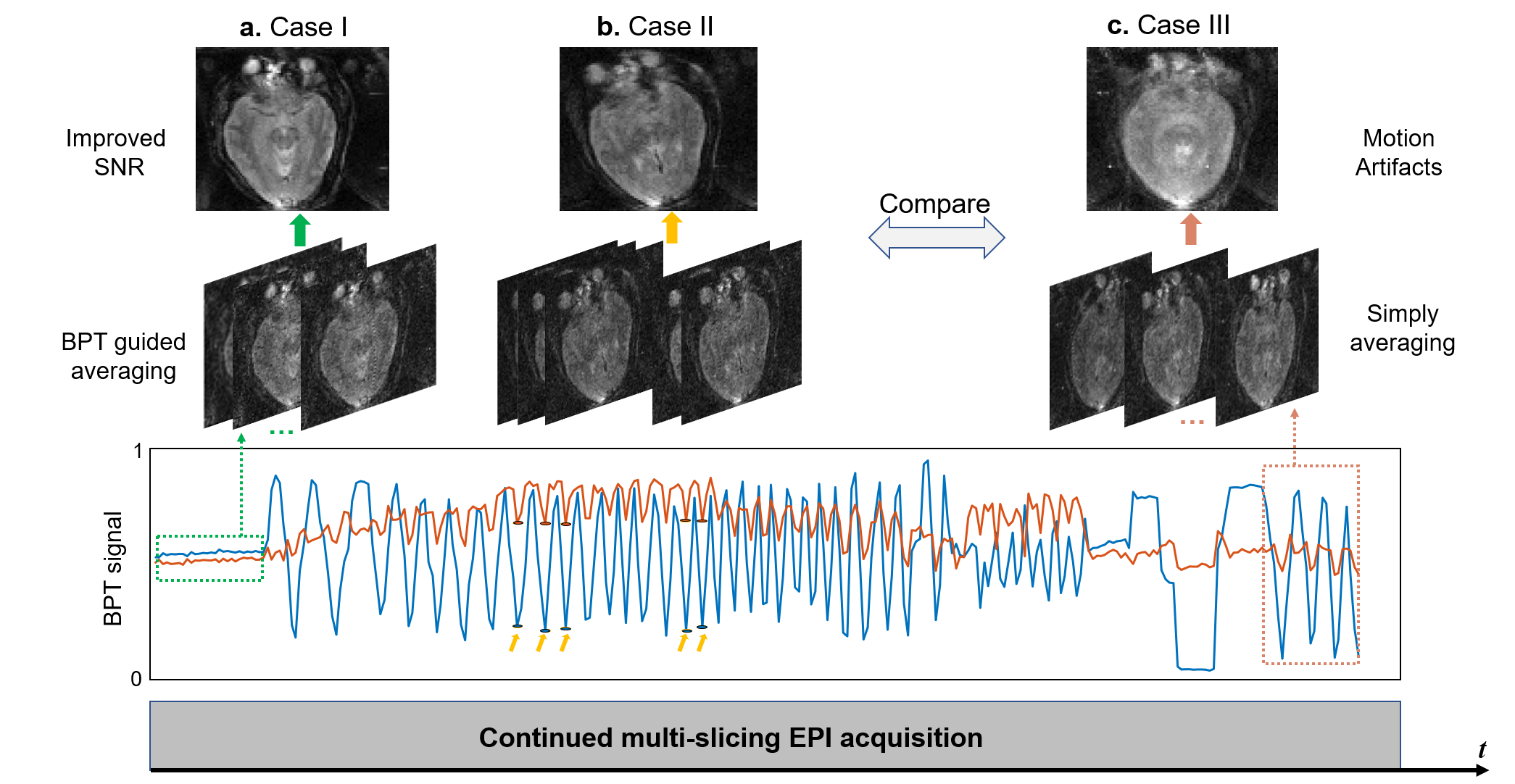
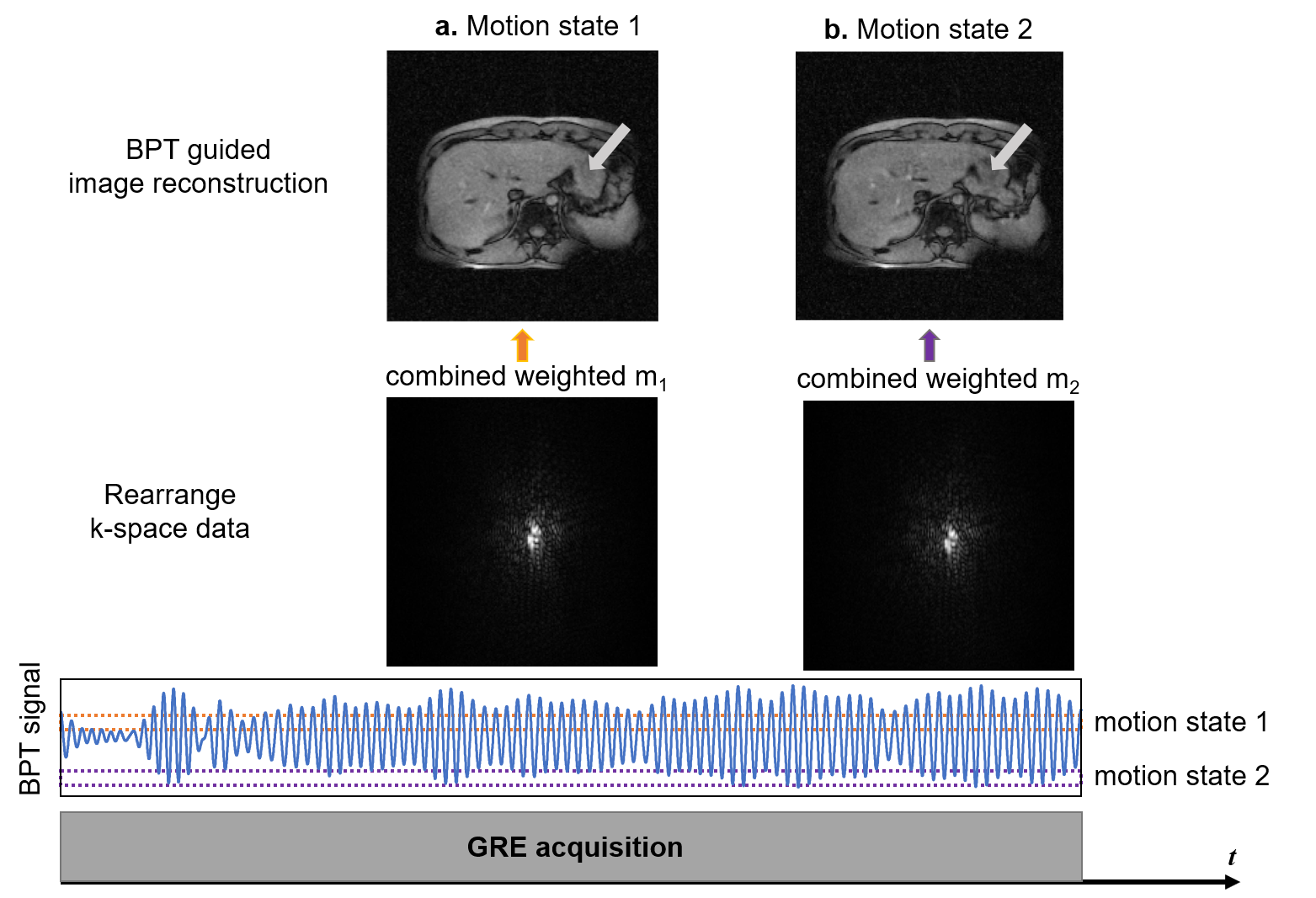
Due to the high acquisition efficiency of EPI, assuming that single slice scan would not be affected by motion, the motion states of different slices and repetition times were different. The images of the same slice with similar BPT signals were averaged directly in the image domain. As for GRE acquisition, there are still non-negligible motions in single-slice phase encodings. BPT signal is used to rearrange the phase-encoding-line data into the corresponding k-space by their motion states and refill them in a motion-solved k space.
Results and Discussion
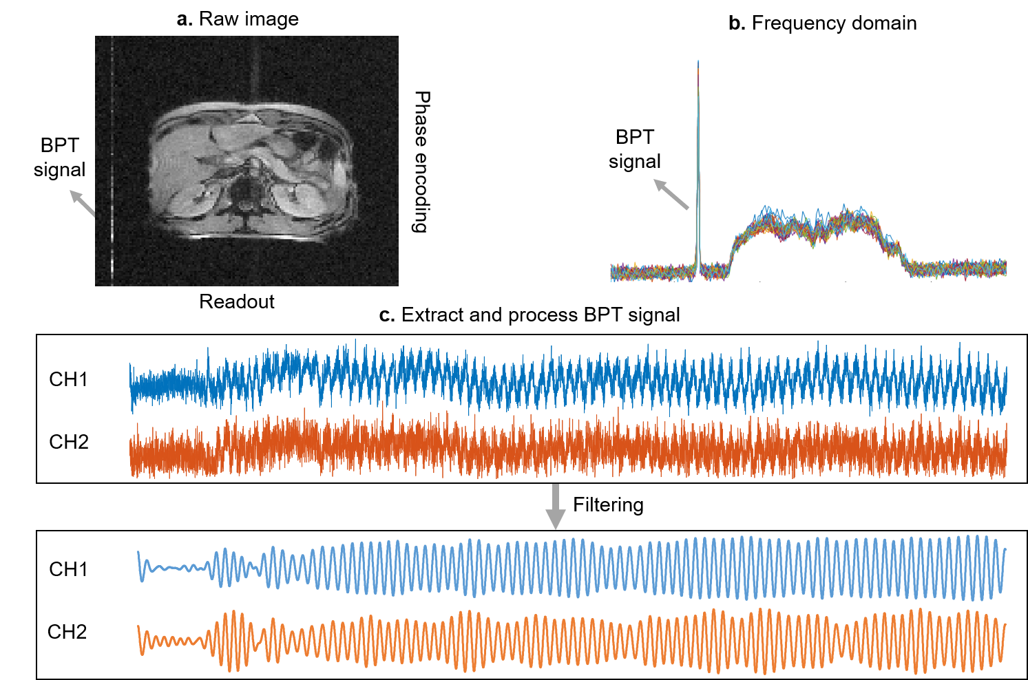
Figure 2a and b show the BPT signal imposed on the image and frequency domain. The received BPT signal was sufficiently spaced from the image signal in the frequency domain and could be easily extracted. Figure 2c lists the extracted BPT signal in the abdominal scan at the 0.25T system, the motion information of the breath-hold state and free-breathing periods can be clearly observed. This demonstrates the feasibility and high sensitivity of BPT in low-field MRI. Figure 3 shows the results of EPI acquisition. The blurring caused by motion severely degraded using the simply averaging image. With BPT-guided averaging, the SNR in the averaged image is significantly improved. This result demonstrates the feasibility of BPT for signal averaging under motion. Figure 4 shows the results of GRE reconstructed with the motion state extracted from BPT. Motion artifacts are not visible in the rearranged images.Conclusions
A contact-free and plug-and-play BPT technique at low-field MRI was proposed. The results demonstrate that BPT keeps its high sensitivity to motion detection in the 0.25T system. Based on EPI and GRE, we improve SNR by averaging BPT-guided images or rearranging k-space signal.Acknowledgements
This work is supported by the National Natural Science Foundation of China National Science Foundation of China (No. 62001290), Shanghai Science and Technology Development Funds (21DZ1100300), and Shanghai Sailing Program (20YF1420900).References
[1] MARQUES J P, SIMONIS F F, WEBB A G. Low‐field MRI: An MR physics perspective. Journal of magnetic resonance imaging, 2019, 49(6): 1528-42.
[2] ANAND S, LUSTIG M. Beat Pilot Tone: Exploiting Preamplifier Intermodulation of UHF/SHF RF for Improved Motion Sensitivity over Pilot Tone Navigators; proceedings of the ISMRM Meeting and Exhibition Abstract, 2021 .
[3] GRAESSLIN I, MENS G, GUILLAUME A, et al. Advancements in contact‐free respiration monitoring using RF pick‐up coils; proceedings of the Proceedings of the 18th Annual meeting of ISMRM, Stockholm, Sweden, 2010 .
[4] LENK M C. Respiratory Motion Tracking in Magnetic Resonance Imaging with Pilot Tone Technology ; The Ohio State University, 2018.
[5] SOLOMON E, RIGIE D S, VAHLE T, et al. Free‐breathing radial imaging using a pilot‐tone radiofrequency transmitter for detection of respiratory motion. Magnetic resonance in medicine, 2021, 85(5): 2672-85.
Figures