1014
Motion Correction for Interleaved EPI Diffusion Imaging using a Markerless Optical Tracking System
Yi Xiao1, Chunyao Wang1, Sisi Li1, Huijun Chen1, and Hua Guo1
1Center for Biomedical Imaging Research, Department of Biomedical Engineering, Tsinghua University, Beijing, China
1Center for Biomedical Imaging Research, Department of Biomedical Engineering, Tsinghua University, Beijing, China
Synopsis
Keywords: Motion Correction, Diffusion Tensor Imaging
Multi-shot interleaved EPI can achieve high-resolution diffusion imaging and effectively reduce geometric distortions. However, multi-shot acquisitions are susceptible to bulk motion which causes artifacts. Optical tracking-based motion correction is an effective method to reduce motion artifacts in DWI. Based on a self-developed Structured Light Optical MOtion tracking (SLOMO) system, a retrospective motion correction method was developed for multi-shot interleaved EPI DWI. The performance was evaluated by in-vivo experiments and compared with software-based correction.Introduction
Multi-shot echo-planar imaging (MS-EPI) is a beneficial technique for high-resolution diffusion imaging. It can effectively reduce geometric distortion by increasing the acquisition bandwidth along the phase-encoding direction. However, MS-EPI DWI is susceptible to macroscopic motion due to its relatively long acquisition time, further leading to severe motion artifacts. The SPIRiT-based simultaneous reconstruction integrating with motion correction algorithm proposed by Dong et al. 1, and the Augmented MUltiplexed Sensitivity Encoding (AMUSE) algorithm proposed by Guhaniyogi et al. 2 provided effective in-plane motion correction for MS-EPI DWI.Recently, optical tracking-based motion detection and correction methods were proposed and proven to be effective in diffusion imaging 3-5. Wang et al 6,7 proposed a Structured Light Optical Motion tracking (SLOMO) system and demonstrated its effectiveness in both brain-rigid and abdominal-respiratory motion correction in MR anatomical imaging. This study proposed a retrospective optical tracking motion correction method for MS-EPI DWI using the SLOMO system. The efficacy of the method was evaluated by in-vivo brain DTI experiments.
Methods
This study was performed on a Philips Ingenia 3.0T MR scanner (Philips Healthcare, Best, The Netherlands) with a 15-channel head-neck coil. The acquisition parameters of the 2D navigated MS-EPI DWI sequence are as follows: 10 transverse slices, 4 shots, FOV = 220×220 mm2, slice thickness = 4 mm, TR/TE = 3000/88ms, in-plane image resolution = 1×1 mm2, 10 diffusion directions, b-value = 800 s/mm2, 2 signal averages.The SLOMO system, consisting of an MR-compatible camera and a parallel-line projector, was a structured light 3D surface measurement system which can reconstruct the whole 3D surface of the head or body. During image acquisitions, the SLOMO system tracked the rigid head motion in real time (30Hz) with parameters of 6 degrees of freedom (Figure 1). The results of motion parameters were then transformed into MR imaging coordinate by a cross-calibration procedure.
Seven healthy volunteers were recruited and scanned using MS-EPI DWI. During four repeated DWI acquisitions, each volunteer was instructed to perform three motion patterns including blip motion, rectangle motion and triangle motion (Figure 2). These motion patterns are all restricted to the transverse plane and the in-plane rotation is controlled within ±15mm/±15°. Besides, a no-motion scan was performed as reference to test the precision of the SLOMO system and motion correction method.
For image reconstruction, the diffusion data acquired under different motion patterns were reconstructed by the SPIRiT-based method 8 to eliminate ghosting artifacts in the multi-shot acquisition. The artifacts from bulk motion were corrected using the motion information of each shot from the SLOMO system. For comparison, The DWI images were also corrected using Dong’s motion correction method 1, in which in-plane translation and rotation were calculated from the sequence navigator. Furthermore, Mean diffusion-weighted images (mDWI) were obtained and color fractional anisotropy maps (FA) were calculated using the FMRIB Software package (FSL) 9.
We also performed statistical analysis to compare the overall qualities between corrected and uncorrected images. The mDWI image quality with and without SLOMO motion correction for three motion patterns were evaluated by subjective scoring (low-high: 1-5). The score differences between corrected and uncorrected groups were compared using a matched-pairs signed-rank Wilcoxon test, with P<0.05 as statistical significance. As a comparison, images corrected by Dong's method 1 were also scored.
Results and Discussion
Figure 3 shows that the rectangle and the triangle motion result in visually obvious blurring and poorer SNR in the mDWI images. After motion correction by the two methods, the severe blurring was distinctly reduced with improved SNR. As shown in the magnified images, the SNR of the SLOMO-corrected images under three motion patterns are comparable to that of the navigator-corrected images.Figure 4 indicates that both navigator and SLOMO motion correction methods can effectively correct the motion-corrupted data and provide the right FA maps. In general, the two correction methods showed no obvious difference in image quality and structure information. It can be seen from the zoomed-in FA images that the white matter structures in the uncorrected image were severely corrupted and the SNR decreased. In comparison, the white matter structures were well restored after motion correction. Moreover, the structures indicated by the white arrows can be better delineated with higher SNR using the SLOMO correction compared with that using the navigator correction.
The comparison of the mean DWI quality scores among images without motion correction, with navigator correction and with SLOMO correction (Figure 5) show that the image qualities were significantly improved (p<0.0001) after both motion correction methods under 3 different motion patterns. There was no significant difference between the two correction methods (p>0.9999).
Conclusion
This study proposed a retrospective motion correction method for interleaved EPI diffusion imaging using a markerless optical tracking system (SLOMO) and demonstrated significant image quality improvement in mean DWI images and color FA maps. The potential of this method in prospective motion correction will be explored in the future.Acknowledgements
This work was supported by the National Key Research & Development (R&D) Program of China (2017YFC0108702).
The authors would like to thank Zijing Dong at Massachusetts General Hospital for helpful discussions about the SPIRiT-based reconstruction of diffusion imaging.
References
- Dong Z, Wang F, Ma X, Dai E, Zhang Z, Guo H. Motion-corrected k-space reconstruction for interleaved EPI diffusion imaging. Magn Reson Med. 2018;79(4):1992-2002.
- Guhaniyogi S, Chu ML, Chang HC, Song AW, Chen NK. Motion immune diffusion imaging using augmented MUSE for high-resolution multi-shot EPI. Magn Reson Med. 2016 Feb;75(2):639-52.
- Aksoy M, Forman C, Straka M, et al. Real-time optical motion correction for diffusion tensor imaging. Magn Reson Med. 2011;66(2):366-378.
- Berglund J, van Niekerk A, Rydén H, et al. Prospective motion correction for diffusion weighted EPI of the brain using an optical markerless tracker. Magn Reson Med. 2021;85(3):1427-1440.
- Chen H, Dai K, Zhong S, Zheng J, Zhang X, Yang S, Cao T, Wang C, Karasan E, Frydman L, Zhang Z. High-resolution multi-shot diffusion-weighted MRI combining markerless prospective motion correction and locally low-rank constrained reconstruction. Magn Reson Med. 2022 Oct 5.
- Wang C, Huang T, Zhang C, Li Y, Wang Y, Wang Y, Li S, Xiao Y, Guo H, Chen H. Structure Light based Optical MOtion Tracking system (SLOMO) for Contact-free respiratory Motion Tracking from Neck in MR Imaging. 29th ISMRM & SMRT Annual Meeting & Exhibition - An Online Experience, 15-20 May 2021. 1380.
- Wang C, Zhang C, Wang Y, Qi H, Huang T, Liu J, Yuan C, Liao H, Chen H. Motion correction in Brain MR imaging using a Structure Light based Optical MOtion Tracking system (SLOMO). ISMRM 27th Annual Meeting & Exhibition, 11-16 May 2019. 4445.
- Dong Z, Wang F, Ma X, et al. Interleaved EPI diffusion imaging using SPIRiT-based reconstruction with virtual coil compression. Magn Reson Med. 2018;79(3):1525-1531.
- Jenkinson M, Beckmann CF, Behrens TE, Woolrich MW, Smith SM. FSL. Neuroimage. 2012;62(2):782-790.
Figures
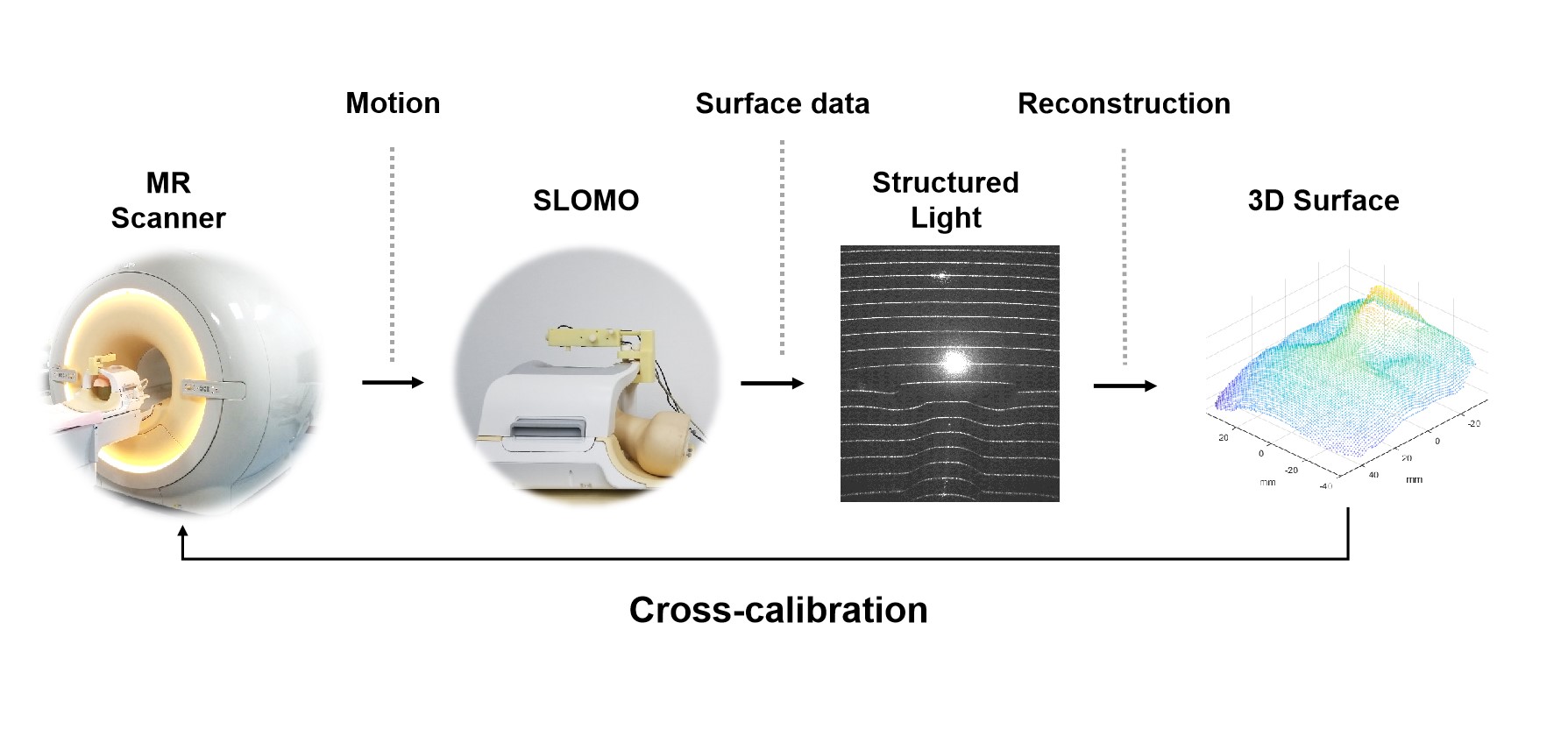
Figure 1: Technical route of MR in-plane motion tracking by SLOMO system.
During the acquisition, the facial surface was reconstructed and registered to
the initial pose in real time, providing rigid motion parameters of 6 degrees
of freedom (DoF). After the cross calibration between the SLOMO and scanner,
the motion information can be transformed to the MR imaging coordinate for
SPIRIT-based DWI reconstruction and motion correction method.
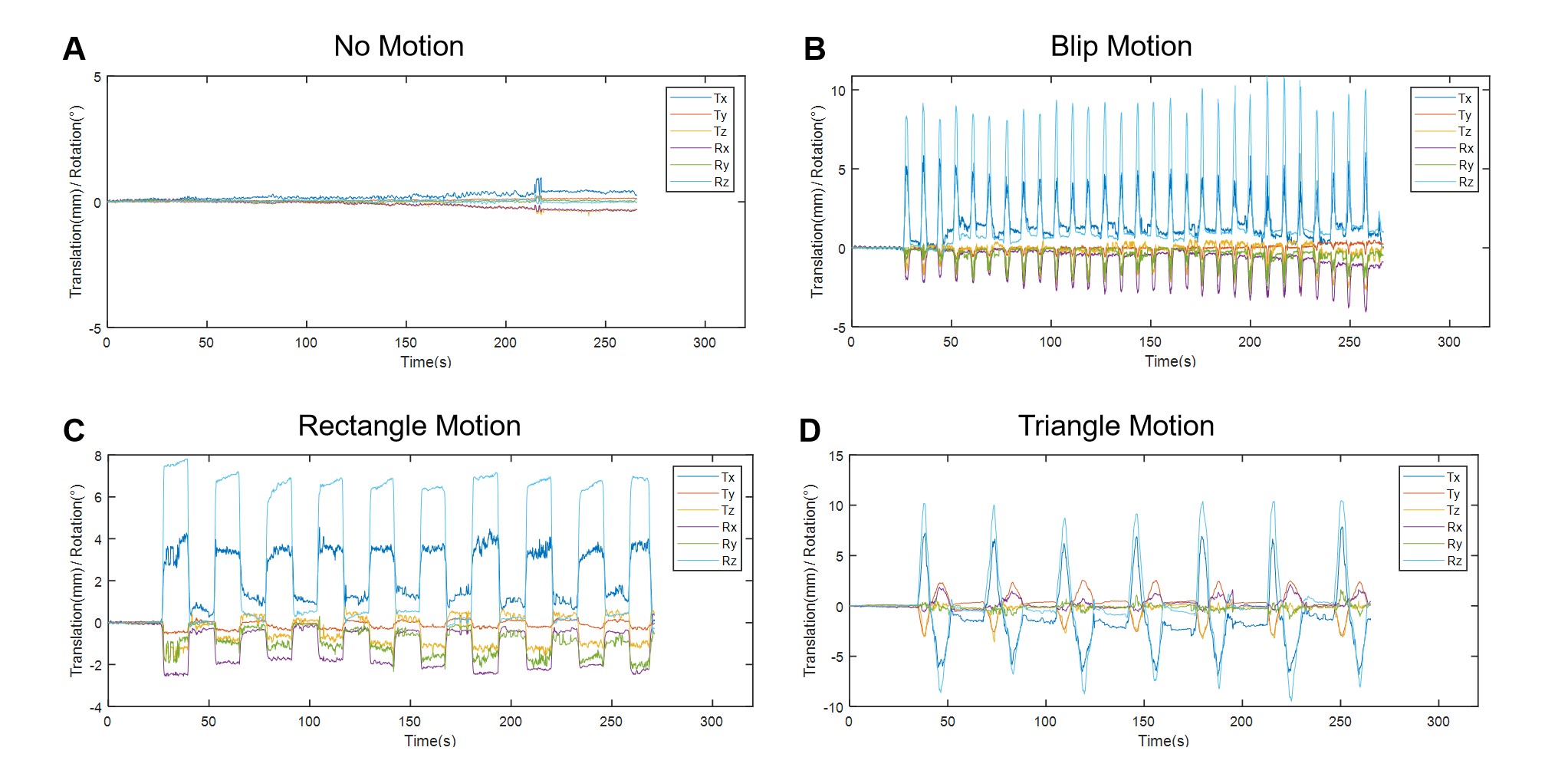
Figure 2: Motion tracking curves of four motion patterns. (A) No Motion.
(B) Blip Motion. (C) Rectangle Motion. (D) Triangle Motion.
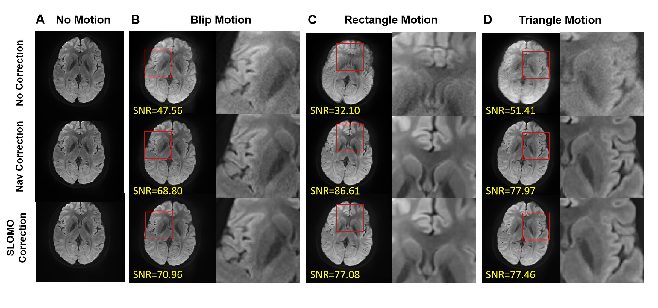
Figure 3: Mean DWI images and zoomed-in views (red boxes) without
correction, with navigator correction and with SLOMO correction for three
motion patterns. (A) No Motion. (B) Blip Motion. (C)
Rectangle Motion. (D) Triangle Motion. Slight motion artifacts in blip
motion are corrected. The overlap artifacts in the images with rectangle motion
and the blur in the images with triangle motion are effectively corrected by both
methods.
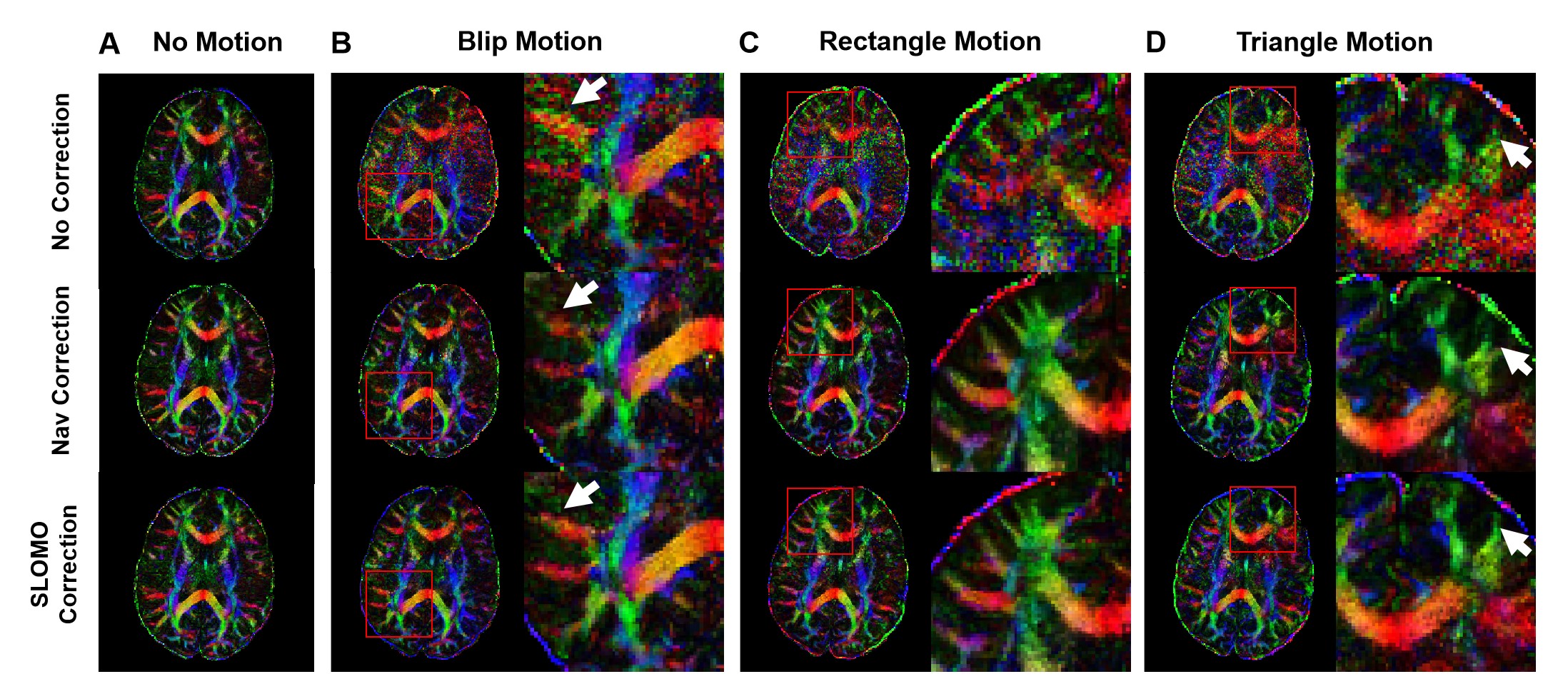
Figure 4: Color
FA maps and zoomed-in images of several regions (red boxes) without correction,
with navigator correction and with SLOMO correction for three motion patterns. (A)
No Motion. (B) Blip Motion. (C) Rectangle Motion. (D) Triangle Motion. Both navigator
and SLOMO correction methods can effectively correct errors in Color FA maps caused
by motion and improve SNR. In the areas indicated by the white arrow, SLOMO
correction can better restore the brain structure than navigator correction.
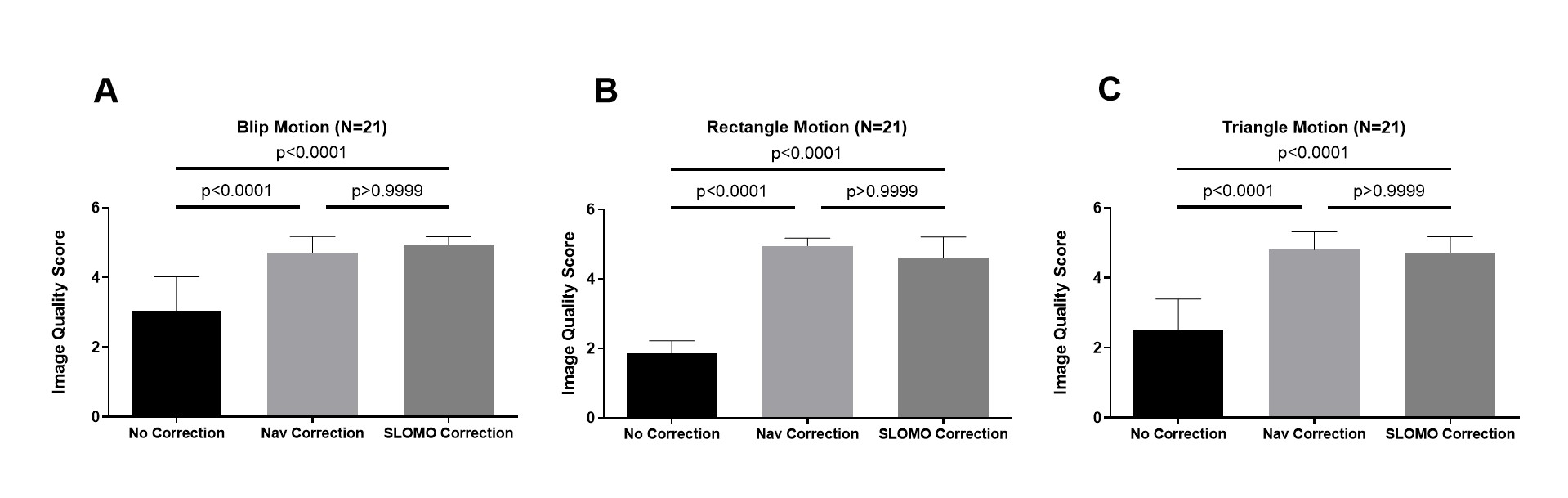
Figure 5: Image quality scores (low-high: 1-5) of mean DWI images without
correction, with navigator correction and with SLOMO correction for three
motion patterns. Three slices from each subject were selected for scoring (N=3 slices
×
7 subjects=21). Both navigator-corrected
and SLOMO-corrected mean DWI images are graded with significantly higher scores
compared with uncorrected images (both P<0.0001). The image quality scores
were comparable between the two correction methods (P>0.9999). The paired
Dunn's multiple comparisons test was used.
DOI: https://doi.org/10.58530/2023/1014