1002
Evaluation of glymphatic system function using DTI-ALPS in patients with temporal lobe epilepsy1Tongji hospital of Tongji medical college, Huazhong university of science and technology, Wuhan, China
Synopsis
Keywords: Epilepsy, Diffusion Tensor Imaging, glymphatic system, asymmetry
The alterations of glymphatic system function in left hemisphere and right hemisphere and the asymmetric features of glymphatic system were not clear in temporal lobe epilepsy (TLE) patients. We investigated the glymphatic system function in TLE patients and evaluated the asymmetric features of glymphatic system by using DTI-ALPS method. Our findings indicated leftward asymmetric tendency of glymphatic system in adult human brain. The abnormality of glymphatic system asymmetry in LTLE was also found. The glymphatic system function was impaired and more severe alterations of glymphatic system in ipsilateral hemisphere than contralateral hemisphere in TLE patients.Introduction
Glymphatic system plays an important role in the interstitial waste clearance of brain1. The glymphatic system dysfunction has been presented in several neurological disorders, including epilepsy2-4. However, the alterations of glymphatic system function in left hemisphere and right hemisphere and the asymmetric features of glymphatic system were not clear in temporal lobe epilepsy (TLE) patients. Recently, diffusion tensor image analysis along the perivascular space (DTI-ALPS), a new non-invasive analysis method, was used to access the activity of the glymphatic system5. Thus, we aimed to investigate the glymphatic system function in TLE patients and to evaluate the asymmetric features of glymphatic system by using DTI-ALPS method.Methods
43 epilepsy patients who were diagnosed as TLE were enrolled and then divided into left TLE (LTLE, n=20) or right TLE (RTLE, n=23) according to the criteria defined by the Commission on Classification and Terminology of the ILAE6. 39 healthy controls (HC) with matched age and gender were included as controls. DTI data were acquired using a 3.0-T MR scanner (Discovery MR750; GE Healthcare, Milwaukee, Wisconsin). The DTI parameters were processed and calculated in FSL. Then, based on the V1 maps with FSLEyes, at the level of the lateral ventricle and adjacent to the medullary veins, a neuroradiologist with over 10 years experience marked two 3 mm spherical ROIs in the region of the projection and association fibers for both hemispheres. The same neuroradiologist drawed the ROIs again in the same way one month later. On the ROIs, the x-, y-, and z-axis diffusivity were measured, and the DTI-ALPS index was calculated using the following formula: DTI-ALPS index = mean (Dxxproj, Dxxassoc) /mean (Dyyproj, Dzzassoc)5. The ALPS index on the left and right hemisphere were named as lALPS_index and rALPS_index respectively. The mean value of ALPS_index of the two measurements were used in final analysis. An asymmetry index (AI) was calculated using the formula AI = (Right − Left)/[(Right + Left)/2] to further quantify the differences between the paired lALPS_index and rALPS_index7. Independent two sample t-test, two-sample paired t-test or univariate ANOVA with Bonferroni correction was conducted to compare ALPS_index differences among the groups. Correlation analysis was conducted with Pearson’s correlation test. All the statistical analysis was conducted by SPSS 24, and statistical significance was defined as a two-tailed p-value of < 0.05.Results
No significant differences were found among the LTLE, RTLE, and HC in age or gender. The intraclass correlation coefficient (ICC) between two measurements were excellent both for rALPS_index (ICC = 0.925) and lALPS_index (ICC = 0.956). Compared to HC, the rALPS_index (p = 0.000) and lALPS_index (p = 0.001) of TLE were both reduced. When comparison within the three groups, lALPS_index of LTLE (p = 0.005) and RTLE (p = 0.040) were reduced, while only rALPS_index of RTLE ( p=0.001) was reduced compared to HC. The two sample paired t-test showed significant differences in HC (p=0.045) and RTLE groups (p=0.009), presenting larger ALPS_index in left hemisphere. In addition, in comparison with contralateral ALPS_index, ipsilateral ALPS_index was reduced in TLE group (p=0.008) as well as RTLE group (p=0.009). HC (AI=-0.024) and RTLE (AI=-0.056) showed leftward asymmetry of glymphatic system by AI analysis, while LTLE presented reduced asymmetric traits compared with RTLE ( p=0.029) . The lALPS_index was negatively correlated with age (r=-0.349, p=0.022) in TLE patients.Discussion
This study showed that both left and right ALPS_index were decreased in patients with TLE compared with HC, indicating glymphatic system dysfunction in TLE. In addition, the ipsiALPS_index was smaller than contraALPS_index in TLE and RTLE group, suggesting more severe alterations of glymphatic system in ipsilateral hemisphere than contralateral hemisphere. It was consistent with previous studies4. The exact underlying mechanisms remain to be elucidated. One hypothesis was the disruption of blood brain barrier, which was characterized by altered permeability across tight junctions and endothelial damage, may contribute to abnormal interstitial fluid (ISF) flow and cerebrospinal fluid (CSF)-ISF exchange, leading to glymphatic system dysfunction8,9. Conversely, the aggregation of neurotoxic solute caused by glymphatic system dysfunction may increase the risk of seizure activity. Interestingly, patients with LTLE and RTLE showed different glymphatic system changes in comparison with HC group. Specifically, ALPS-index in both hemisphere in RTLE were reduced while only lALPS_index in LTLE showed significant lower than HC, which supported that LTLE and RTLE are etiologically distinct and pathologically different syndromes from the outset.Furthermore, the rALPS_index was smaller than lALPS_index in HC group, indicating the leftward asymmetric tendency of glymphatic system in human brain. The asymmetric function of the glymphatic system may be related to the structural and functional asymmetries in the human, which should be further confirmed. AI of LTLE was reduced, indicating the abnormality of glymphatic system asymmetry in patients with LTLE.
The lALPS_index was negatively correlated with age in TLE patients, but no significant correlation between ALPS_index and epilepsy clinical variances were found. Thus, the clinical implication of ALPS_index in TLE should be elucidated with caution.
Conclusion
Our findings indicated leftward asymmetric tendency of glymphatic system in adult human brain. The glymphatic system function was impaired and more severe alterations of glymphatic system in ipsilateral hemisphere than contralateral hemisphere in TLE patients.Acknowledgements
The authors are grateful to Suiqiang Zhu, Shanshan Huang, and Xiaoyan Liu, Department of Neurology, Tongji Hospital, Tongji Medical College of Huazhong University of Science and Technology, for helpful in diagnosing of TLE. The authors are also grateful to the support by the National Natural Science Foundation of China (Nos. 82202130).
References
1. Iliff JJ, Wang M, Liao Y, Plogg BA, Peng W, Gundersen GA, Benveniste H, Vates GE, Deane R, Goldman SA, Nagelhus EA, Nedergaard M. A paravascular pathway facilitates CSF flow through the brain parenchyma and the clearance of interstitial solutes, including amyloid β. Sci Transl Med. 2012 Aug 15;4(147):147ra111.
2. Lee HJ, Lee DA, Shin KJ, Park KM. Glymphatic system dysfunction in patients with juvenile myoclonic epilepsy. J Neurol. 2022 Apr;269(4):2133-2139.
3. Lee DA, Lee J, Park KM. Glymphatic system impairment in patients with status epilepticus. Neuroradiology. 2022 Jul 15.
4. Lee DA, Park BS, Ko J, Park SH, Lee YJ, Kim IH, Park JH, Park KM. Glymphatic system dysfunction in temporal lobe epilepsy patients with hippocampal sclerosis. Epilepsia Open. 2022 Jun;7(2):306-314.
5. Taoka T, Masutani Y, Kawai H, Nakane T, Matsuoka K, Yasuno F, Kishimoto T, Naganawa S. Evaluation of glymphatic system activity with the diffusion MR technique: diffusion tensor image analysis along the perivascular space (DTI-ALPS) in Alzheimer's disease cases. Jpn J Radiol. 2017 Apr;35(4):172-178.
6. Berg AT, Berkovic SF, Brodie MJ, Buchhalter J, Cross JH, van Emde, Boas W, et al. Revised terminology and concepts for organization of seizures and epilepsies: report of the ILAE Commission on Classification and Terminology, 2005-2009. Epilepsia. 2010;51:676–685.
7. Carper RA, Treiber JM, DeJesus SY, Muller RA. Reduced hemispheric asymmetry of white matter microstructure in autism spectrum disorder. J Am Acad Child Adolesc Psychiatry. 2016;55:1073–1080.
8. Janigro D. Are you in or out? Leukocyte, ion, and neurotransmitter permeability across the epileptic blood-brain barrier. Epilepsia. 2012 Jun;53 Suppl 1(0 1):26-34.
9. Marchi N, Banjara M, Janigro D. Blood-brain barrier, bulk flow, and interstitial clearance in epilepsy. J Neurosci Methods. 2016 Feb 15;260:118-24.
Figures
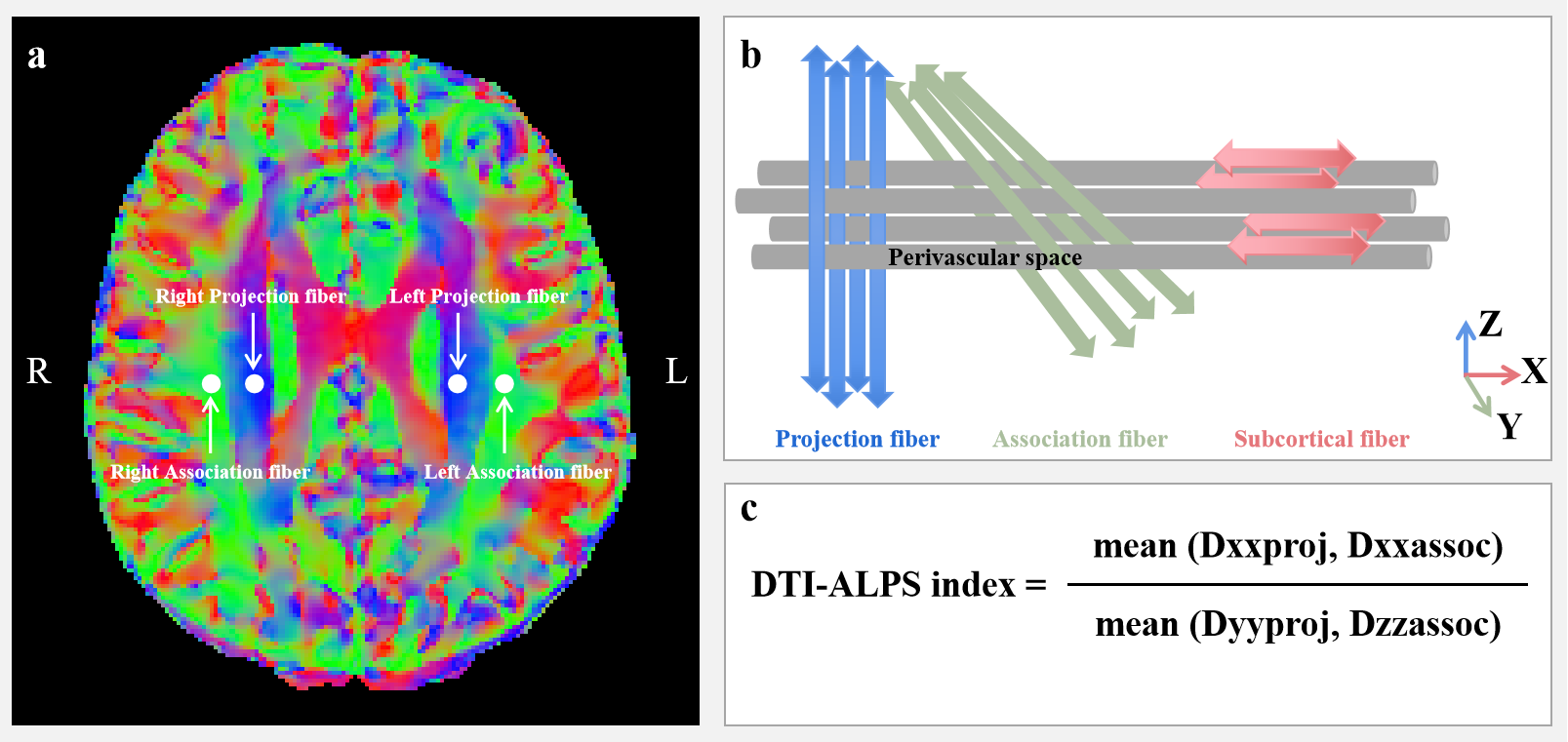
Figure 1. (a) DTI V1 color map shows the direction of the projection fibers (blue; z-axis), association fibers (green; y-axis), and the subcortical fibers (red; x-axis). Three ROIs are placed to measure diffusivities of the three fibers. (b) Schematic diagram presents the relationship between the direction of the perivascular space (gray cylinders) and the fibers. The direction of the perivascular space is perpendicular to both projection and association fibers. (c) The formula to measure values of the DTI-ALPS index.
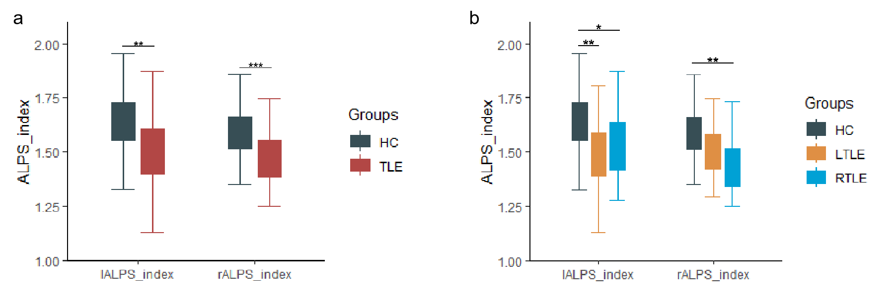
Figure 2. (a) The differences of lALPS_index and rALPS_index between HC and TLE. (b) The differences of lALPS_index and rALPS_index among HC, LTLE, and RTLE. (*P < 0.05; **P < 0.01; ***P < 0.001).
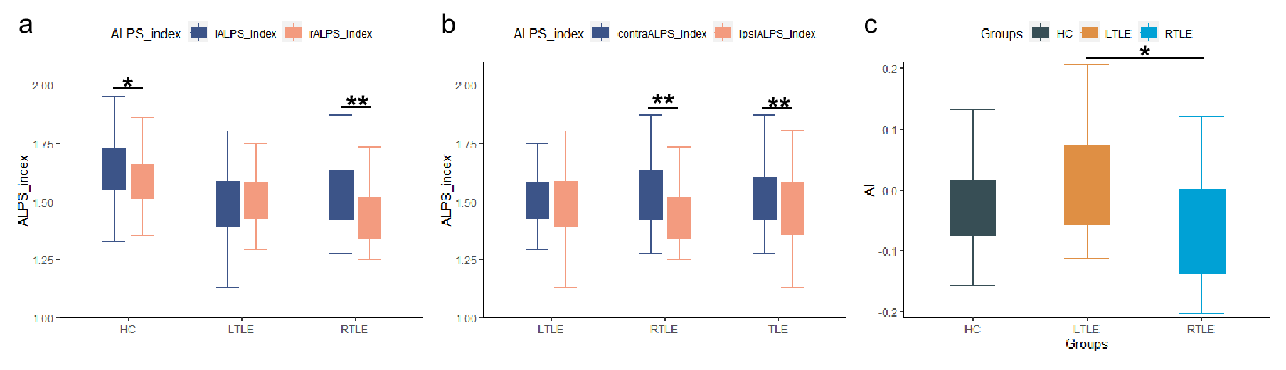
Figure 3. (a) The differences of paired lALPS_index and rALPS_index in HC, LTLE, and RTLE respectively. (b) The differences of paired contraALPS_index and ipsiALPS_index in LTLE, RTLE, and TLE respectively. (c) The asymmetry index (AI) differences of the paired ALPS_index in bilateral hemispheres among HC, LTLE, and RTLE (*P < 0.05; **P < 0.01; ***P < 0.001).
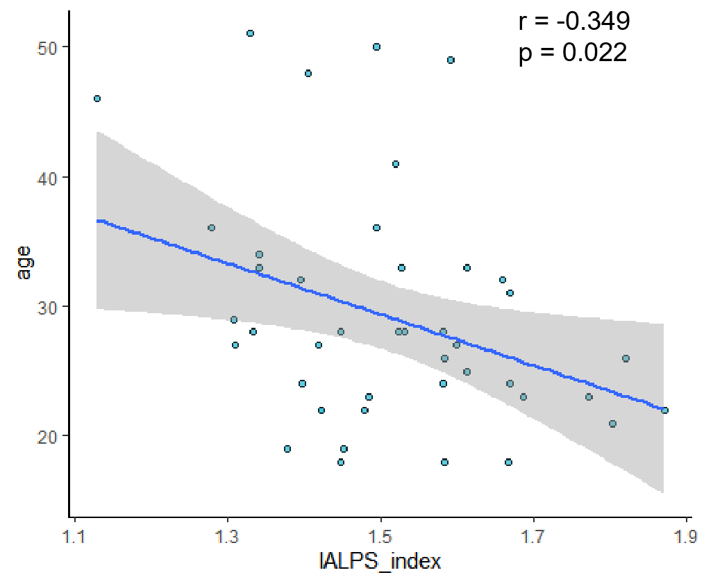
Figure 4. Represent a negative correlation between the lALPS_index and age in patients with TLE.