0991
Robust Electromagnetic Interference (EMI) Elimination for RF Shielding-Free MRI via Active EMI Sensing and Deep Learning MRI Signal Prediction1Laboratory of Biomedical Imaging and Signal Processing, The University of Hong Kong, Hong Kong, China, 2Department of Electrical and Electronic Engineering, The University of Hong Kong, Hong Kong, China
Synopsis
Keywords: System Imperfections: Measurement & Correction, New Signal Preparation Schemes
MRI scans are commonly performed inside a fully-enclosed RF shielding room, posing stringent installation requirement and unnecessary patient discomfort. This study develops a strategy of active EMI sensing and deep learning MR signal prediction using residual U-Net for RF shielding-free MRI. We implemented it on an ultra-low-field 0.055T head MRI scanner. Our experimental results demonstrated that this strategy could directly and accurately predict EMI-free MRI signals from the signals acquired by MRI receive coil and EMI sensing coils. It worked robustly with strong and dynamically varying EMI sources, yielding significantly improved brain image quality.Introduction
Clinical MRI scanners all require bulky and enclosed RF shielding cage to prevent EMI during MRI scanning. Several methods have been recently proposed to remove this shielding requirement for portable ultra-low-field (ULF) MRI1-7. An analytical approach was proposed to estimate EMI signal in MRI receive coil from EMI signals detected by EMI sensing coils based on frequency domain transfer functions (TFs) among coils1,2, and further extended for time domain implementation with an adaptive procedure4. Our recent study proposed a deep learning method using a CNN model to better characterize the relationships among EMI signals detected by EMI sensing coils and MRI receive coils, producing improved EMI reduction for shielding-free MRI at 0.055T5. We also demonstrated its applicability at 1.5T6,7. However, EMI environments can be extremely complex in practice. For example, EMI sources can be very strong, and vary temporally and spatially. Patient body position change during scanning may also alter EMI behaviours since body can act as an antenna8,9. Such practical issues can degrade the performance of these newly developed EMI removal methods. For shielding-free MRI to become a reality, it is imperative to develop more robust strategies.In this study, a new EMI elimination strategy is developed for RF shielding-free MRI. We propose direct prediction of EMI-free MRI signals from the signals detected by MRI receive coil and EMI sensing coils through a residual U-Net based deep learning. We demonstrated its robust performance for 0.055T brain MRI under complex EMI conditions.
Methods
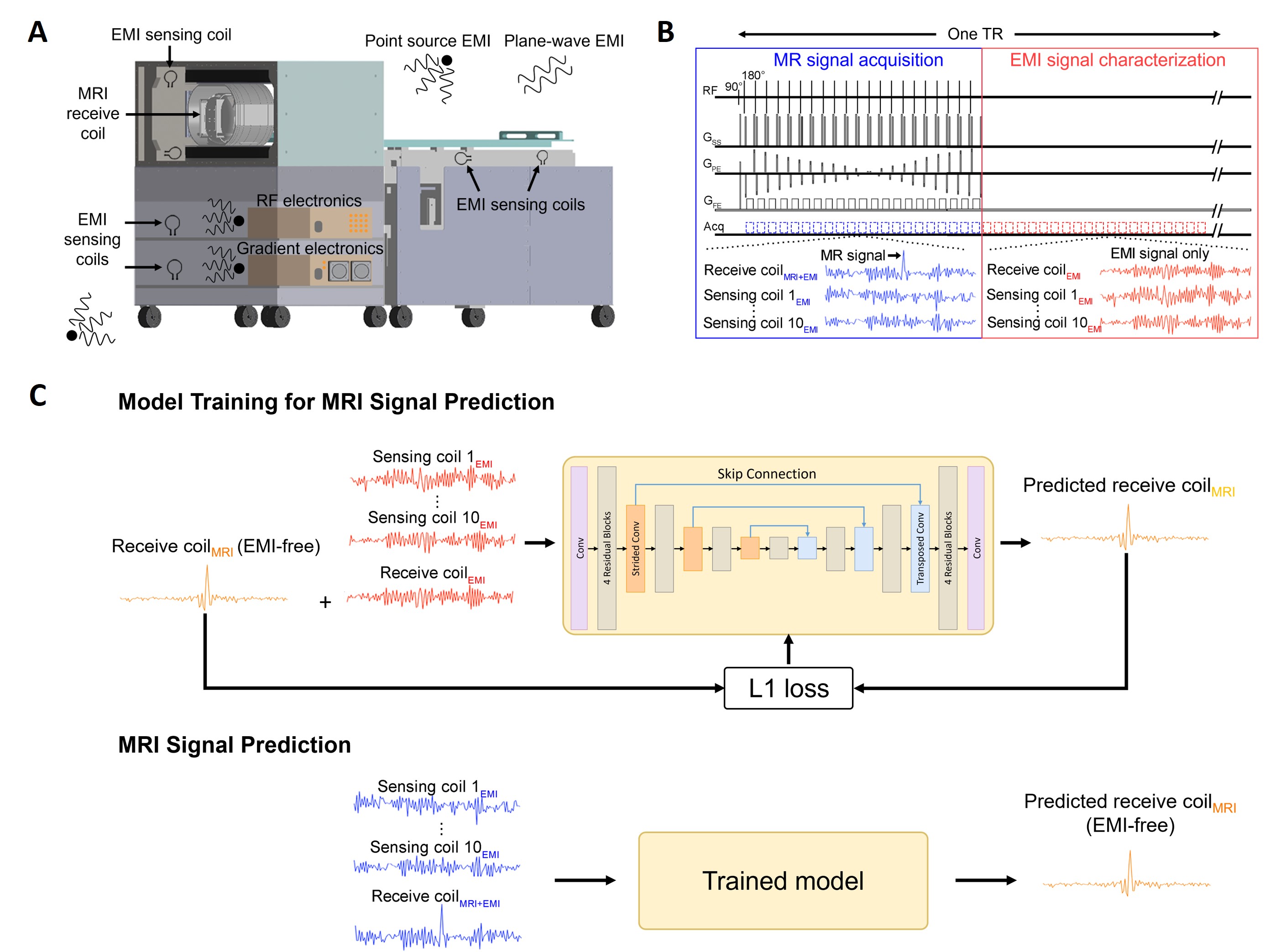
Active EMI Sensing and Direct Deep Learning MRI Signal PredictionFig. 1 illustrates the proposed EMI elimination strategy. EMI sensing coils are strategically placed around and within scanner to actively detect radiative EMI signals from both external environments and internal electronics (Fig. 1A). Within each TR, MRI receive coil and EMI sensing coils are used to simultaneously sample data within two windows, one for conventional MRI signal acquisition, and the other for acquiring EMI characterization data (i.e., EMI signals only) (Fig. 1B).
After each scan, we train a residual U-Net model to directly predict the 1D temporal MRI signals (i.e., frequency encoding or FE lines) from the signals acquired by both MRI receive coil and EMI sensing coils during EMI characterization window (Fig. 1C). Specifically, EMI-free k-space MRI signals from 3T brain MRI data (or any publicly available data) are first added into EMI-only signals received by MRI receive coil. Such synthesized EMI-contaminated MRI signals and signals received by EMI sensing coils are used as model inputs, while EMI-free MRI signals serve as model target. The trained model is then applied to predict EMI-free MRI signals from the signals collected during MRI signal acquisition window, creating EMI-free k-space data prior to any averaging or/and image reconstruction.
Evaluation and Model Implementation
Brain imaging experiments were conducted on a shielding-free 0.055T MRI scanner with one MRI receive coil and ten EMI sensing coils. T1W datasets were acquired using 3D GRE with BW = 6.25kHz (acquisition matrix = 128×128×32 and NEX = 2). T2W and FLAIR-like datasets were acquired using 3D FSE with BW = 10kHz (T2W: acquisition matrix = 128×126×32 and NEX = 2; FLAIR: acquisition matrix = 128×117×32 and NEX = 4).
A residual U-Net architecture10 containing 4 scales was employed. Four successive residual blocks were used in each scale (Fig. 1C). 1D temporal signals detected by MRI receive coil and EMI sensing coils were treated as separate channels, and the number of channels in each layer from the first to the fourth scale was 12, 32, 64, and 128, respectively. For each dataset, a model was trained by minimizing L1 loss using Adam optimizer11 with batch size = 64 for 40 epochs and initial learning rate = 0.0002. Typical training time for each model (i.e., each dataset) was around 8 mins on a Quadro RTX 8000 GPU and Intel Core i9-10900X CPU.
Results
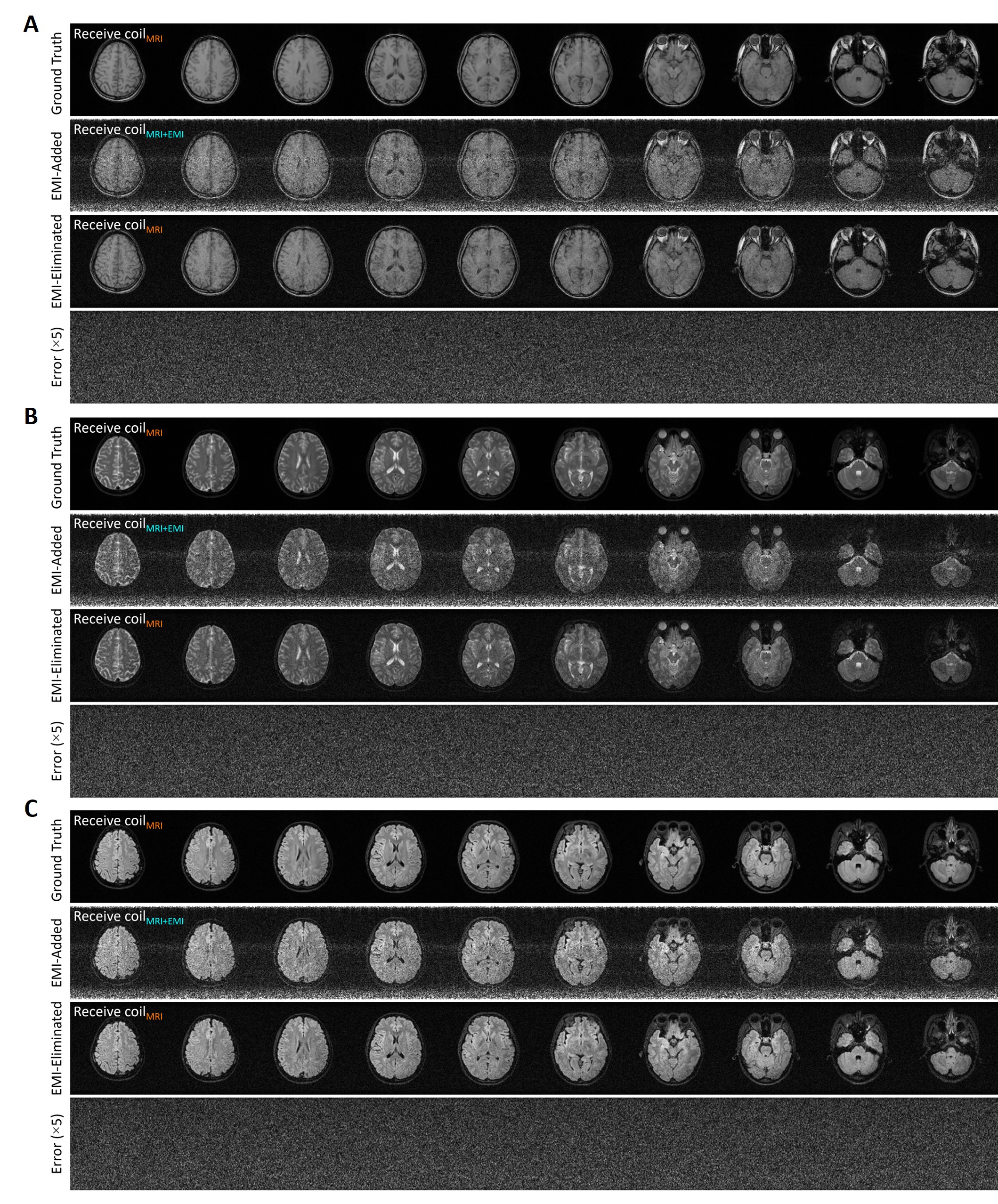
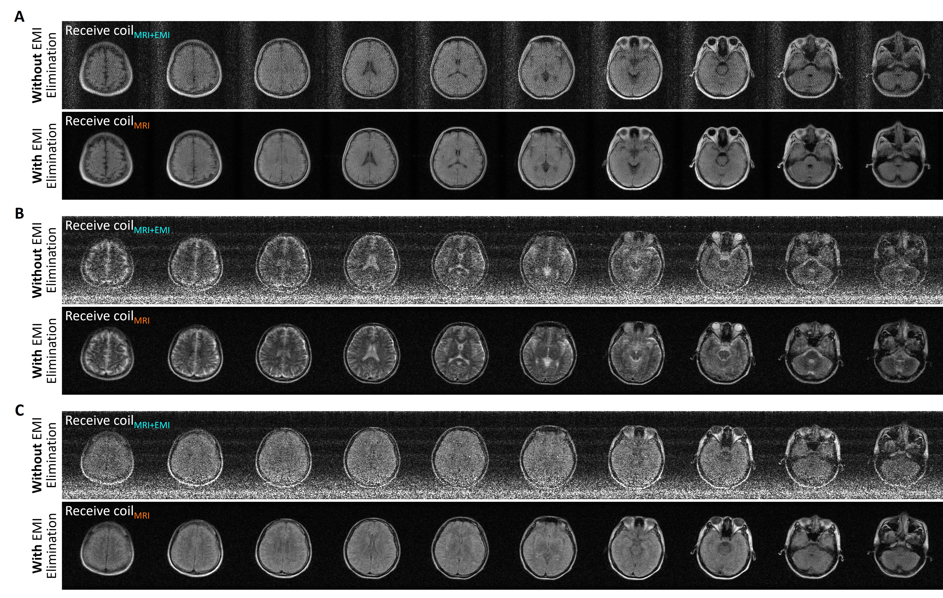
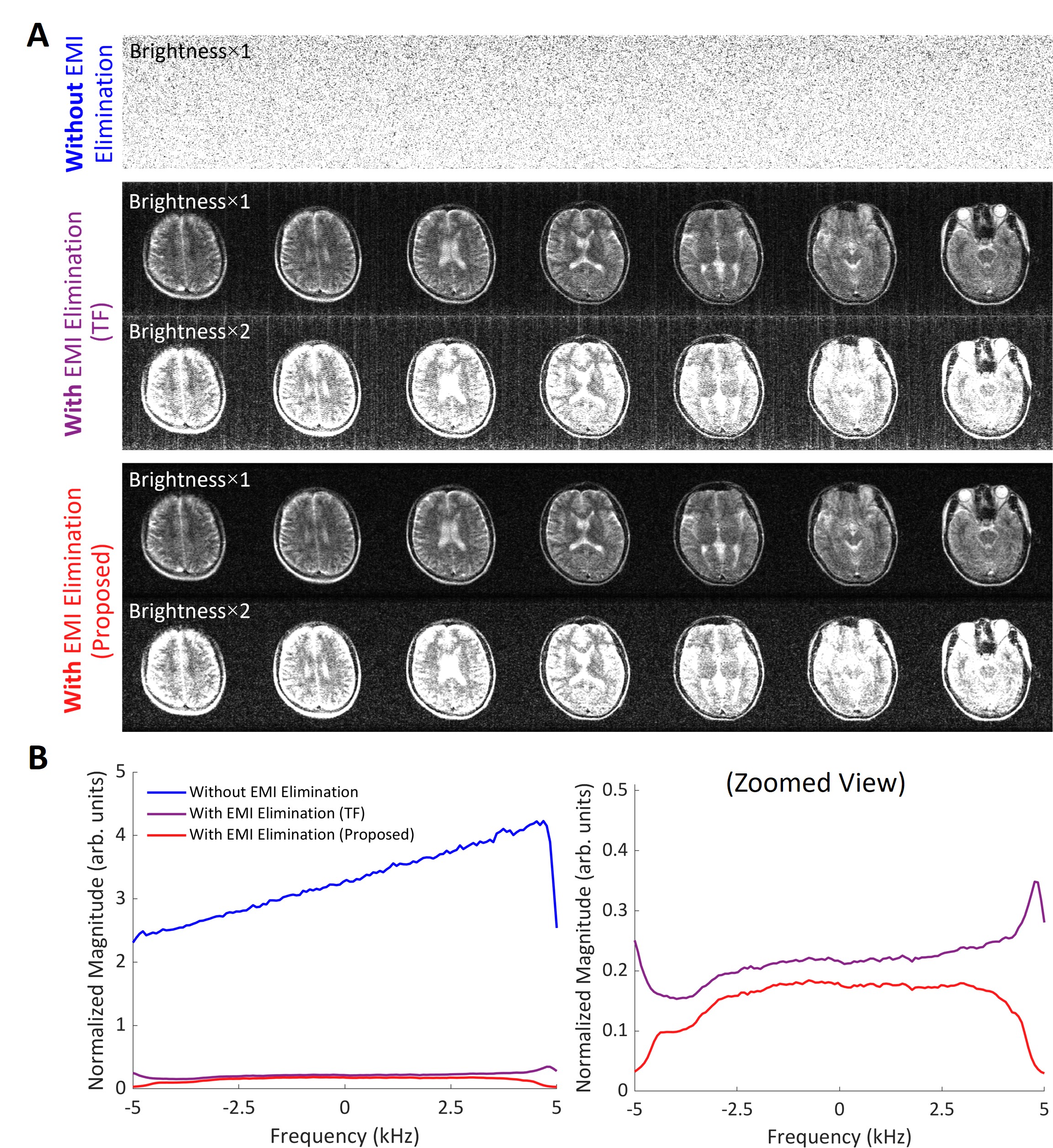
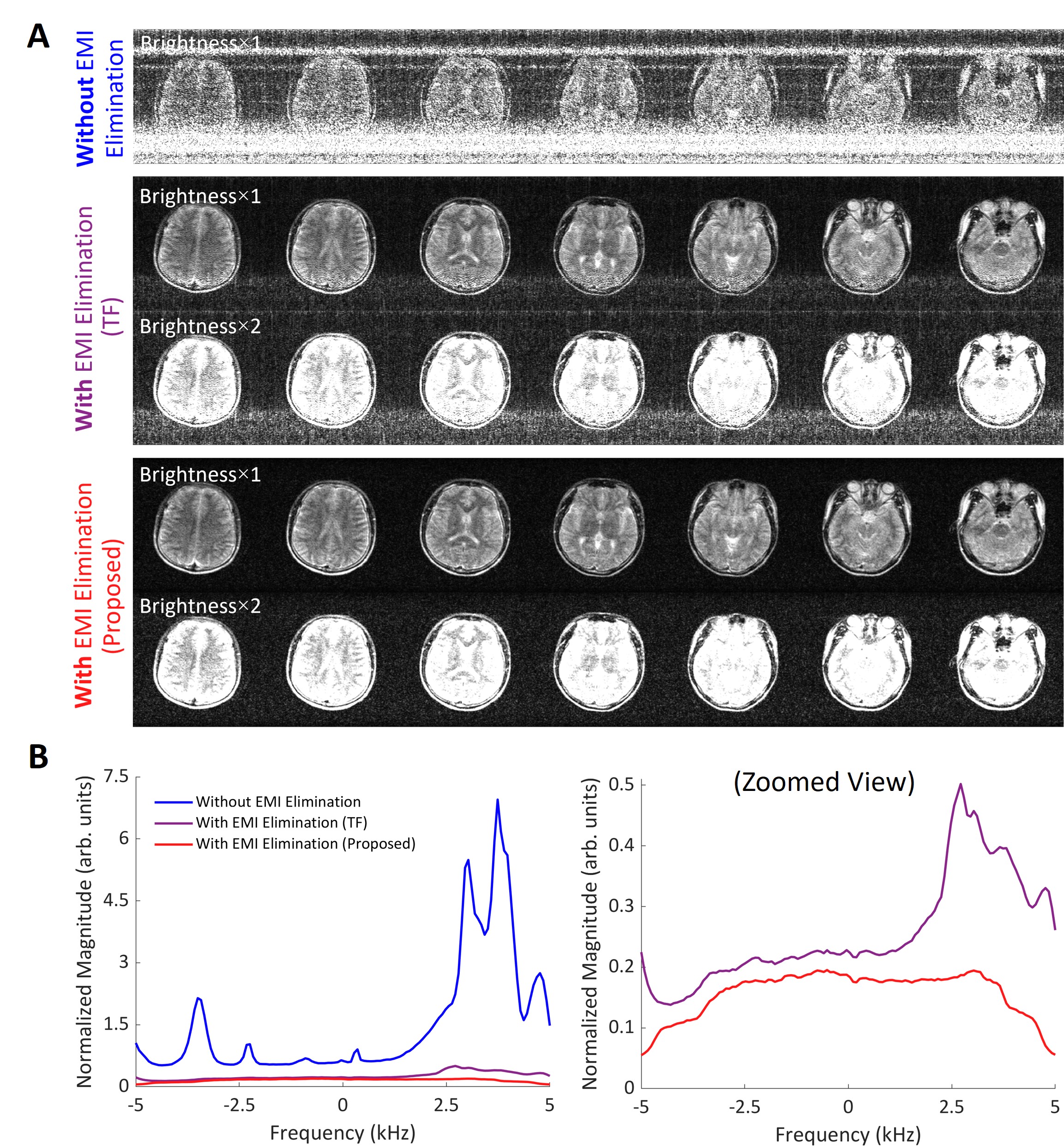
Fig. 2 presents the performance of the proposed EMI elimination method for simulated brain datasets, indicating nearly complete removal of EMI signals. Moreover, no pseudo-structures were observed in error maps, supporting that the MRI signals were accurately predicted. Figs. 3 to 5 show EMI elimination results for human brain datasets acquired on a shielding-free 0.055T MRI platform. The proposed strategy reliably eliminated all undesirable EMI signals in all cases as seen in both images and spectral analyses, yielding significantly improved image quality. Figs. 4 and 5 demonstrate the robust performance of the proposed method when EMI sources were extremely strong and dynamically varied during scanning. In contrast, the existing transfer function (TF) method2 produced incomplete EMI signal removal, leading to residual EMI artifacts and increased noise level in the images.Discussion and Conclusions
This study presents a new deep learning EMI elimination strategy for RF shielding-free MRI. It exploits the coupling relationships among MRI receive coil and EMI sensing coils. In contrast to direct EMI signal prediction as in recently developed methods1-7, our new method directly predicts MRI signals from acquired MRI receive coil and EMI sensing coil signals, leading to minimal noise or bias propagation. The experimental results demonstrate its robust performance even in presence of strong and complex EMI sources.Acknowledgements
This work was supported in part by Hong Kong Research Grant Council (R7003-19F, HKU17112120, HKU17127121 and HKU17127022 to E.X.W., and HKU17103819, HKU17104020 and HKU17127021 to A.T.L.L.), Lam Woo Foundation, and Guangdong Key Technologies for AD Diagnostic and Treatment of Brain (2018B030336001) to E.X.W..References
[1] Rearick T, Charvat GL, Rosen MS, Rothberg JM; Noise suppression methods and apparatus patent US Patent No. 9,797,971. 2017.
[2] Dyvorne H, Rearick T, Poole M, Lazarus C, Weiss P, Sacolick L, Jordan J, Hugon C, Mileski W, Chen G, O'Halloran R, McNulty C, Lowthert J, Nelson A, Lorenzo A, Zwart N, Kundu P, Martin S, Loutchouk A, Welch EB, By S, Cahn B, Yuen M, Mazurek M, Pranhat A, Rosen M, Sheth K, Rothberg J. Freeing MRI from its Faraday cage with Interference Rejection. In: Proceedings of the 29th Annual Meeting of ISMRM, 2021, p 0749.
[3] SA; S, CZ; C, JP; S, PC; M, LL. W. Retrospective electromagnetic interference mitigation in a portable low field MRI system. In: Proceedings of the 28th Annual Meeting of ISMRM, 2020, p 1269.
[4] Srinivas SA, Cauley SF, Stockmann JP, Sappo CR, Vaughn CE, Wald LL, Grissom WA, Cooley CZ. External Dynamic InTerference Estimation and Removal (EDITER) for low field MRI. Magn Reson Med 2022;87(2):614-628.
[5] Liu Y, Leong ATL, Zhao Y, Xiao L, Mak HKF, Tsang A, Lau GKK, Leung GKK, Wu EW. A Low-cost and shielding-free ultra-low-field brain MRI scanner. Nat Commun 2021;12(1):1-14.
[6] Zhao Y, Xiao L, Liu Y, Leong ATL, Wu EX. Deep Learning Driven EMI Prediction and Elimination for RF Shielding-Free MRI at 0.055T and 1.5T. In: Proceedings of the 31st Annual Meeting of ISMRM, 2022, p 3864.
[7] Zhao Y, Xiao L, Lau V, Liu Y, Leong AT, Wu EX. Robust Electromagnetic Interference (EMI) Elimination via Simultaneous Sensing and Deep Learning Prediction for RF Shielding-free MRI. arXiv preprint arXiv:221006730 2022.
[8] Kibret B, Teshome AK, Lai DT. Analysis of the human body as an antenna for wireless implant communication. IEEE Transactions on antennas and propagation 2016;64(4):1466-1476.
[9] Sen S, Maity S, Das D. The body is the network: to safeguard sensitive data, turn flesh and tissue into a secure wireless channel. IEEE Spectrum 2020;57(12):44-49.
[10] Zhang K, Li Y, Zuo W, Zhang L, Van Gool L, Timofte R. Plug-and-play image restoration with deep denoiser prior. IEEE Trans Pattern Anal Mach Intell 2022;44(10):6360-6376.
[11] Kingma DP, Ba J. Adam: A method for stochastic optimization. In: 3rd International Conference on Learning Representations ICLR, San Diego, CA, USA, 2015.
Figures