0951
Deep learning for quantitative evaluation of motion correction in dynamic contrast enhanced (DCE) MRI1Physikalisch - Technische Bundesanstalt (PTB), Braunschweig and Berlin, Germany, 2Charité Universitätsmedizin Berlin, Berlin, Germany
Synopsis
Keywords: Liver, DSC & DCE Perfusion
Respiratory motion can impair the accurate estimation of physiological parameters in DCE-MR of the liver. A Deep learning network is proposed to quantitatively investigate the impact of respiratory motion on the estimation of physiological parameter maps. The proposed network provides quantitative parameters for DCE-MR and uncertainty estimates for these parameters. Here we could show that the estimated epistemic uncertainty of k_trans is sensitive to motion. This could provide important information about how well motion correction worked and how reliable the obtained quantitative DCE parameters are.INTRODUCTION
DCE-MRI enables to quantitatively measure tissue perfusion and microvascular parameters, which serve as biomarkers in oncology 1,2. The quantification of perfusion parameters is usually performed by analysis of the concentration time curves (CTCs)3. However, the analysis of the temporal enhancement pattern of a tissue is challenged by respiratory motion during the acquisition of T1-weighted images at different time points4,5. This affects the accuracy of physiological parameter estimation as the motion induces variation in the signal intensity curve of a voxel. Several motion correction techniques have been proposed in the literature. However, the quantitative evaluation of the performance of motion correction method is a challenge. We propose a deep learning network to quantitatively evaluate the effect of motion correction on physiological parameter estimation by estimating not just the quantitative parameters but also the associated uncertainties.METHODS
Simulated DataHigh quality 3D DCE-MR data in the liver is difficult to acquire due to motion. Hence, CTCs were simulated by applying the extended Tofts (eTofts) model (equation 1)6 on physiological parameters, $$$\theta = \{k_{trans}, ve$$$ and $$$vp\}$$$. $$$k_{trans}$$$ is the volume transfer constant from the blood plasma to extravascular extracellular space (EES), $$$ve$$$ is the fractional volume of interstitial space, $$$vp$$$ is the fractional plasma volume. $$$C(t)$$$ and $$$C_p(t)$$$ are tissue and plasma concentration curves, respectively.
$$C(t) = v_pC_p(t) + k_{trans}\int_{0}^{t} \ C_p(t^\prime-\Delta t) e^{-(\frac{k_{trans}}{v_e})(t-t^{\prime}-\Delta t)} dt^{\prime} $$
Gaussian noise was added and this simulated data was used for training a deep learning (DL) network.
In-vivo Data
We applied the trained network to the DCE-MR data of a patient with hepatic tumors to evaluate parameter estimation on motion corrected and uncorrected images. The DCE-MR images were acquired continuously during free breathing for a duration of 5 minutes. A bolus of 0.01 mmol/kg of hepato-specific contrast agent (gadoxeate disodium) was administered during the acquisition. For the motion corrected images, a motion‐corrected image reconstruction method was applied 4.
Deep learning (DL) network
The architecture of the DL network employs a Bayesian neural network with six fully connected layers. The network transforms the CTCs into physiological parameters of the eTofts model and their associated aleatoric and epistemic uncertainties for each voxel. Aleatoric uncertainty is the inherent ambiguity in the data and cannot be reduced by increasing the size of the training data7, 8. Epistemic uncertainty arises from a mismatch between the data distribution used for training (e.g. without motion artefacts) and the distribution of data used during application (e.g. with motion artefacts).
ROI analysis
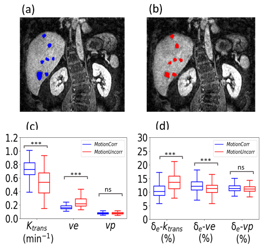
We chose seven ROIs within tumors for the motion corrected and uncorrected data separately, because respiratory motion can lead to a shift of the anatomy compared to the corrected data. The parameter estimates and relative epistemic uncertainties of these regions were investigated to evaluate the effect of motion.
RESULTS
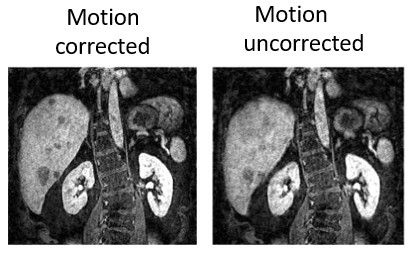
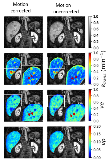
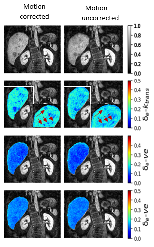
Figure 1 shows an exemplary DCE-MR slice of a liver for motion corrected and uncorrected data. The estimated physiological parameters and relative epistemic uncertainty maps of each parameter for motion corrected and uncorrected images are shown in Figure 2 and Figure 3, respectively. Tumors are characterized by high $$$k_{trans}$$$ and $$$vp$$$ and low $$$ve$$$ values. The epistemic uncertainties for motion uncorrected data increased especially in tumor regions for $$$k_{trans}$$$ as shown in Figure 3. The arrows show the increase in the epistemic uncertainty in corresponding regions of tumor for the motion corrected and uncorrected data. Figure 4a,b shows the different ROIs selected for tumor for motion corrected and uncorrected data, respectively. $$$k_{trans}$$$ was 39.59 ± 32.16% higher for the motion corrected than the uncorrected data. $$$ve$$$ was smaller by 24.85 ± 10.57% for the motion corrected than the uncorrected data with p-value <0.001. In addition, the epistemic uncertainty for $$$k_{trans}$$$ increased by 15.13 ± 11.18% when motion was not corrected. The epistemic uncertainty for $$$ve$$$ was also statistically significant different between corrected and uncorrected data but the difference was very small (10.30 ± 7.13%). There was no significant difference for parameter and uncertainty estimate of $$$vp$$$ (p-value >0.05) in both motion corrected and uncorrected data.DISCUSSION
The physiological parameter maps for motion uncorrected data illustrated the impact of motion, which led to an underestimation of $$$k_{trans}$$$ and overestimation of $$$ve$$$, especially for small tumors4. In the motion uncorrected images, the epistemic uncertainty increased due to motion. Motion altered the shape of the CTCs and because the network was not trained with motion impaired data, the epistemic uncertainty increased. In particular, this was shown with an increase in the epistemic uncertainty of $$$k_{trans}$$$ in regions of tumors for motion uncorrected data. Unlike $$$ve$$$ and $$$vp$$$, $$$k_{trans}$$$ was more sensitive to motion.CONCLUSION
This work for the first time demonstrated a link between uncertainty estimates obtained by DL and motion artefacts for DCE-MR of the liver. Motion correction improved estimation of the physiological parameters in the liver. This is confirmed by the additional epistemic uncertainty estimates which provided sensitive markers for motion artefacts.Acknowledgements
We would like to acknowledge funding from German Research Foundation, project number GRK2260, BIOQIC.References
1. Türkbey, B., Thomasson, D., Pang, Y., Bernardo, M. & Choyke, P. L. The role of dynamic contrast-enhanced MRI in cancer diagnosis and treatment. Diagn. Interv. Radiol. 16, 186–192 (2010).
2. Wake, N. et al. Accuracy and precision of quantitative DCE-MRI parameters: How should one estimate contrast concentration? Magn. Reson. Imaging 52, 16–23 (2018).
3. Jerosch-Herold, M. Quantification of myocardial perfusion by cardiovascular magnetic resonance. J. Cardiovasc. Magn. Reson. Off. J. Soc. Cardiovasc. Magn. Reson. 12, 57 (2010).
4. Ippoliti, M. et al. 3D nonrigid motion correction for quantitative assessment of hepatic lesions in DCE-MRI. Magn. Reson. Med. 82, 1753–1766 (2019).
5. Jansen, M. J. A. et al. Evaluation of motion correction for clinical dynamic contrast enhanced MRI of the liver. Phys. Med. Biol. 62, 7556 (2017).
6. Tofts, P. S. et al. Estimating kinetic parameters from dynamic contrast-enhanced t1-weighted MRI of a diffusable tracer: Standardized quantities and symbols. J. Magn. Reson. Imaging 10, 223–232 (1999).
7. Valdenegro-Toro, M. & Mori, D. S. A Deeper Look into Aleatoric and Epistemic Uncertainty Disentanglement. in 2022 IEEE/CVF Conference on Computer Vision and Pattern Recognition Workshops (CVPRW) 1508–1516 (2022). doi:10.1109/CVPRW56347.2022.00157.
8. Barbano, R., Arridge, S., Jin, B. & Tanno, R. Chapter 26 - Uncertainty quantification in medical image synthesis. in The MICCAI Society book Series (eds. Burgos, N. & Svoboda, D. B. T.-B. I. S. and S.) 601–641 (Academic Press, 2022). doi:https://doi.org/10.1016/B978-0-12-824349-7.00033-5.
9. Peressutti, D. et al. A novel Bayesian respiratory motion model to estimate and resolve uncertainty in image-guided cardiac interventions. Med. Image Anal. 17, 488–502 (2013).
Figures