0920
Blood volume based functional MRI using velocity selective pulses and SERIOS readout1Radiology, University of Michigan, Ann Arbor, MI, United States
Synopsis
Keywords: New Signal Preparation Schemes, Brain, fmri
We investigate a new fast readout sequence to collect blood volume image time series in using two different velocity selective pulses. We compare two classes of pulses that target the blood or the stationary tissue. The two approaches are demonstrated and compared in a simple viso-motor blood volume based functional MRI experiment.Introduction
Layer specific FMRI typically uses arterial volume changes to detect and differentiate activity at different neuronal layers. Previous approaches to dynamic blood volume imaging include VASO and VAPER [Lu 2012, Huber 2017]. These methods are challenged by speed and SNR, particularly at clinical field strengths. Previous work by Qin et al [Qin 2019, Li 2021] has shown that velocity selective pulses are effective for blood volume imaging at rest. Here, we combine this approach with a new fast readout scheme to demonstrate that two different types of velocity selective pulses can be leveraged to achieve the necessary arterial blood volume contrast for functional MRI. We demonstrate their feasibility in a custom built flow phantom and in healthy volunteers undergoing a visuo-motor stimulation paradigm.Methods
All experiments were conducted at 3 tesla, using a UHP scanner (GE medical systems, Waukesha , WI) and a 32-channel coil (Nova Medical Systems). In all experiments, a time series of images was collected preceded by a spin preparation pulse. The readout portion of the pulse sequence consists of a fast Spin Echo with Rotating In-Out Spiral (SERIOS) trajectory.Image reconstructions were carried out using an iterative model-based method, taking into account coil sensitivities, using routines in the MIRT reconstruction toolbox ( Fessler 2022)
Image time series were acquired preceded by one of the following preparation pulses 100 ms before readout: (1) FTVSS: Fourier Transform velocity selective saturation similar to (Qin 2019) followed by a crusher gradient. This pulse aims to saturate signals from stationary tissue, and minimally affect spins moving faster than 1 cm/s (based on FWHM of velocity profile). (2) BIR-8: Velocity Selective B-field Insensitive Rotation pulses with 8 lobes (BIR-8) pulses (Guo 2014) to saturate signals from spins moving faster than 1 cm/s (based on zero crossing of velocity profile).
Importantly, no control/tag image pairs were collected as in previous velocity selective ASL experiments. The whole time series were collected under ‘labeling’ conditions in both cases.
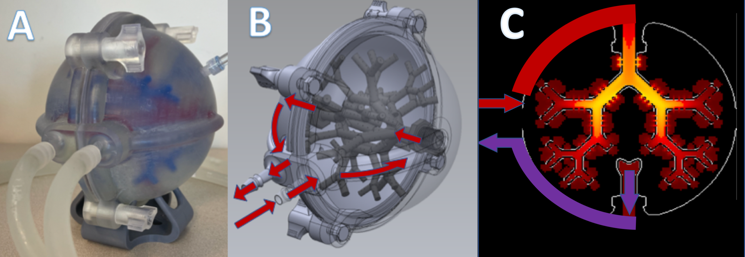
Perfusion phantom Experiment: A total of four time series of images were collected from a 3D printed perfusion phantom (see figure 1) to test the feasibility and effectiveness of each of the two schemes. Input flow velocity was controlled using a peristaltic pump set to zero and 20cm/s (?). The readout used four trains of 14 echoes acquired after the preparation pulse. (TR = 5 s, TE = 29ms, matrix size = 64 x 64 x 64, FOV = 24 cm, readout time = 420.5 ms per segment). A total of 10 frames (20 seconds per frame) were acquired per time series. The first two frames were acquired without any VS pulses.
Human activation Experiment: Healthy volunteers (N=1) were scanned using both methods, while they performed a visuo-motor activation paradigm for 5 minutes (30 seconds off, 30 seconds on. Complex finger tapping during flashing (8Hz) checkerboard visual stimulus. ). The SERIOS readout used a single train of 17 echoes 100ms after the VS preparation pulse (TR = 3 sec, TE = 48.5 ms, matrix size = 64 x 64 x 64, FOV = 24 cm, total readout time = 849 ms)
Results
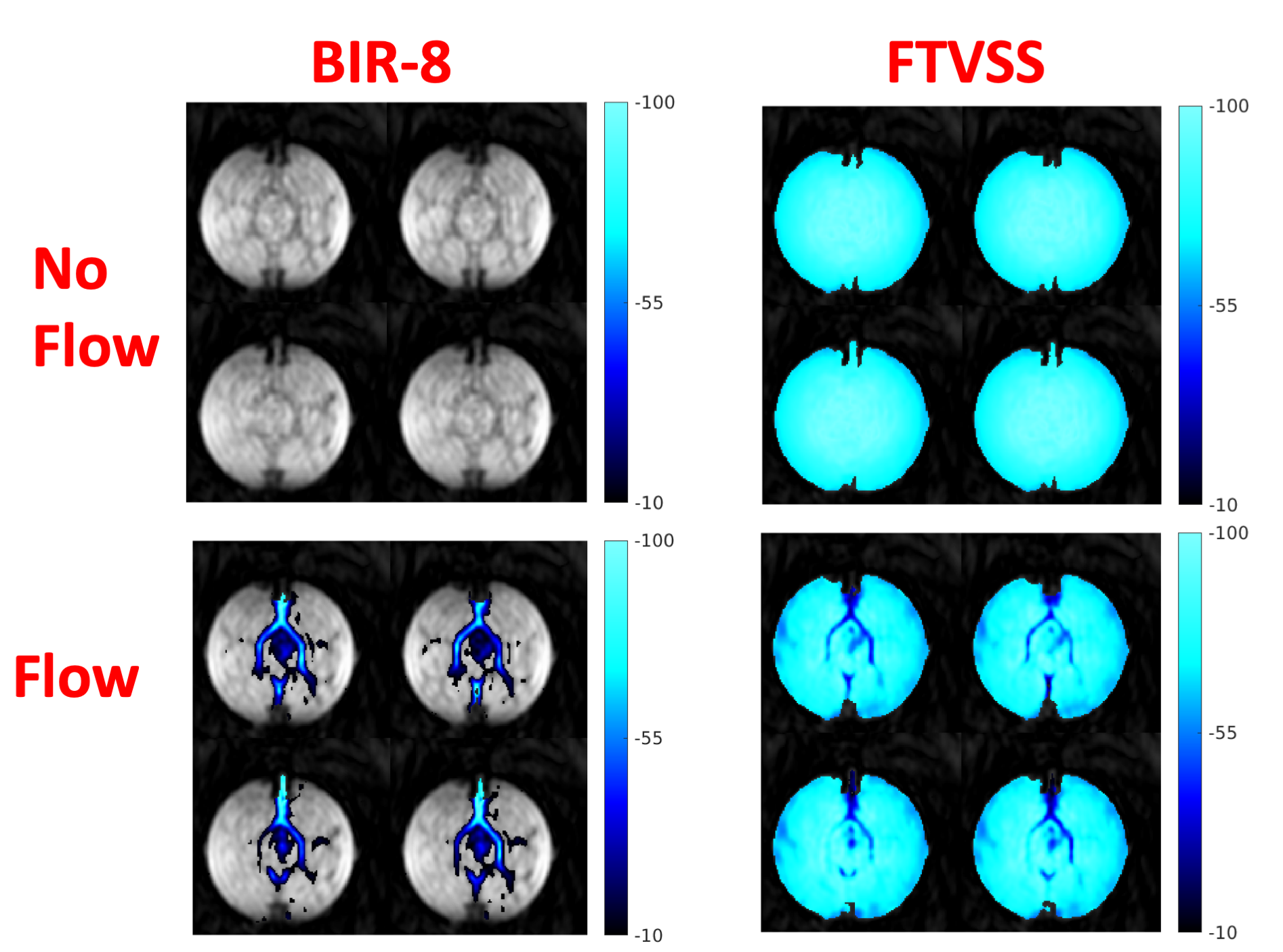
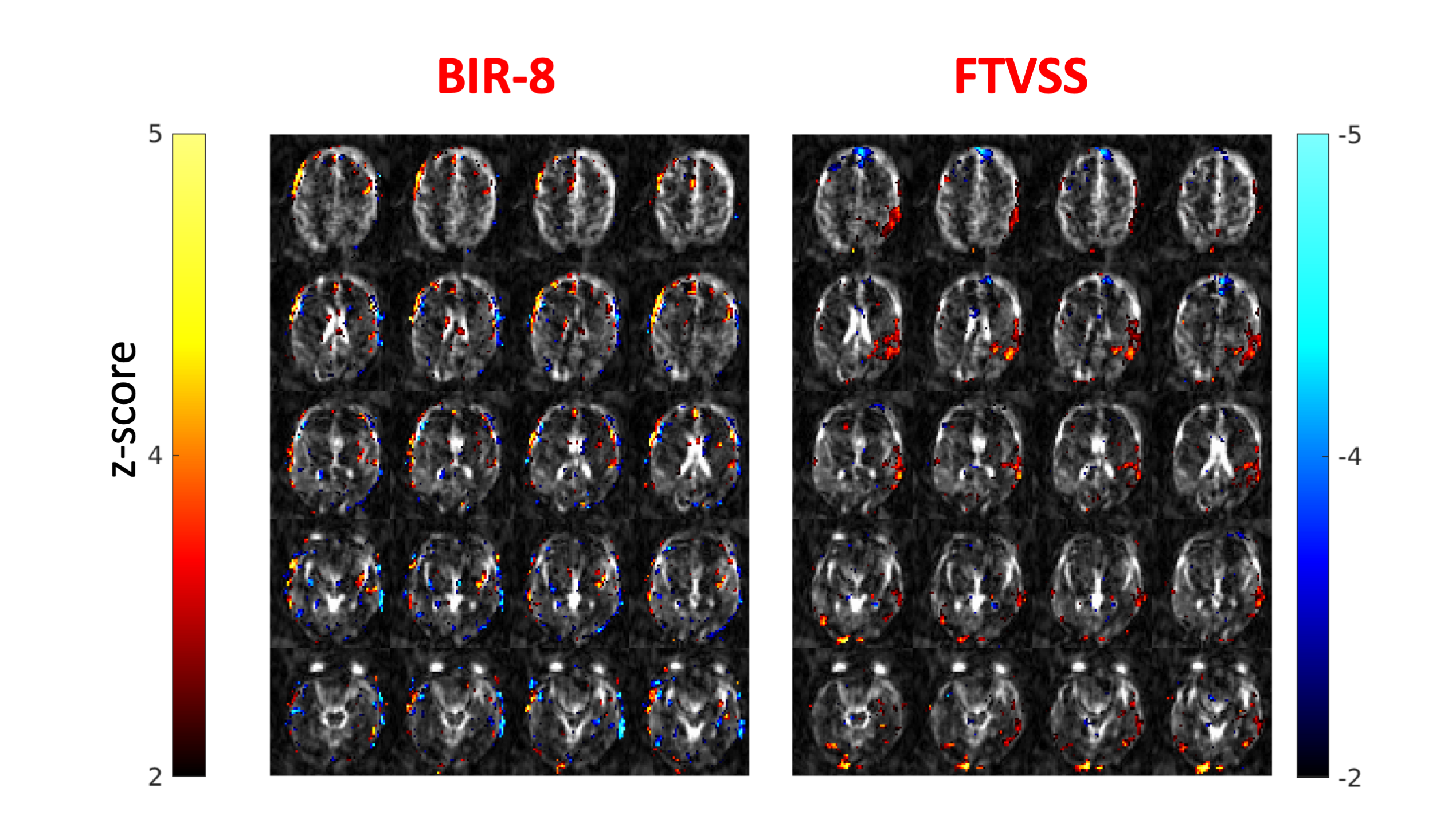
Figure 2 shows the effect of each type of VS pulse on the phantom. In the case of the BIR-8 pulses, the signal in the phantom’s ‘arteries’ is attenuated when the flow is turned on. In the case of the FT-VSS pulses the stationary spins are saturated, while spins moving along the encoding direction (top to bottom in the image) experience much less attenuation.Figure 3 shows results of activation studies. Image quality using a single shot SERIOS readout within 800 ms was excellent, considering the isotropic 64 x 64 x 64 coverage . FTVSS pulses significantly produced increase in signal during activation. Unexpectedly, BIR-8 pulses did not produce consistent signal changes during activation.
Discussion
While the phantom studies produced the expected signal attenuations. Only the FTVSS pulses produced consistent activation maps. We hypothesize that this is in part due to the background suppression that results from saturating stationary and slow moving spins. More research is needed to optimize preparation pulses and timing parameters in the FMRI acquisition. However, this method is a promising approach to blood volume based FMRI, which is an important tool for layer specific FMRI studies.Acknowledgements
This work is supported NIH grant R01 NS 112233References
Lu H, van Zijl PCM (2012) A review of the development of Vascular-Space-Occupancy (VASO) fMRI. Neuroimage 62:736–742. https://doi.org/10.1016/j.neuroimage.2012.01.013
Huber L, Handwerker DA, Jangraw DC, et al (2017) High-Resolution CBV-fMRI Allows Mapping of Laminar Activity and Connectivity of Cortical Input and Output in Human M1. Neuron 96:1253-1263.e7. https://doi.org/10.1016/j.neuron.2017.11.005
Qin Q, Qu Y, Li W, et al (2019) Cerebral blood volume mapping using Fourier-transform–based velocity-selective saturation pulse trains. Magn Reson Med 81:3544–3554. https://doi.org/10.1002/mrm.27668
Li W, Liu D, van Zijl PCM, Qin Q (2021) Three-dimensional whole-brain mapping of cerebral blood volume and venous cerebral blood volume using Fourier transform–based velocity-selective pulse trains. Magn Reson Med 86:1420–1433. https://doi.org/10.1002/mrm.28815
Guo J, Meakin J a., Jezzard P, Wong EC (2014) An optimized design to reduce eddy current sensitivity in velocity-selective arterial spin labeling using symmetric BIR-8 pulses. Magn Reson Med 73:1085–1094. https://doi.org/10.1002/mrm.25227
Fessler, JA, et al. "Michigan Image Reconstruction Toolbox https://github.com/JeffFessler/mirt, downloaded 2022.
Figures