0898
Semiautomated Pelvic Lymph Node Treatment Response Evaluation for Patients with Advanced Prostate Cancer: Based on MET-RADS-P Guidelines1peking university first hospital, Beijing, China
Synopsis
Keywords: Cancer, Prostate, metastases
This retrospective study aims to develop and evaluate a deep learning-based algorithm for semiautomated treatment response assessment of pelvic lymph nodes. A total of 162 patients who had undergone at least two scans for follow-up assessment after advanced prostate cancer metastasis treatment were enrolled. A previously reported deep learning model was used to perform automated segmentation of pelvic lymph nodes. Our results showed that the accuracies of automated segmentation-based response assessment were high for all the target lesions, nontarget lesions and nonpathological lesions according to MET-RADS-P criteria and achieved good consistency with the attending radiologist and fellow radiologist.introduction
Introduction: The evaluation of treatment response according to METastasis Reporting and Data System for Prostate Cancer (MET-RADS-P) criteria is an important but time-consuming task for patients with advanced prostate cancer. A deep learning-based algorithm has the potential to assist with this assessment.Objective
Objective: To develop and evaluate a deep learning-based algorithm for semiautomated treatment response assessment of pelvic lymph nodes.Methods
Methods: A total of 162 patients who had undergone at least two scans for follow-up assessment after APC metastasis treatment were enrolled. A previously reported deep learning model was used to perform automated segmentation of pelvic lymph nodes. The performance of the deep learning algorithm was evaluated using the Dice similarity coefficient (DSC) and volumetric similarity (VS). The consistency of the short diameter measurement with the radiologist was evaluated using Bland-Altman plotting. Based on the segmentation of lymph nodes, the treatment response was assessed automatically with a rule-based program according to the MET-RADS-P criteria. Kappa statistics were used to assess the accuracy and consistency of the treatment response assessment by the deep learning model and two radiologists [attending radiologist (R1) and fellow radiologist (R2)].Results
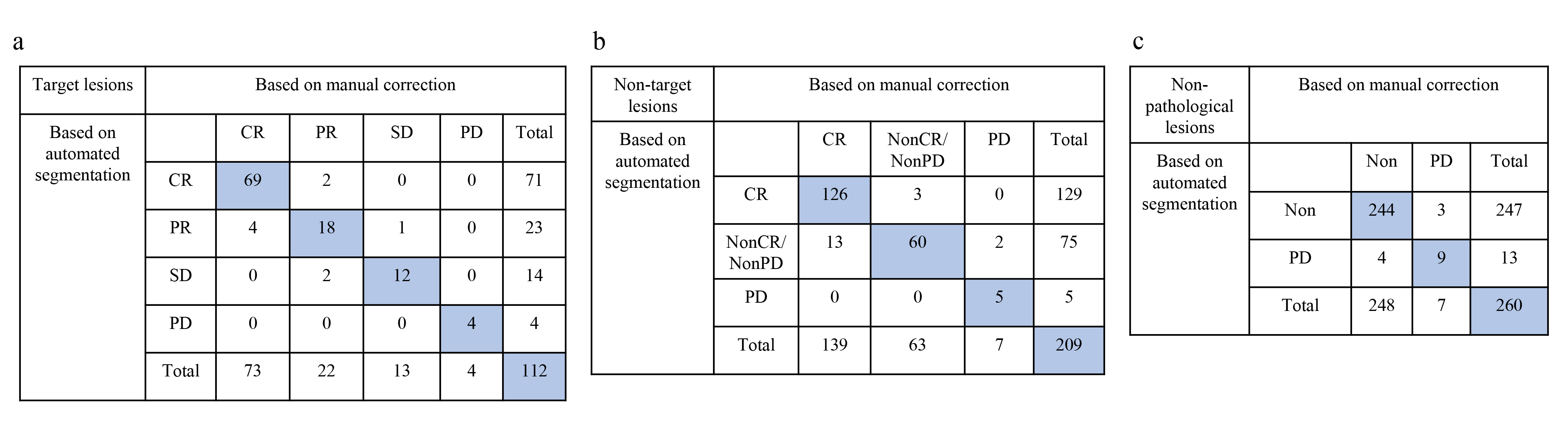
Results: The mean DSC and VS of the pelvic lymph node segmentation were 0.82 ± 0.09 and 0.88 ± 0.12, respectively. Bland-Altman plotting showed that most of the lymph node measurements were within the upper and lower limits of agreement (LOA). The accuracies of automated segmentation-based assessment were 0.92 (95% CI: 0.85-0.96), 0.91 (95% CI: 0.86-0.95) and 75% (95% CI: 0.46-0.92) for target lesions, nontarget lesions and nonpathological lesions, respectively. The consistency of treatment response assessment based on automated segmentation and manual annotation was excellent for target lesions [K value: 0.92 (0.86-0.98)], good for nontarget lesions [0.82 (0.74-0.90)] and moderate for nonpathological lesions [0.71 (0.50-0.92)].Discussion
Discussion:MET-RADS-P is a guideline for the treatment response evaluation of systemic metastases of patients with APC, which involves the evaluation of primary focus, bone metastases, lymph node metastases and organ metastases [1]. Analysis of lymph node metastases in the pelvis is crucial for clinical practice and drug studies in patients with APC, which is the most common metastatic site [2]. In this study, the established semiautomatic pelvic lymph node treatment response evaluation process according to MET-RADS-P criteria included two parts. First, a previously established pelvic lymph node segmentation model was used to perform the automatic segmentation of lymph nodes. The model achieved good segmentation performance here, which is similar to the segmentation results reported in previous literature, especially the target lesions, further highlighting its potential usefulness.Second, based on the quantitative measurements obtained from the automated segmentation, we can directly evaluate the treatment response according to MET-RADS-P criteria, which can be more practical in clinical settings. A clinical radiology report provides a qualitative narrative, but does not provide standardized, quantitative information about the patient's progress or response to treatment [3]. Natural language processing and deep learning models have been employed in previous studies to estimate responses from clinical text [4-5]. These approaches can be feasible for quantitative assessment related to MET-RADS-P criteria but can be indirect.Conclusion
Conclusion: The deep learning-based semiautomated algorithm showed high accuracy for the treatment response assessment of pelvic lymph nodes and demonstrated comparable performance with radiologists.Acknowledgements
The authors gratefully acknowledge the technical support of Yaofeng Zhang and Jialun Li from Beijing Smart Tree Medical Technology Co. Ltd.References
1. Padhani AR, Lecouvet FE, Tunariu N, Koh DM, De Keyzer F, Collins DJ, et al. METastasis Reporting and Data System for Prostate Cancer: Practical Guidelines for Acquisition, Interpretation, and Reporting of Whole-body Magnetic Resonance Imaging-based Evaluations of Multiorgan Involvement in Advanced Prostate Cancer. Eur Urol. 2017,71(1):81-92.
2. Kim YJ, Song C, Eom KY, Kim IA, Kim JS. Lymph node ratio determines the benefit of adjuvant radiotherapy in pathologically 3 or less lymph node-positive prostate cancer after radical prostatectomy: a population-based analysis with propensity-score matching. Oncotarget. 2017,8(66):110625-34.
3. Arbour KC, Luu AT, Luo J, Rizvi H, Plodkowski AJ, Sakhi M, et al. Deep Learning to Estimate RECIST in Patients with NSCLC Treated with PD-1 Blockade. Cancer Discov. 2021,11(1):59-67.
4. Chen MC, Ball RL, Yang L, Moradzadeh N, Chapman BE, Larson DB, et al. Deep Learning to Classify Radiology Free-Text Reports. Radiology. 2018,286(3):845-52.
5. Bozkurt S, Alkim E, Banerjee I, Rubin DL. Automated Detection of Measurements and Their Descriptors in Radiology Reports Using a Hybrid Natural Language Processing Algorithm. J Digit Imaging. 2019,32(4):544-53.
Figures
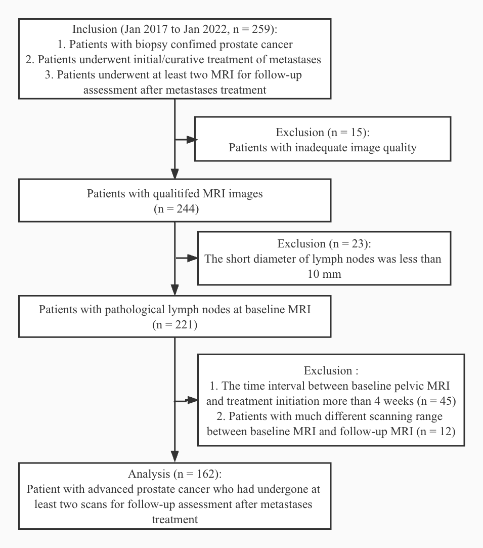
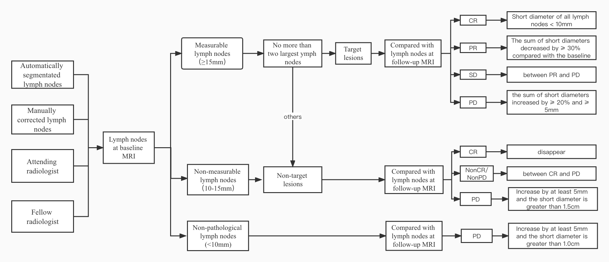
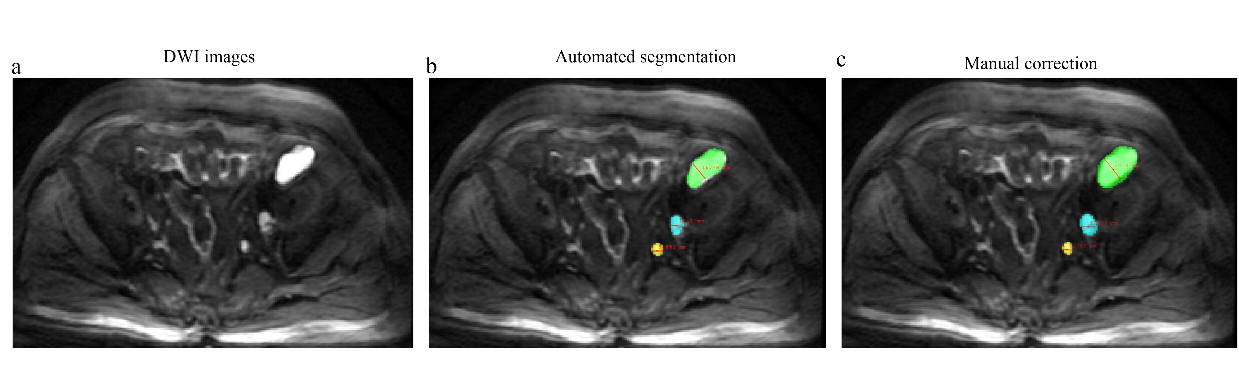
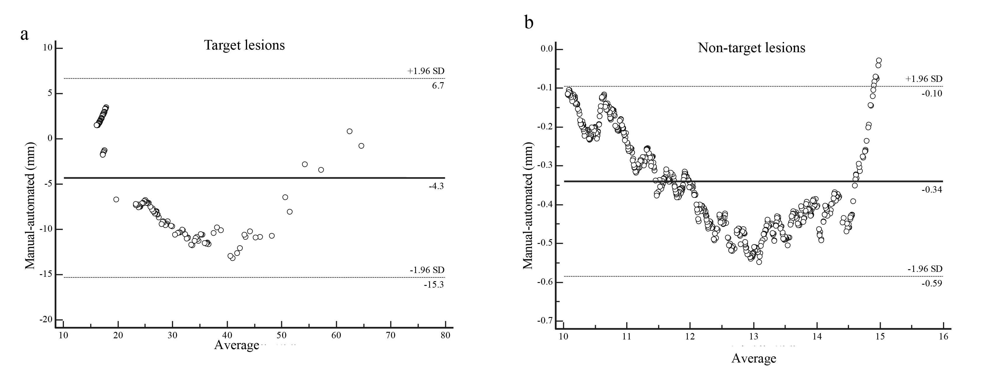
Figure 4. Agreement between the automatically segmented and manually segmented lymph nodes
(a) target lesions; (b) nontarget lesions.