0895
In-Vivo Simultaneous Hyperpolarized 129Xe MRI and [15O]-water PET Multi-Modal Imaging: A Proof of Concept Study
Ramanpreet K. Sembhi1, Matthew S. Fox1,2, Heeseung Lim2,3, Justin W. Hicks2,4, Shawn N. Whitehead5, Jonathan D. Thiessen2,4, Grace Parraga4,6, and Alexei V. Ouriadov1,2,7
1Department of Physics and Astronomy, The University of Western Ontario, London, ON, Canada, 2Lawson Health Research Institute, London, ON, Canada, 3Siemens Healthcare Limited, London, ON, Canada, 4Department of Medical Biophysics, The University of Western Ontario, London, ON, Canada, 5Department of Anatomy and Cell Biology, The University of Western Ontario, London, ON, Canada, 6Robarts Research Institute, London, ON, Canada, 7School of Biomedical Engineering, The University of Western Ontario, London, ON, Canada
1Department of Physics and Astronomy, The University of Western Ontario, London, ON, Canada, 2Lawson Health Research Institute, London, ON, Canada, 3Siemens Healthcare Limited, London, ON, Canada, 4Department of Medical Biophysics, The University of Western Ontario, London, ON, Canada, 5Department of Anatomy and Cell Biology, The University of Western Ontario, London, ON, Canada, 6Robarts Research Institute, London, ON, Canada, 7School of Biomedical Engineering, The University of Western Ontario, London, ON, Canada
Synopsis
Keywords: PET/MR, Hyperpolarized MR (Gas)
In this proof-of-concept study, simultaneous 129Xe-based MRI and [15O]-water PET images were collected and compared from rat brain. For initial validation phase in phantoms, we have dissolved 129Xe in [15O]-water to simultaneously use both imaging modalities and confirm that PET/MRI images reflect the true density of the 15O/129Xe. The comparison results show similarity between both imaging modalities and tracers, moving towards the next step in validating the Xenon imaging technique as a potential for brain perfusion measurement. The acquisitions were carried out using a 3T PET/MRI (Siemens Biograph mMR).INTRODUCTION
Inhaled hyperpolarized (HP) 129Xe MRI is a non-invasive imaging method, currently used to measure lung structure and function.1,2 This MRI approach helps to obtain simultaneous ventilation/perfusion lung measurements of functional gas-exchange within the lungs due to the high-natural-solubility of xenon (Ostwald solubility of 0.173) in lung-tissue compared to other imaging gases. A variety of physical properties of 129Xe isotope makes it beneficial for brain imaging, thus, it can serve as a new probe for brain blood flow, grey and white matter mapping4,5 and functional measurements. These measurements are possible due to a distinct and large range of chemical shifts (~200ppm) of 129Xe when residing within lung tissue, brain tissue and red blood cells compared to the gas phase. 129Xe-based imaging aims to improve sensitivity where traditional MRI may fall short, and by reaching beyond the resolution limitations of positron emission tomography (PET). [15O]-water PET is the gold-standard imaging method for determining cerebral-perfusion.6-8 The half-life of 15O isotopes is short (~2min). These are freely-diffusible (i.e., [15O]-water) and like 129Xe, is metabolically inert. These properties make it highly-attractive for non-invasive imaging-based perfusion measurements. In this proof-of-concept study, we propose to use the gold-standard [15O]-water PET and 129Xe MRI to demonstrate in-vivo feasibility of a single-shot, multi-modal-imaging approach utilizing simultaneous PET/MRI. This will serve as a PET-Xenon MRI imaging platform for validation work performing 129Xe-based brain perfusion techniques, directly and simultaneously with [15O]-water PET.METHODS
Double Tracer Phantom setup: We used the [15O]-water solution (30mL) contained in a 60mL plastic syringe to dissolve 30mL of the hyperpolarized 129Xe gas. After dissolving, all leftover xenon gas was removed from the syringe.Double Tracer in-vivo setup: Rats were induced with 5% isoflurane and oxygen and maintained at 2%. A 24g tail vein catheter was inserted for delivery of the [15O]- water / 129Xe mixture. Once prepared, rats were transferred to a custom animal bed that interfaces with the PET insert animal positioning system and couples with the inserted RF coils, co-localizing the rat brain for all modalities.
Hyperpolarized 129Xe MRI: Hyperpolarized 129Xe gas was obtained from a turn-key, spin-exchange polarizer system (Polarean 9800 129Xe polarizer). The initial 129Xe polarization was 15%. 129Xe dissolved phase images were acquired in a 3T PET/MRI (Siemens Biograph mMR, Siemens Healthineers, Erlangen, Germany) scanner using whole-body gradients and a homebuilt rat-sized RF coil tuned to 34.09MHz. A Fast-Gradient-Recalled-Echo sequence was utilized for phantom (Matrix Size=64x64; Slice thickness=250mm; TE/TR=2.04/20ms; BW=660Hz/pixel; Flip angle=11o FOV=150x150mm2) and rat (Matrix Size=64x64; Slice thickness=250mm; TE/TR=3.44/100ms; BW=660Hz/pixel; Flip angle=11o FOV=70x70mm2) scans. Three consecutive axial images were acquired during the PET scan. The total scan time was 1sec per image for the phantom and 14sec for rat.
[15O]-water PET phantom Scan: [15O]-water PET data (half-life-time of 2 min) were acquired simultaneously with 129Xe MRI for 600 sec using the integrated PET system in the 3T PET/MRI. PET data were reconstructed into ten 60sec frames using a 3D iterative routine (OSEM) with three iterations, 21 subsets, in-plane resolution of 1.04 x 1.04 mm2 and a slice thickness of 2.03mm.
[15O]-water PET in-vivo Scan: PET imaging was obtained using a small animal MRI compatible PET insert (Cubresa Inc.) (Fig.1) with an FOV of 58.9mm (trans-axial) by 67.2mm (axial). We waited until the 0.5mL xenon/water mixture was 25MBq, then injected into the tail vein. PET data were reconstructed into ten 120sec frames using a 3D iterative routine (OSMAPOSL) with eight iterations, 25 subsets, in-plane resolution of 1.04 x 1.04 mm2 and a slice thickness of 2.03mm.
RESULTS
Fig.2 shows 2D axial 129Xe MRI images and [15O]-water PET images acquired simultaneously. 129Xe/PET images indicate that the diameter of the phantom from both PET and MRI images are similar. The 129Xe image demonstrates a sufficient SNR level (80) suggesting that 3D 129Xe imaging is possible. Figs. 3 and 4 show the anatomical-proton and [15O]-water-PET-perfusion images of rat-brain.DISCUSSION AND CONCLUSIONS
The results of this proof-of-concept study clearly indicate the feasibility of simultaneous hyperpolarized 129Xe MRI and [15O]-water PET measurements. To our knowledge, for the first time ever, we have demonstrated that hyperpolarized 129Xe dissolved in [15O]-water can be used in multi-modal imaging. The in-vivo demonstration did not bring the desired 129Xe image quality because of various reasons. In general, it is a very complicated measurement which is hard to execute considering a synchronization of the two tracers preparation mixing and injection. There was a time delay between the usage of both tracers leading to the significant polarization decay of the 129Xe gas from the initial 15% polarization to 7% at the time of mixing and injecting. We plan to significantly increase the initial xenon polarization (up to 50%) and minimize the xenon-waiting-time. With all improvements we should be able to use 129Xe as a non-radioactive tracer, providing similar and complimentary information as 15O PET may be a much more cost-effective alternative to PET for imaging stroke, brain cancer and other brain diseases and will significantly increase the number of 129Xe MRI clinical applications. Keeping in mind that 129Xe MRI should be FDA approved any moment from now and this opens the door for better diagnoses, treatment planning and treatment assessment of patients with chronic-lung, cardiac and neurodegenerative diseases.Acknowledgements
The authors would like to thank the research and financial supports received from BrainsCAN-Accelerator-Program, R5942A02 and the Natural-Sciences-and-Engineering-Research-Council of Canada, R5942A04.References
- Kaushik, S. S. et al. Single-breath clinical imaging of hyperpolarized (129)Xe in the airspaces, barrier, and red blood cells using an interleaved 3D radial 1-point Dixon acquisition. Magn Reson Med 75, 1434-1443, doi:10.1002/mrm.25675 (2016).
- Kaushik, S. S. et al. Probing the regional distribution of pulmonary gas exchange through single-breath gas- and dissolved-phase 129Xe MR imaging. J Appl Physiol (1985) 115, 850-860, doi:10.1152/japplphysiol.00092.2013 (2013).
- Moller, H. E. et al. MRI of the lungs using hyperpolarized noble gases. Magn Reson Med 47, 1029-1051, doi:10.1002/mrm.10173 (2002).
- Kershaw, J. et al. Confirming the existence of five peaks in 129Xe rat head spectra. Magn Reson Med 57, 791-797, doi:10.1002/mrm.21186 (2007).
- Wakai, A., Nakamura, K., Kershaw, J. & Kanno, I. In vivo MR spectroscopy of hyperpolarized Xe-129 in rat brain. International Congress Series 1265, 139-143, doi:10.1016/j.ics.2004.04.063 (2004).
- Fan, A. P., Jahanian, H., Holdsworth, S. J. & Zaharchuk, G. Comparison of cerebral blood flow measurement with [15O]-water positron emission tomography and arterial spin labeling magnetic resonance imaging: A systematic review. J Cereb Blood Flow Metab 36, 842-861, doi:10.1177/0271678X16636393 (2016).
- Ssali, T., Anazodo, U. C., Thiessen, J. D., Prato, F. S. & St Lawrence, K. A Noninvasive Method for Quantifying Cerebral Blood Flow by Hybrid PET/MRI. J Nucl Med 59, 1329-1334, doi:10.2967/jnumed.117.203414 (2018).
- Raichle, M. E., Martin, W. R. W., Herscovitch, P., Mintun, M. A. & Markham, J. Brain Blood Flow Measured with Intravenous H215O.: II. Implementation and Validation. Journal of Nuclear Medicine 24, 790-798 (1983).
- Wild, J. M. et al. P283 Hyperpolarised Gas MRI – a pathway to Clinical Diagnostic Imaging. Thorax 70, A220.223-A221, doi:10.1136/thoraxjnl-2015-207770.419 (2015).
- 129Xe MRI Clinical Trials Consortium. https://cpir.cchmc.org/XeMRICTC.
Figures
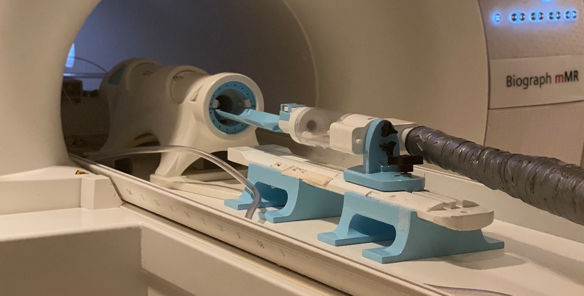
PET insert used for carrying out in-vivo scans placed on the 3T PET/MRI scanner patient table.
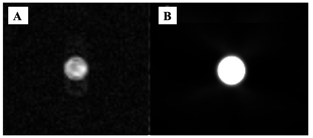
A) 2D axial 129Xe MRI image obtained for xenon dissolved in [15O]-water inside the syringe phantom. The SNR of the 129Xe image is 80. B) 2D axial [15O]-water PET image obtained with the syringe phantom. The PET acquisition was synchronized with the 129Xe MRI acquisition.
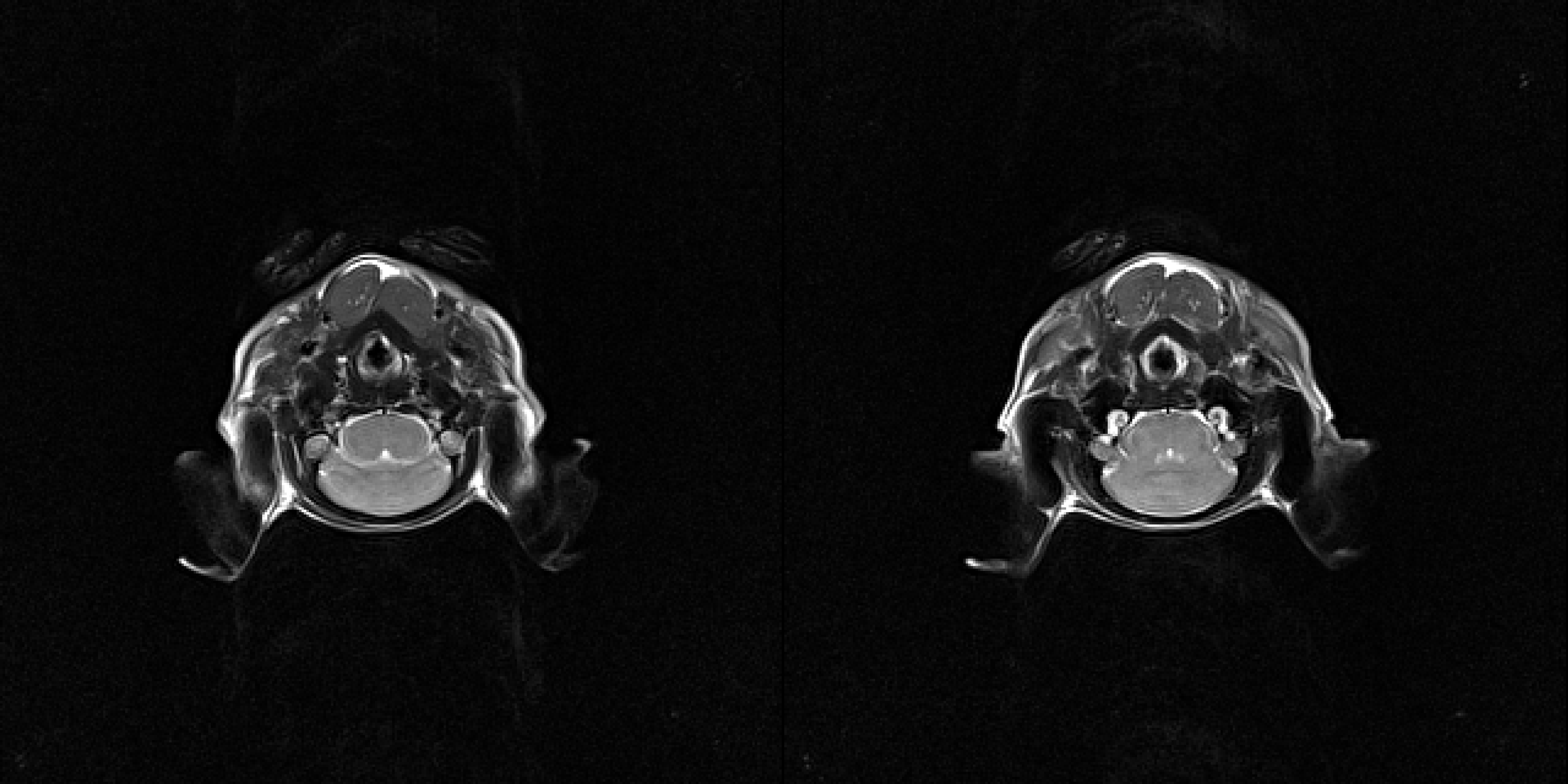
Two representative consequenced 1H images for rat brain were acquired using the T2-weighted Spin Echo Sequence. The FOV was 70x70mm2, Matrix size=128x128, TE/TR=97/2930ms and Slice thickness=1.5mm. Images obtained in the axial view.
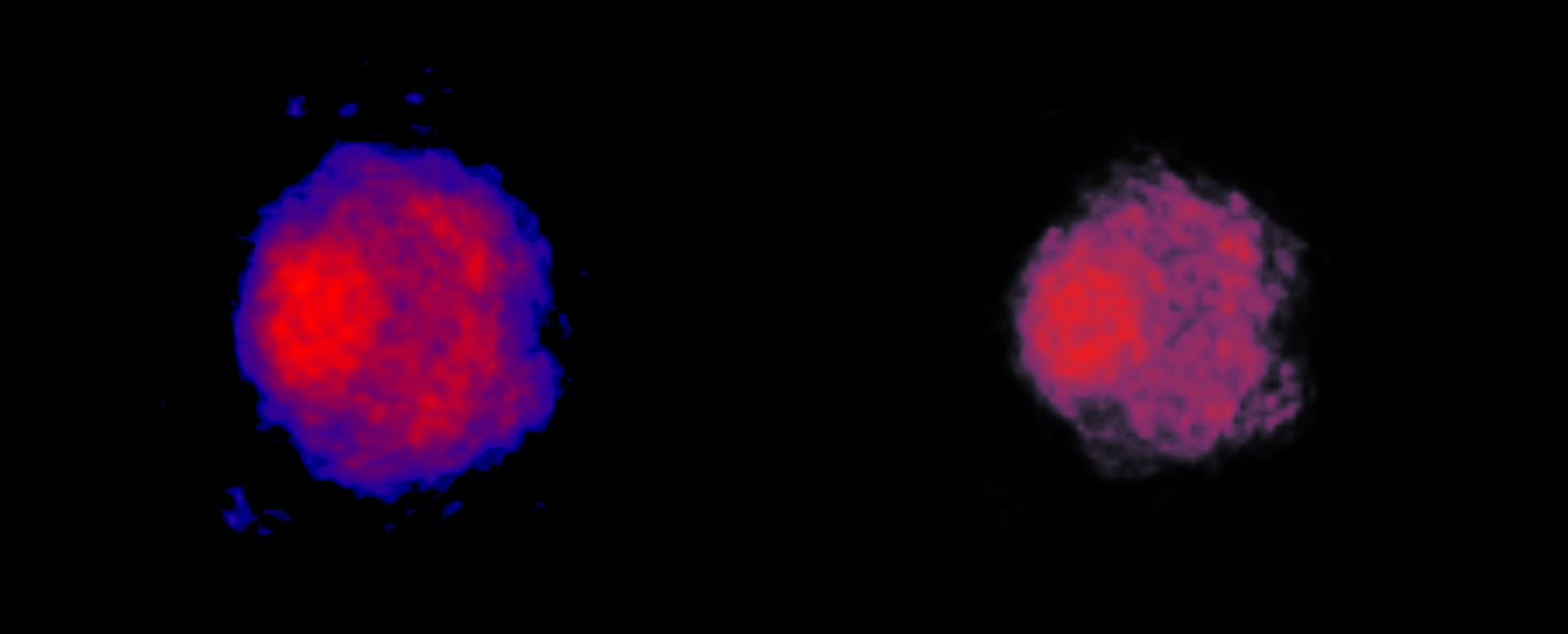
Two representative consequenced [15O]-water PET perfusion images obtained for rat brain.The total acquisition time was 20 minutes. A 3D iterative method (OSMAPOSL) with eight iterations, 25 subsets, in-plane resolution of 1.04 x 1.04 mm2, and a slice thickness of 2.03 mm was used to reconstruct the PET data into ten 120-second frames.
DOI: https://doi.org/10.58530/2023/0895