0851
Multi-Voxel PRESS of Hyperpolarized 13C-labelled Agents In Vivo1TUM School of Medicine, Department of Nuclear Medicine, Klinikum Rechts der Isar, Technical University of Munich, Munich, Germany, 2TUM School of Medicine, Institute of Diagnostic and Interventional Radiology, Klinikum Rechts der Isar, Technical University of Munich, Munich, Germany, 3German Cancer Consortium (DKTK, partner Site Munich), Munich, Germany
Synopsis
Keywords: Hyperpolarized MR (Non-Gas), Hyperpolarized MR (Non-Gas)
A newly developed multi-voxel point resolved spectroscopy (MV-PRESS) sequence was evaluated for the study of murine pancreatic ductal adenocarcinoma (PDAC) using hyperpolarized (HP) 13C-labelled molecules. First, the sequence was compared to standard 2D free-induction decay chemical shift imaging (2D FID-CSI) in vivo using HP [1-13C]pyruvate (PA). Then, multiple ROIs in mice with endogenous PDAC were selected based on anatomical 1H imaging. From those voxels, high resolution spectra using the MV-PRESS sequence were acquired to evaluate differences in pyruvate-to-lactate conversion. Finally, the sequence was used to determine the extracellular pH of tumor lesions and kidneys in the same animal model.
Introduction
Using hyperpolarization the signal of magnetic resonance based molecular imaging agents such as [1-13C]pyruvate or [1,5-13C2]Z-OMPD can be increased by a factor of up to 10.000[1]. Thereby, metabolic activity and pH can be determined non-invasively in vivo[2]. However, as the signal enhancement decays, efficient sequences to collect the spectra are needed. For high-spectral resolution the most commonly used method is a 2D FID-CSI[3]. Its limitation to a single slice represents a drawback, especially if multiple lesions are present or multiple organs need to be studied. For example, PDAC is a highly heterogonous disease and often distributed throughout the abdomen. In this work, the feasibility of a previously presented newly developed MV-PRESS sequence[4] for the study of endogenous PDAC in mice was evaluated.Methods
MR System: Preclinical 7T magnet, 1H/13C volume resonator (ID 31mm) or volume resonator (ID 72 mm) with a 13C surface receiver coil.Hyperpolarization: 22-24mg 14M [1-13C]pyruvate or 36mg 7M [1,5-13C2]Z-OMPD (Z-OMPD) polarized in a HyperSense DNP and then rapidly dissolved in buffer solution (for Z-OMPD deuterated).
Animal Subjects: Eight Ptf1aCre/wt;KRASLSL-G12D/wt;Trp53fl/fl mice[5] with endogenously grown PDAC tumors, anesthetized with isoflurane and injected with 80mM PA or 84mM Z-OMPD via a tail vein catheter.
Proton Imaging: T2-weighted RARE.
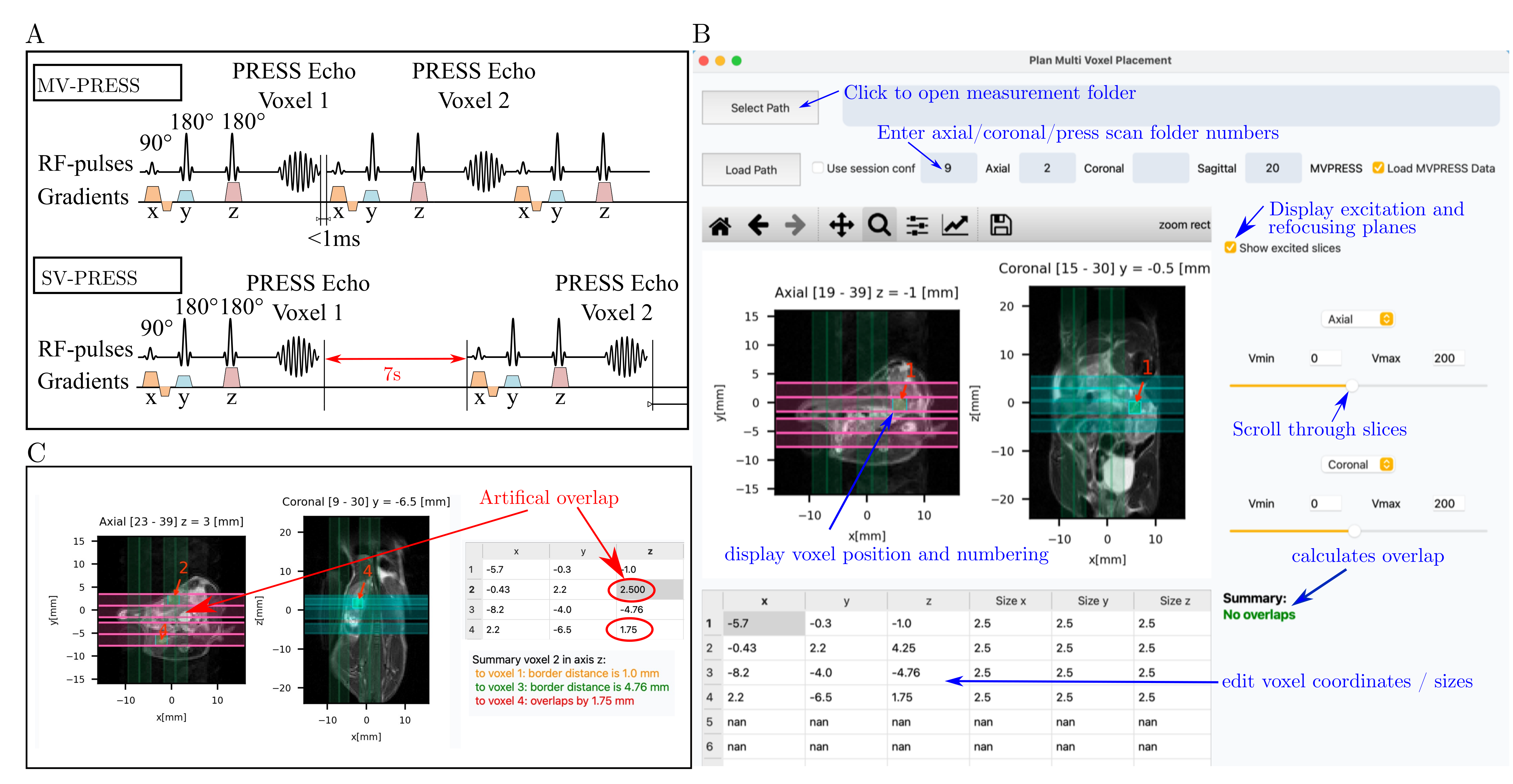
13C Spectroscopy: The MV-PRESS sequence consists of consecutive single PRESS voxels (Fig. 1 A). Scan time of the sequence could be decreased to less than 1.5s for 4 voxels, only limited by the duration of the collected spin echo. Planning of excitation and refocusing slices for voxels was done manually first leading to difficulties in voxel placement and overlap in initial experiments. To monitor this issue, a custom software was developed (Fig. 1B/C)[6].
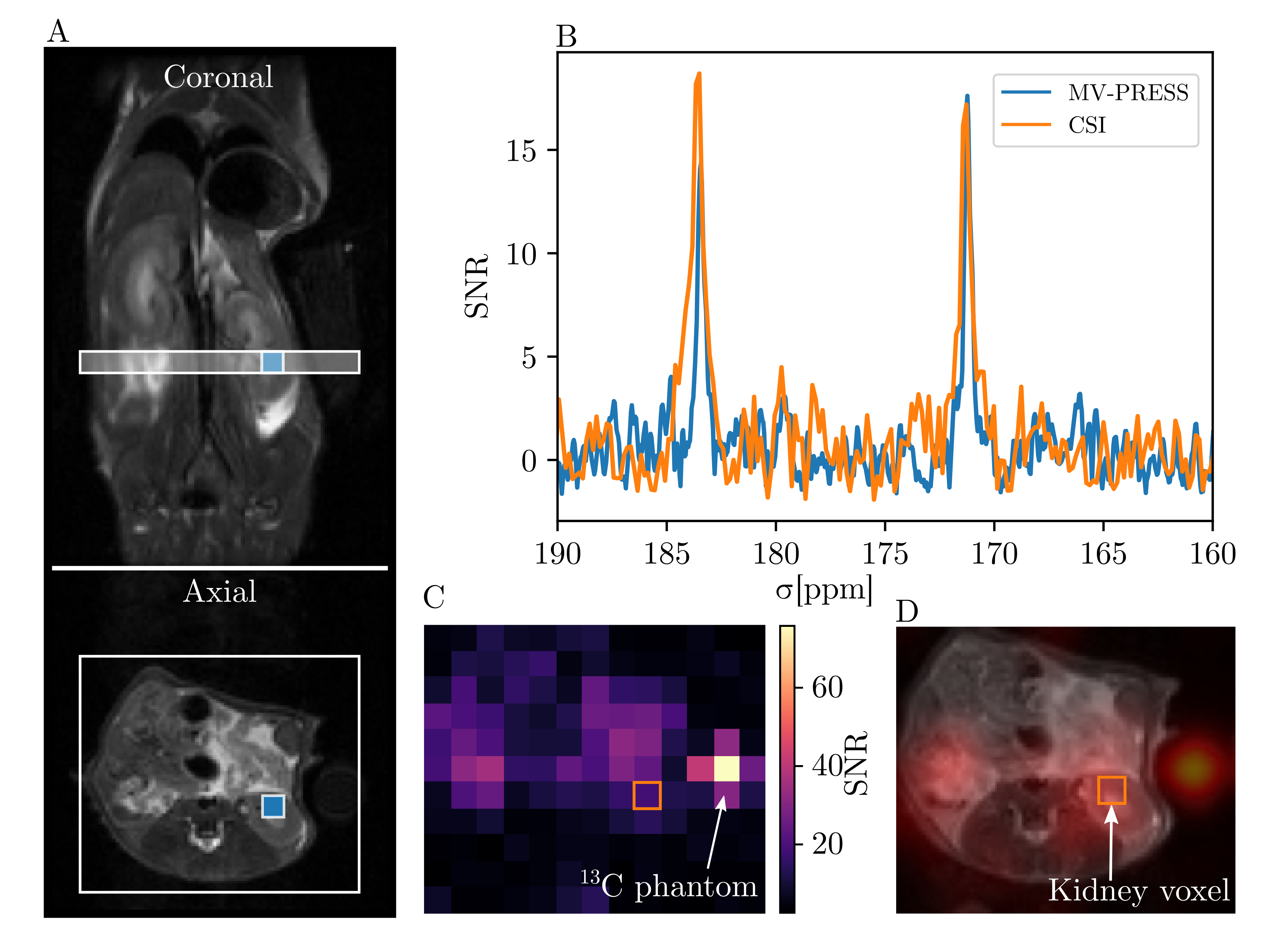
For the in vivo SNR comparison, spectra of hyperpolarized [1-13C]pyruvate were acquired using a 2D FID-CSI sequence (TR=90ms, TE=2.7ms, FA=12°, matrix size=13x11, in plane resolution=2x2mm3, slice thickness = 2mm, spec.acq.pts=256, RxBW=40ppm, spec.res=5.9 Hz/Pt, acq.time=12.9s) and a same-voxel-size MV-PRESS sequence (TR=1000ms, TE=16ms, 4.2 and 3.4kHz SLR pulses for excitation and refocusing, acq.pts=1024, RxBW=40ppm, acq.time/voxel=339ms and spec.res=1.5Hz/Pt). A 2° non-selective excitation preceded both sequences to compare different initial signal intensities affected by relaxation and polarization level. T1 decay constants of HP samples were measured in a 1T tabletop NMR after injection. For in vivo measurements to determine pyruvate to lactate conversion and pH, 2x2x2mm3 and 2.5x2.5x2.5mm3 voxels were measured starting 15-29s (PA) and 13-18s (Z-OMPD) after the end of the injection.
Data Analysis: Scan data was analysed using Python[7,8]. MRS Spectra were fitted to a Lorentzian model, and integrated for pyruvate/lactate ratios while for Z-OMPD, inter peak distances were calculated and translated to pH values using a logarithmic calibration function[2].
Results
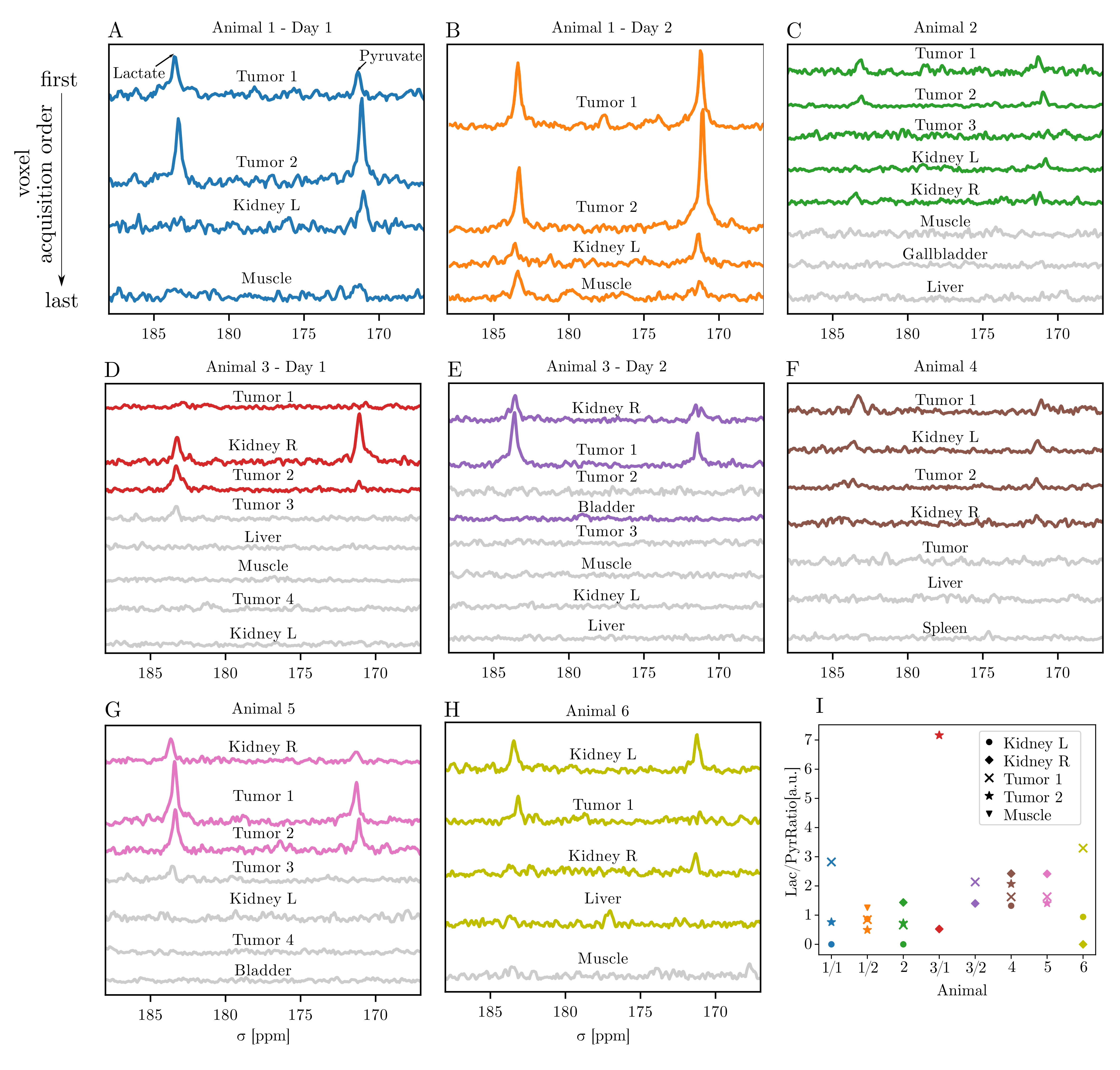
For SNR comparison between 2D FID-CSI and MV-PRESS two injections were performed within the same animal. First a spectrum for a MV-PRESS sequence with a voxel placed in the kidney was recorded (Fig. 2A/B). After 1 hour a second injection was given and a 2D FID-CSI with a slice covering the same location as the kidney voxel was recorded (Fig. 2A/D). Using the measured T1 value of each sample, timing of dissolution, injection and start of experiments as well as the 2° non-selective excitation, spectra show similar SNR (Fig. 2B).Next, the sequence was evaluated in 6 animals by placing voxels in tumor nodules, kidneys and other tissues depending on the anatomy. Spectra (Fig.3A-H) were fitted and lactate to pyruvate values calculated (Fig. 3I) showing an increase in lactate production for tumor nodules and muscles when compared to kidneys (mean lac/pyr: tumor=0.15 (n=13), kidney=0.12 (n=8), muscle=0.7 (n=2)) . Differences in lactate production for lesions within one animal were found (see Figure 3A). An overlap of slices was evaluated for voxels measured after the first interesting ROIs leading to signal loss (greyed out spectra).
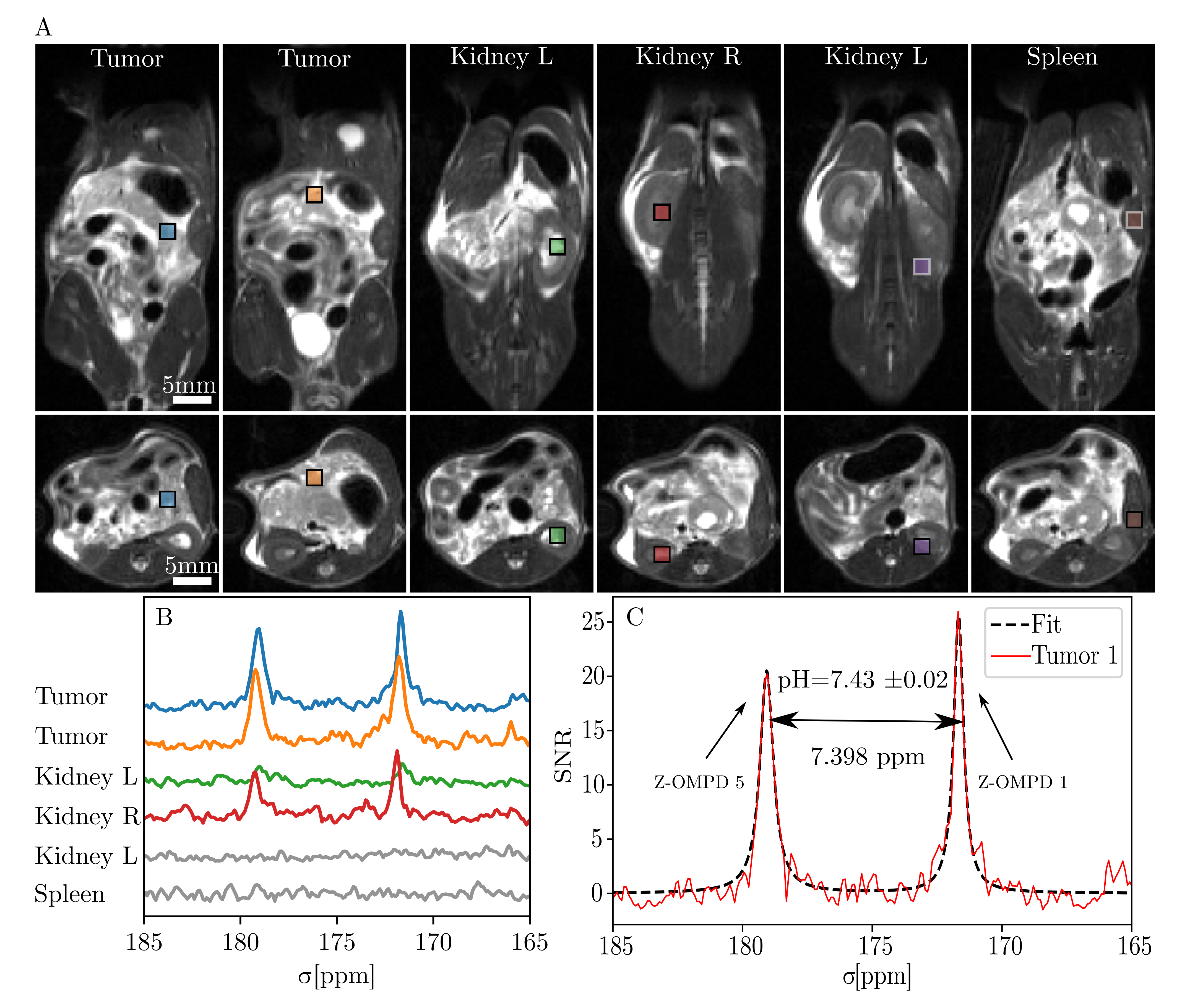
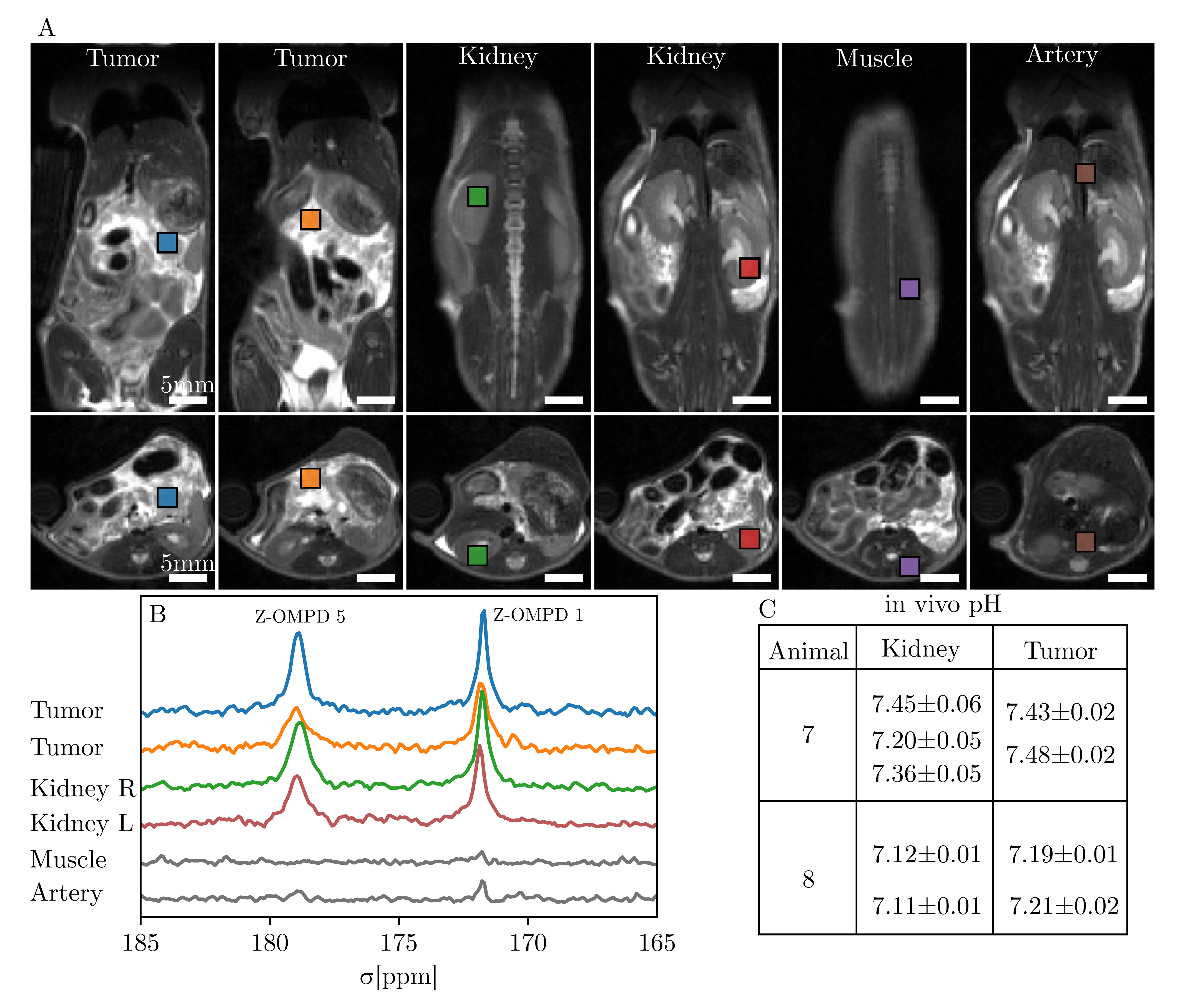
Finally, the pH dependency of the Z-OMPD peaks[2] was utilized to detect changes in pH in different tumor nodules and kidneys of two animals (Fig. 4-5). Lesions in Animal 7 showed pH in or close to the physiological range (7.43-7.48), while voxels placed in cortex/medulla region of kidneys are compatible with previous studies[9]. Animal 7 was scanned again after 4 days, showing similar pH values as before. Animal 8 shows an overall lower pH when compared to Animal 7 (tumor 7.21, kidney 7.11), possibly due to placed voxels covering multiple kidney compartments where individual peaks are masked by broad lineshapes.
Conclusion
A MV-PRESS sequence was used to detect 13C labelled molecules in mouse PDAC with a SNR comparable to a 2D CSI. The main advantages over CSI are its adaptability to image lesions that cannot be covered by a single CSI slice as well as a reduction in partial volume effects and a higher spectral resolution. Here voxel volumes from 8-18 µl were evaluated, trading off between signal and limited space due to slice overlap between voxels. Pyruvate to lactate ratios showed an increase in lactate production for tumors with differences between lesions within an animal. In vivo pH differed between the tumors of two animals while kidney mean pH values concur with literature.Acknowledgements
This research was funded by the Deutsche Forschungsgemeinschaft (DFG, German Reasearch Foundation, Emmy Noether program, grant number 391523415) and German Federal Ministry of Education and Research (BMBF 13N16450/QuE-MRT).References
1. Ardenkjær-Larsen, J. H., Fridlund, B., Gram, A., Hansson, G., Hansson, L., Lerche, M. H., ... & Golman, K. (2003). Increase in signal-to-noise ratio of> 10,000 times in liquid-state NMR. Proceedings of the National Academy of Sciences, 100(18), 10158-10163.
2. Grashei M, Wodtke P, Skinner JG, et al. Simultaneous Magnetic Resonance Imaging of pH, Perfusion and Renal Filtration using Hyperpolarized 13C-labelled Z-OMPD. Manuscript submitted for publication
3. Topping, G. J., Hundshammer, C., Nagel, L., Grashei, M., Aigner, M., Skinner, J. G., ... & Schilling, F. (2020). Acquisition strategies for spatially resolved magnetic resonance detection of hyperpolarized nuclei. Magnetic Resonance Materials in Physics, Biology and Medicine, 33(2), 221-256.
4. Gottwald, W., Nagel, L., Topping, G.J. and Schilling, F. Multi-voxel PRESS for fast and sensitive static MRS of hyperpolarized nuclei. ISMRM 2022 Abstract and Poster. 6-13.2022. London
5. Heid, I., Steiger, K., Trajkovic-Arsic, M., Settles, M., Eßwein, M. R., Erkan, M., ... & Braren, R. F. (2017). Co-clinical assessment of tumor cellularity in pancreatic cancer. Clinical Cancer Research, 23(6), 1461-1470.
6. Bauer, S., 2022. 3D-Visualization of Metabolic Magnetic Resonance Spectroscopy and Anatomical MRI. Munich. Bachelor Thesis.
7. NMR group of the Institute of Scientific Instruments of the Czech Academy of Sciences (2020), GitHub Repository. https://github.com/isi-nmr/brukerapi-python
8. Döpfert. J (2014), GitHub Repository. https://github.com/jdoepfert/brukerMRI
9. Düwel, S., Hundshammer, C., Gersch, M., Feuerecker, B., Steiger, K.,
Buck, A., ... & Schilling, F. (2017). Imaging of pH in vivo using
hyperpolarized 13C-labelled zymonic acid. Nature communications, 8(1), 1-9.
Figures