0845
Novel Structure-guided Design of Amino Acid based Hyperpolarized 13C Probes for Metabolic Imaging1National Cancer Institute, National Institutes of Health, Bethesda, MD, United States, 2The University of Tokyo, Bunkyo-ku, Japan
Synopsis
Keywords: Hyperpolarized MR (Non-Gas), Molecular Imaging
Profiling the metabolic phenotypes of tumors by molecular imaging is a promising approach in treatment planning and response monitoring. Here, dissolution Dynamic Nuclear Polarization (dDNP) is an emerging technique to detect site-specific enzymatic activities noninvasively. While drastic dDNP sensitivity enhancements are advantageous for real-time measurements, the limited number of applicable probes with longer T1 relaxation times continues to be a major drawback particularly for in vivo use. In this presentation, we will demonstrate a framework to design novel dDNP probes, detecting APN enzymatic activities which is a cancer therapeutic target, and its applications to in vivo pancreatic cancer.Purpose
Metabolic MRI, an imaging methodology monitoring the key metabolic processes that are critical to diseases, is becoming increasingly an important technique to visualize metabolic processes in real-time and offers powerful insight into pathologies non-invasively. Hyperpolarized MRI is considered as a promising emerging technology to provide such detailed information. However, the limited number of practically applicable dissolution Dynamic Nuclear Polarization(dDNP) probes is still a major bottleneck for in vivo applications. In order to obtain optimum sensitivities, spectral resolutions, and T1 relaxation times for the dDNP approach, it is mandatory to label isotopically specific positions with 13C , 12C, 15N and/ or 2H, which can limit dDNP applications as there are cases that cannot easily isotopically label desired positions. Therefore, there is considerable interest in developing a framework to design novel dDNP probes particularly with rational designs for in vivo use, complying with the physical and physiological requirements. Here, we have recently designed Ala-[1-13C]Gly-d2-NMe2 probe to detect aminopeptidase N (APN) activities in vivo based on atomic-level structure-guided approaches, including structural biology, computational molecular dynamics, and enzymology.1 Aminopeptidase N(CD13) has a variety of essential roles physiologically, highly expressed in various tumors related to progression in malignancy, angiogenesis and metastasis, where it is considered as a therapeutic target and biomarker,2,3 however, robust molecular imaging probes that can interrogate its enzymatic activity effectively in vivo have not been reported previously.4 In this presentation, we demonstrate application of this APN probe for in vivo applications on 13C metabolic MRI with/without an inhibitor, phebestin, and cancer therapeutics, sunitinib. Advantages and limitations of our approaches on developing novel probes will also be discussed.Methods
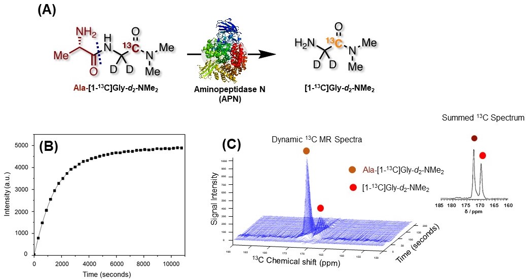
Hyperpolarized 13C MRI: MiaPaCa-2 pancreatic tumor bearing mice were formed subcutaneously into the right hind legs of mice. Hyperpolarized 13C MRI experiments were performed on a 3T MRI scanner(MR solutions Inc.) using a 17 mm diameter home built 13C solenoid coil with a saddle 1H coil. A solution of 80 μl of 5M Ala-[1-13C]Gly-d2-NMe2, containing 19mM of OX063, was polarized at 3.35T and 1.4 K in a Hypersense DNP polarizer(Oxford Instruments). Each hyperpolarized sample was rapidly dissolved in 4.0 ml of a superheated DPBS dissolution buffer, containing 0.68mM ethylenediaminetetraacetic acid. 400 ml of hyperpolarized solution was injected intravenously into the tumor xenografts.Results and Discussion
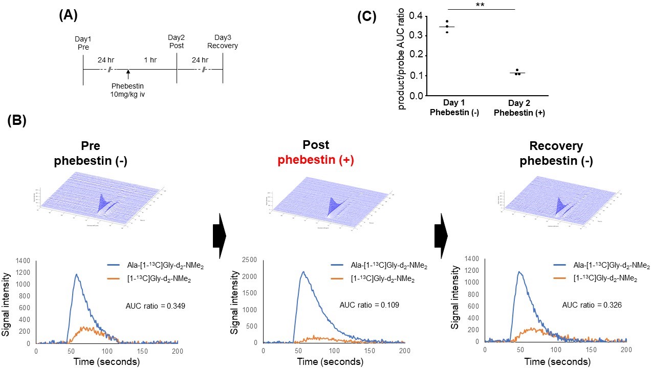
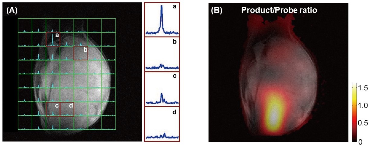
The previously reported APN probe [1-13C]Ala-NH2 was required to modified significantly for in vivo use.4 A newly designed probe, Ala-[1-13C]Gly-d2-NMe2 were developed as summarized following;1 firstly, the probe candidates were screened using a quantum mechanical/molecular mechanical computational calculations to optimize the enzymatic affinities(Km) and turnover rate(kcat). Then, these results were experimentally validated based on abovementioned enzymatic kinetic parameters. The T1 relaxation times as well as chemical shift dispersions of final candidates were examined while actual levels of APN enzymatic activities were measured on 9.4 T NMR spectrometers as well as 3T MRI. Finally, the structure of Ala-Gly-NMe2 that has the enzymatic affinities(Km) of 1.9±0.1 mM and turnover rates(kcat) of 255±20 s-1, whereas the wider 13C chemical shift changes between probe and product peaks of 2.8 ppm. T1 relaxation time of deuterated Ala-[1-13C]Gly-d2-NMe2 drastically improved to 35.4±0.3 seconds at 9.4T and 56.7±8.7 seconds at 3T, although the protonated version of Ala-[1-13C]Gly-NMe2 has T1 of 27.3±0.3 seconds at 9.4T. This rationally designed APN probe exhibited excellent polarization efficiencies for dDNP as shown in the solid-state build up curve Figure1(A, B), and it produced time-dependent 13C MR spectra with sufficient generations of product peaks at 170.0 ppm from the probe Ala-[1-13C]Gly-d2-NMe2 at 172.6ppm. These in vitro results were further validated in vivo without/with an inhibitor of APN enzymatic activities on MiaPaCa-2 tumor xenografts noninvasively as shown in Figure2. We confirmed that the APN probe successfully reports the selectivity of APN enzymatic activities(Figure2C). Encouraged by these results, we demonstrated the monitoring the responses towards cancer treatments including applications of sunitinib, which inhibits receptors that play a role in both tumor angiogenesis and tumor cell proliferation. After 6 hours of these therapeutic applications, the inhibitions of the APN activities in vivo MiaPaCa-2 tumor xenografts were observed(Figure3). This newly developed APN probe is also applicable to magnetic resonance spectroscopic imaging(MRSI) to report site-specific enzymatic activities of APN, what indicates the enhanced APN activities in the tumor region as shown in Figure4.Conclusions
To our knowledge this is the first study rationally designed an exogenous dDNP probe that (1) was optimized based on atomic-level structural evaluations and enzymology of a specific enzyme, which is a therapeutic target in cancer, (2) satisfied the physical/physiological requirements for its in vivo use, and (3) applied effectively on the pancreatic cancer xenografts to monitor their cancer treatments. In particular, the successful in vivo demonstrations of this novel probe were owing to satisfying following characteristics; (i) extended T1 relaxation times, (ii) higher selectivity to detect APN enzymatic activities, (iii) speeding up the enzymatic reactions at the physiological concentrations, (iv) larger chemical shift dispersions between probe and product peaks, (v) its water solubility, (vi) its biocompatibility, (vii) its stability in blood, and (viii) higher hyperpolarization efficiency.1 This structural-guided approach to design in vivo dDNP probes can be a powerful methodology that can be widely used to pave the way for the limited numbers of available dDNP MR probes.Acknowledgements
This study was supported by intramural research program at NCI/NIH.References
[1] Saito Y et al., Structure-guided design enables development of a hyperpolarized molecular probe for the detection of aminopeptidase N activity in vivo., Sci. Adv. 2022; 8:eabj2667.
[2] Dixon J et al., Expression of aminopeptidase-N (CD13) in normal tissues and malignant neoplasms of epithelial and lymphoid origin., J. Clin. Pathol. 1994; 47:43-47.
[3] Guzman-Rojas L et al., Cooperative effects of aminopeptidase N (CD13) expressed by nonmalignant and cancer cells within the tumor microenvironment., Proc. Natl. Acad. Sci. U.S.A. 2012; 109:1637-1642.
[4] Hata R et al., Design of a hyperpolarized molecular probe for detection of aminopeptidase N activity., Angew. Chem. Int. Ed. 2016; 55:1765-1768.
Figures