0779
Detection of Age-related Cerebral Microvessel Tortuosity and Its Association with Dilated Perivascular Space Using USPIO-enhanced 7T MRI1Department of Radiology, New York University Grossman School of Medicine, New York, NY, United States, 2Vilcek Institute of Graduate Biomedical Sciences, New York University Grossman School of Medicine, New York, NY, United States, 3Department of Radiology, Wayne State University, Detroit, MI, United States
Synopsis
Keywords: Blood vessels, Aging
Microvessel tortuosity, which is lack detection on clinical imaging, may have direct detrimental effects on capillary flow and nutrient supply which is critical in maintaining neural functions. A high-resolution USPIO-enhanced 7T MRI demonstrated corkscrew appearing medullary arterioles (about 50 µm) in white matter with some enclosed in dilated perivascular space (PVS). The number of tortuous arterioles increased with aging and more tortuous vessels with dilated PVS were found in elderly people (P<0.05). The dynamic interactions between vulnerable tortuous arteries and PVS dilation may be the basis of age-related hypoperfusion, white matter pathology, and waste clearance dysfunction in the elderly.INTRODUCTION
With aging, the vascular wall is multi-factorial impacts, including repetitive mechanical forces of pulsation, collagen deposition, and elastin degradation. As a result, tortuosity of large arteries is commonly seen in the elderly and has been extensively reported in neuroimaging studies. However, morphological changes (e.g., corkscrew appearance) of cerebral small arteries or arterioles have only been described in postmortem studies [1, 2]. In some cases, the tortuous artery is surrounded by a dilated perivascular space (PVS), which may imply a dynamic interplay between vulnerable tortuous arteries and PVS enlargement [3]. Arterial tortuosity has been shown to be associated with increased flow resistance and elongation of arteries, which have direct effects on the distal capillary flow and nutrient supply, leading to neuronal degeneration. Conventional MRI often failed to detect small arterial tortuosity due to inadequate imaging resolution and contrast. High-resolution 7T T2*-weighted imaging with the ultrasmall superparamagnetic iron oxide (USPIO) is a new tool that allows the observation of morphological changes in small arterioles [4]. This study assessed the feasibility of in vivo detection of age-related microvessel morphological changes within deep white matter (WM) as well as their association with dilated PVS using USPIO-enhanced 7T MRI.METHODS
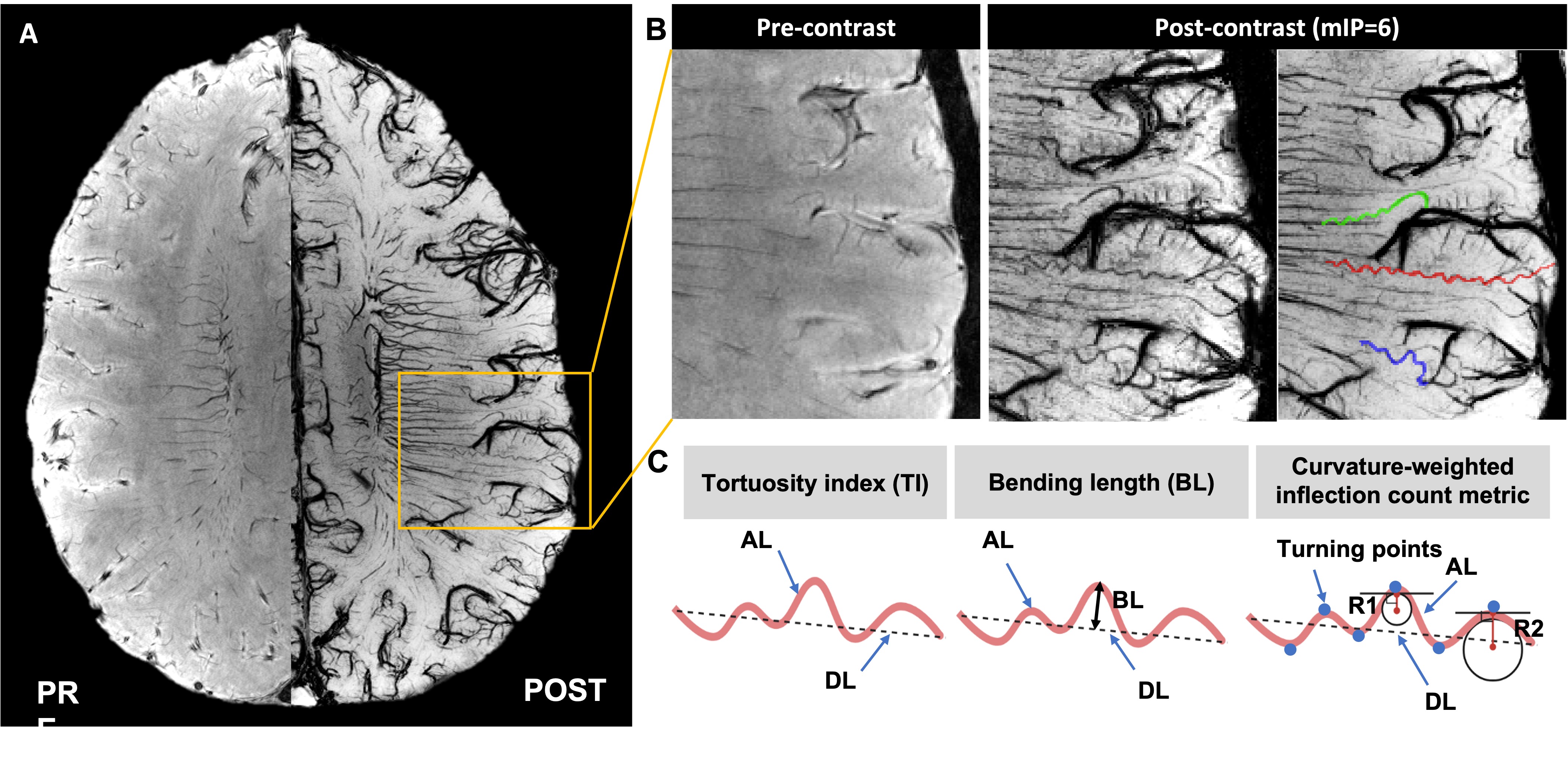
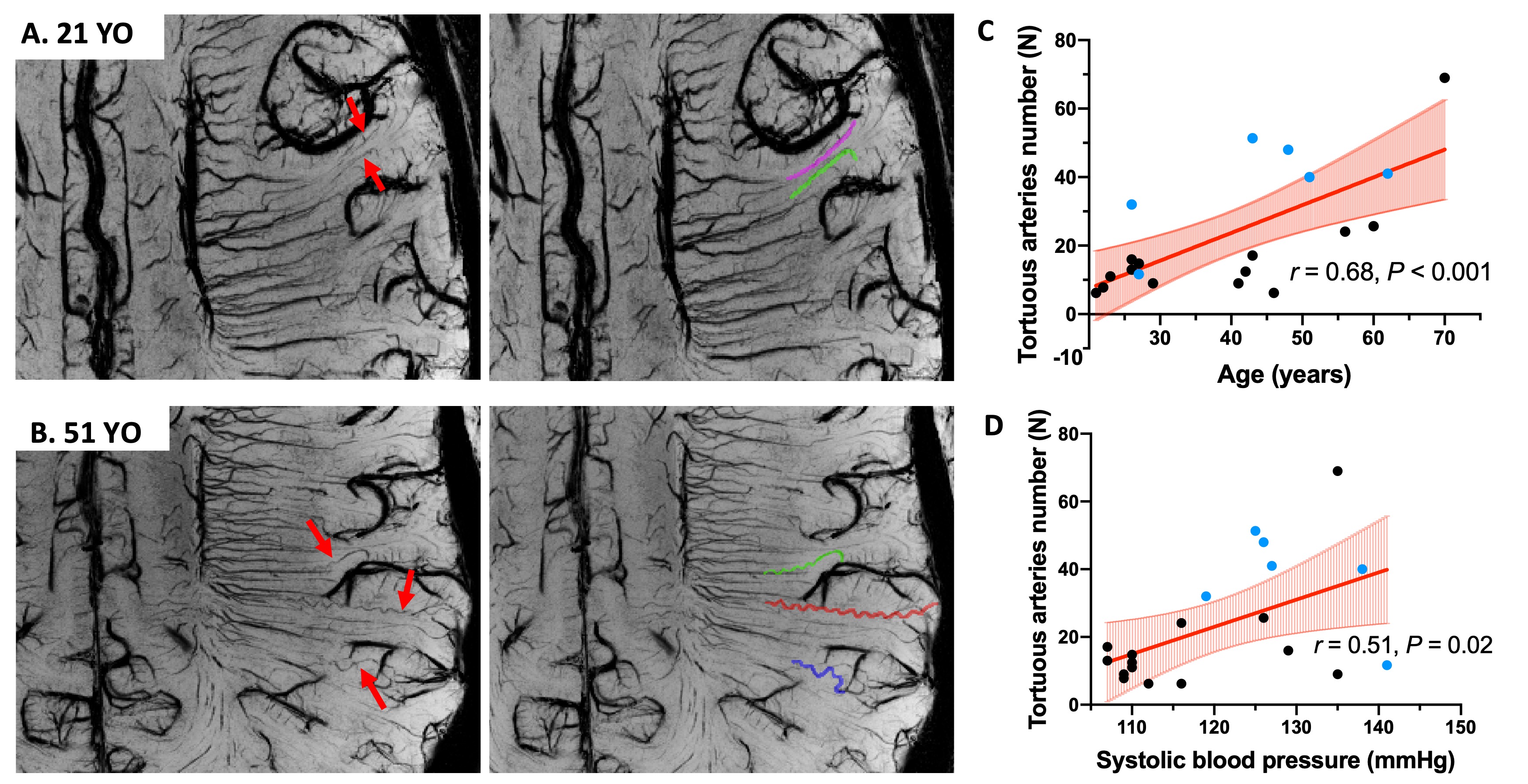
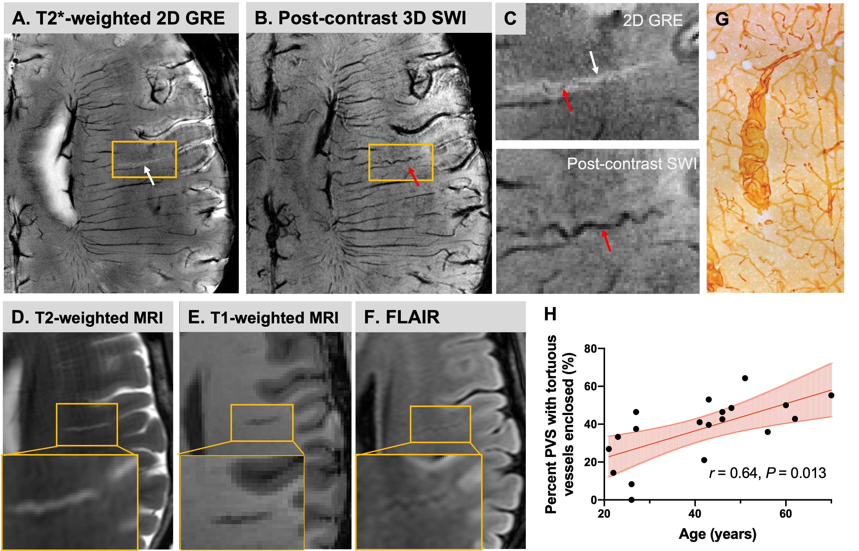
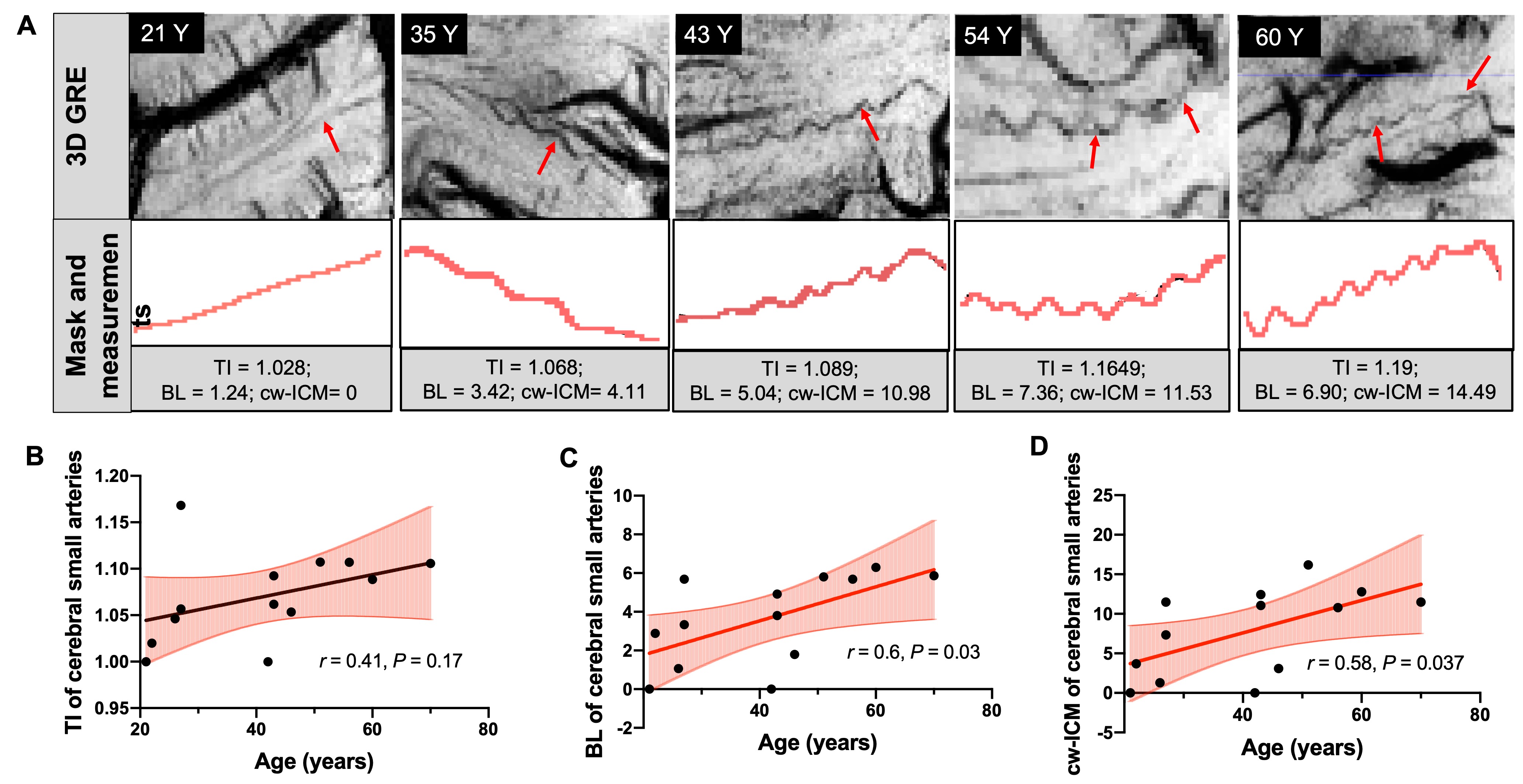
Twenty healthy participants (age: 21-70 years old, 39. 5±15.0 years; 10 males/10 females) were scanned at 7T using a 32-channel head coil. The imaging protocol included: (1) T1-weighted MPRAGE (spatial resolution = isotropic 1mm3) before USPIO (Ferumoxytol), (2) 2D gradient echo (GRE) sequence (TR/TE=1250/25ms, voxel size=0.25*0.25*2mm3) after 5-minute USPIO infusion and (3) dual-echo 3D susceptibility weighted imaging (SWI) (TR=22ms, TE1/TE2=7.5/15ms, voxel size = 0.25*0.25*1mm3) before and after USPIO [5]. SWI images were processed using a high-pass filter to generate an unwrapped phase mask [6]. We focused on medullary arteries (MAs) (Figure 1A-B), a group of long-end small arteries from pial arteries penetrating the cerebral cortex to supply deep WM. Combining these criteria, tortuous arteries were differentiated and counted on high-resolution T2*-weighted 2D GRE images, which provide unique contrast between vessels and tissues as well as PVS CSF. The fast-marching algorithm was implemented to extract the centerline after manual segmentation. Several tortuosity measurements were calculated, including 1) tortuosity index (TI), the ratio between actual length (AL) and direct length (DL); 2) bending length (BL), the maximum perpendicular distance between AL and DL; 3) curvature-weighted inflection count metric (cw-ICM), the product of turning points number (N), TI, and maximum curvature (Figure 1C). Pearson's correlation was performed to reveal the relationship between age and MA tortuosity as well as between age and the percent dilated PVS that has tortuous MA enclosed.RESULTS
USPIO-enhanced 7T MRI can detect MAs as small as 50 μm with a characteristic corkscrew appearance in the centrum semiovale and periventricular WM, which are invisible on pre-contrast images. These tortuous MAs run from the cortical surface to deep WM, and are invisible in the pre-contrast SWI, whereas visible in post-USPIO images (Figure 1A-B), in contrast to medullary veins, which have a parallel arrangement around the lateral ventricle and drain blood from deep WM to the ependymal veins. The number of tortuous arteries was positively correlated with age after correcting for gender, BMI, and blood pressure (BP) (r=0.68, P<0.001). The tortuous artery number was also positively correlated with systolic BP; however, this effect disappeared after correcting for age and gender as covariates (Figure 2). Compared with conventional MRI, 7T 2D GRE data provided excellent contrast for the in vivo visualization of dilated PVS (hyperintense) and enclosed tortuous arteries (hypointense), consistent with ex vivo histological studies [7] (Figure 3). Percent dPVS with enclosed tortuous arteries was positively correlated with age (r=0.64, P=0.013). Pearson's correlation between age and quantitative tortuosity measurements also revealed that BL, cw-ICM are positively correlated with age (r=0.6, P<0.05; r=0.58, P<0.05, respectively), indicating cerebral small arteries become more tortuous with aging (Figure 4).DISCUSSION AND CONCLUSION
While the histological studies strongly suggest age-related vessel tortuosity, few imaging studies have shown such microvessel changes. Using USPIO-enhanced 7T MRI, small corkscrew-appearing tortuous arteries/arterioles are clearly shown in the subcortical WM and increase with normal aging. This technique offers excellent contrast and details to visualize the sub-voxel small arteries in live human brains, which are invisible in the conventional MRI. The number of dilated PVS with enclosed tortuous arteries detected on high-resolution 2D T2*-weighted MRI increases with aging as well. These findings are consistent with previous histological studies [8]. Quantitative morphological measurements (e.g., TI, BL, and cw-ICM) also showed an age-related tortuosity increase in the microvasculature. Our results indicate this technique can detect early microvessel tortuosity changes (before symptom onset), which may contribute to age-related tissue hypoperfusion, white matter hyperintensities (WMHs), and neuronal dysfunction in the elderly.Acknowledgements
This study was funded by RF1 NS 110041, R01 NS108491, R01 AG077422 from National Institutes of Health.References
1. Brown, W.R., et al., Microvascular changes in the white mater in dementia. Journal of the Neurological Sciences, 2009. 283(1): p. 28-31.
2. Thore, C.R., et al., Morphometric analysis of arteriolar tortuosity in human cerebral white matter of preterm, young, and aged subjects. J Neuropathol Exp Neurol, 2007. 66(5): p. 337-45.
3. Brown, W.R., et al., Venous collagenosis and arteriolar tortuosity in leukoaraiosis. J Neurol Sci, 2002. 203-204: p. 159-63.
4. Buch, S., et al., Subvoxel vascular imaging of the midbrain using USPIO-Enhanced MRI. Neuroimage, 2020. 220: p. 117106.
5. Chen, Y., et al., An interleaved sequence for simultaneous magnetic resonance angiography (MRA), susceptibility weighted imaging (SWI) and quantitative susceptibility mapping (QSM). Magn Reson Imaging, 2018. 47: p. 1-6.
6. Haacke, E.M., et al., Susceptibility-Weighted Imaging: Technical Aspects and Clinical Applications, Part 1. American Journal of Neuroradiology, 2009. 30(1): p. 19-30.
7. Brown, W.R. and C.R. Thore, Review: cerebral microvascular pathology in ageing and neurodegeneration. Neuropathol Appl Neurobiol, 2011. 37(1): p. 56-74.
8. Spangler, K.M., et al., Arteriolar Tortuosity of the White Matter in Aging and Hypertension. A Microradiographic Study. Journal of Neuropathology & Experimental Neurology, 1994. 53(1): p. 22-26.
Figures