0778
Utilizing High Performance Gradients to Image Human Brain Arterial Vasculature with High Resolution 7T Time-of-flight MR Angiography1Siemens Medical Solutions USA, Inc., Berkeley, CA, United States, 2Helen Wills Neuroscience Institute, University of California, Berkeley, Berkeley, CA, United States, 3Advanced MRI Technologies, Sebastopol, CA, United States, 4Dr. Laub Consulting, LLC, San Mateo, CA, United States
Synopsis
Keywords: Blood vessels, High-Field MRI
Time of flight (TOF) 3D MR Angiography (MRA) has been shown to effectively image small (50-300um diameter) pial vessels. However, such high resolution requires strong flow compensation (flow-comp) gradient pulses and short echo times which are limited by peripheral nerve stimulation and hardware capability in most scanners. We demonstrate high resolution TOF MRA acquisition using an advanced head gradient system (200mT/m, 900 T/m/s) at ultrahigh field 7T.Purpose
Time of flight (TOF) MR angiography (MRA) is a classic technique for non-invasively imaging brain vasculature and plays a major role in understanding brain physiology and vessel alterations in the presence of disease. Especially when used at ultrahigh field 7T, the increased signal-to-noise ratio (SNR) and the longer T1 relaxation times of blood and gray matter (Von Morze 2007) enable spectacular imaging of the neurovasculature. . High resolution TOF has recently been shown to effectively visualize very small pial vessels on the order of 50-300um diameter with complex branching patterns when pushed to extremely high resolutions (Bollmann 2022). However, these high resolution acquisitions require the playing of strong flow compensation (flow-comp) gradients in a very short amount of time to achieve the short echo times needed to retain SNR. The practical implementation is limited by peripheral nerve stimulation (PNS) and maximum gradient slew rates (Mansfield and Harvey 1993) on commonly available MR systems. In this study, we evaluate the benefits of the high performance (“Impulse” Siemens) head gradients with slew rate 900 T/m/s and capable of maximum gradient 200 mT/m, which were specially designed to mitigate PNS (Davids 2021) at ultrahigh field 7T. Utilizing the higher Gmax and faster slew rate specifications of the Impulse gradients as well as the advanced RF hardware (96 channel receiver) enables the shortening of echo times to reduce flow dephasing while improving acceleration to preserve SNR at higher resolutions.Methods
Two healthy adults (1 male, ages 19-38) were scanned after obtaining written informed consent in accordance with local IRB guidelines. Experiments were conducted on an investigational Siemens (Siemens Healthcare, Erlangen, Germany) MAGNETOM Terra Impulse Edition NexGen 7T scanner with advanced head gradient and RF hardware (Feinberg 2021). Imaging data were acquired using a custom built 16Tx/96Rx head coil (MrCoilTech, Glasgow, UK, Gunamony 2022). The product TOF sequence was utilized to acquire 0.25 mm isotropic MRA data with the following sequence parameters: TE=3.07ms with asymmetric echo, TR=23ms, flip angle=24deg, FOV=204mm x 178.5mm, matrix size=728x832, slab thickness=15mm, slice oversampling=20%, R>>L phase encoding direction, readout bandwidth=188Hz/px, GRAPPA=4, no. reference lines=32, no partial Fourier, 3D centric reordering of the phase encoding steps, read and slab-select flow compensation for a total acquisition time of 5:46min. Tilted optimized non-saturating excitation (TONE) pulses (Atkinson 1994) were not used because the blood flow of the small vessels was not always exactly perpendicular to the imaging slab. The use of flow compensation in the readout and slab-select gradients was evaluated, and the flip angle was optimized within specific absorption rate (SAR) limitations to achieve higher SNR. Images were viewed and evaluated with maximum intensity projections (MIP).Results and Discussion
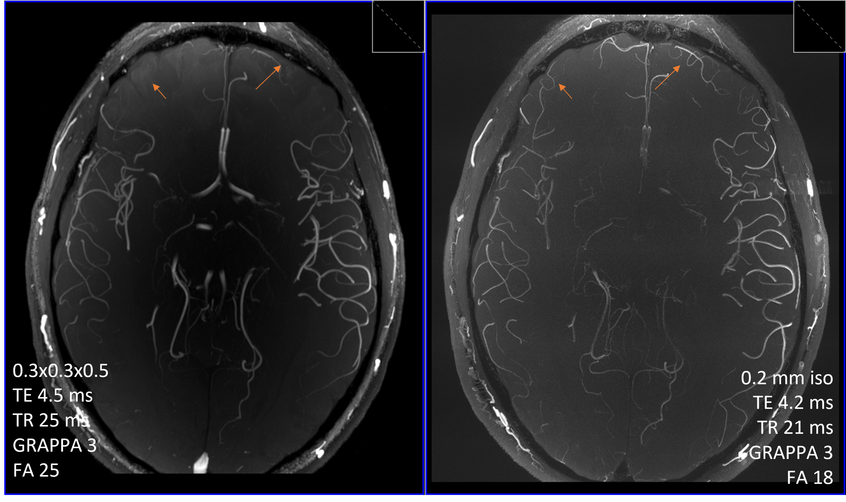
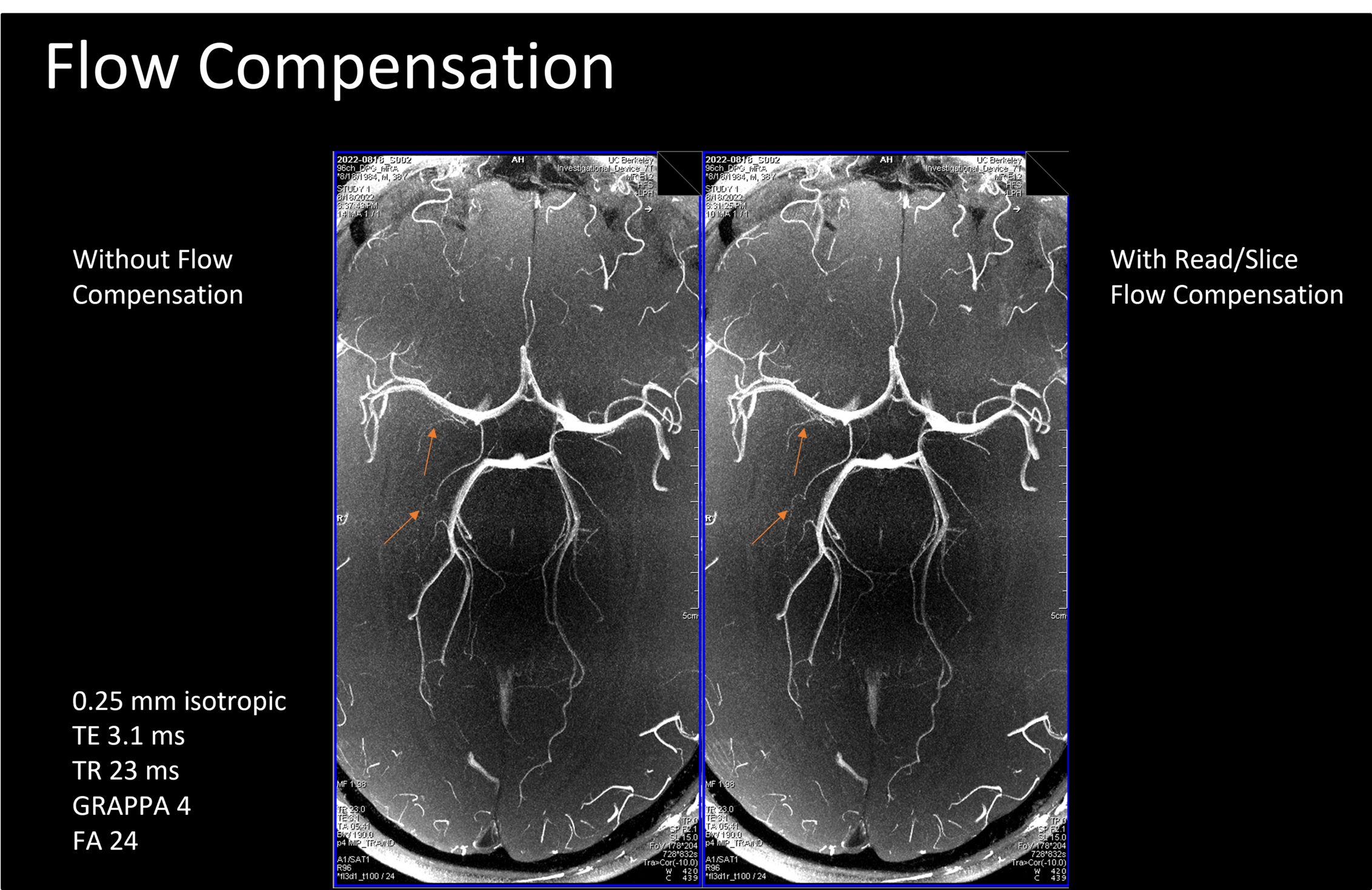
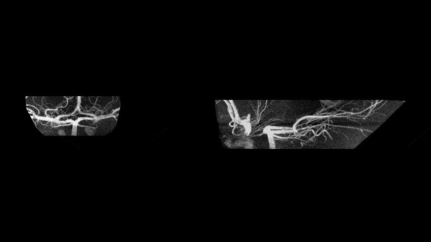
Figure 1 shows, at the cost of signal in some of the larger vessels, the reduction in voxel volume better resolves additional small branches, especially near the cortical surface.Figure 2 demonstrates the benefits of using flow compensation in the readout and slab-select gradients, by improving visualization of several of the smaller branches. This is especially true for pial arteries, but there is also a clear benefit at small arteries branching from the Circle of Willis.
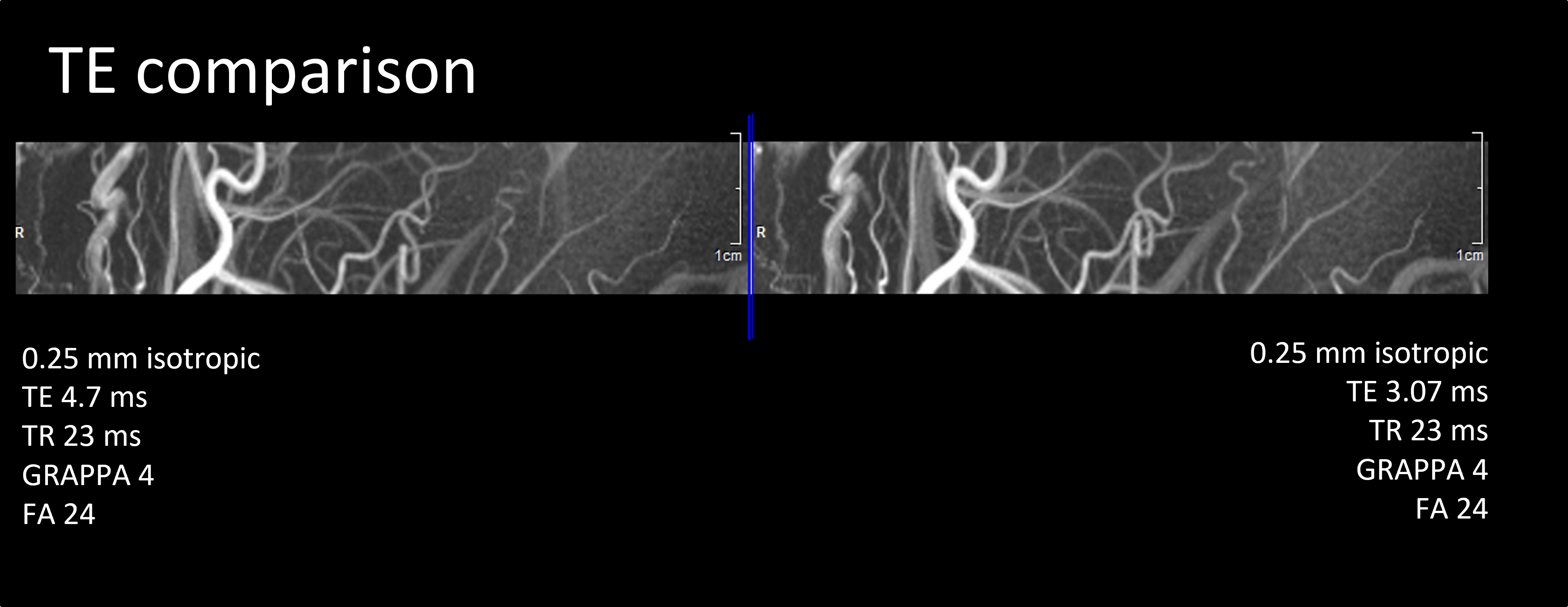
Figure 3 shows the high resolution TOF with the shorter TE of 3.07ms achievable with the Impulse gradients compared to TE of 4.34ms on standard SC72 gradients (80mT/m, 200T/m/s). Slight improvement is observed in the image quality and vessel signal, and this short TE and faster gradient ramping did not produce PNS effects in subjects. Previous reports on a standard body gradient system have discussed PNS as a limiting factor in achieving high resolution (Bollmann 2022).
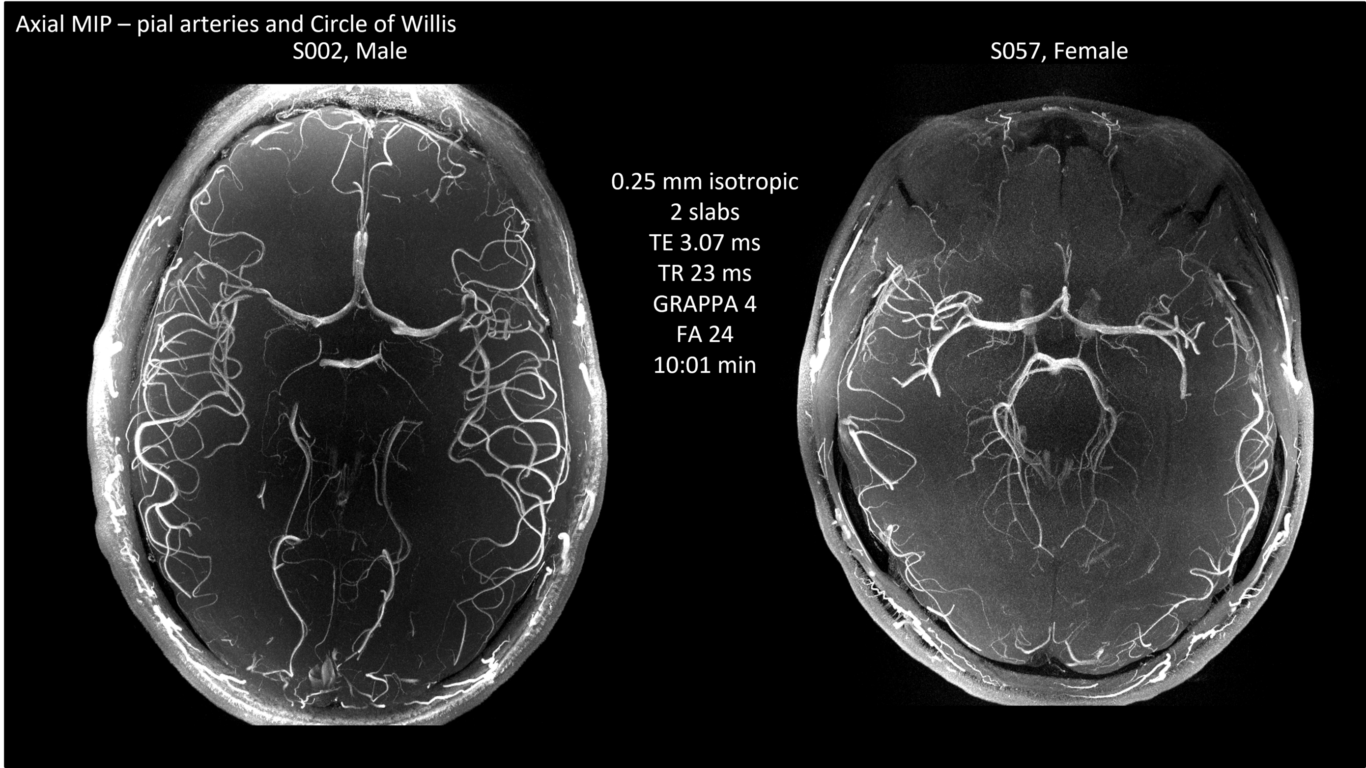
Figure 4 displays two slabs of 0.25mm isotropic MIPs from two subjects, in which the higher in-plane acceleration of GRAPPA=4 enabled the high-resolution acquisition within 10 min.
Figure 5 shows tumbling views focused on the Circle of Willis to demonstrate the level of detail that can be observed with high resolution TOF, including the branching characteristics of the lenticulostriate arteries from the MCA. There is potential for detailed exploration of vascular morphometry in applications such as lacunar infarct and microaneurysms among others (Barisano, 2019).
This preliminary study demonstrated that the Impulse edition MAGNETOM Terra is capable of relatively fast, high quality, and high resolution TOF MRA. With sequence optimization, we expect to further improve acceleration using advanced RF hardware and achieve shorter TE without producing PNS in subjects. Faster high resolution MRA has potential applicability in querying the relationship of arterial morphology with functional organization of the brain. This will complement the goal of the Impulse edition 7T to explore brain hemodynamics at an unprecedented resolution (Feinberg 2021).
Summary
The higher performance gradients enabled shorter TE in flow compensated MRA and enabled high quality, high resolution image acquisitions without peripheral nerve stimulation. This high resolution MRA has great potential for characterizing small vessel morphometry as it relates to the functional organization and hemodynamics of the brain.Acknowledgements
This project is supported by the NIH BRAIN Initiative (R01MH111444, U01EB025162), 1R44MH129278.References
Von Morze C, Xu D, Purcell DD, Hess CP, Mukherjee P, Saloner D, Kelley DAC, Vigneron DB. Intracranial time-of-flight MR angiography at 7T with comparison to 3T. Journal of Magnetic Resonance Imaging: JMRI. 2007;26:900–904. doi: 10.1002/jmri.21097.
Saskia Bollmann, Hendrik Mattern, Michaël Bernier, Simon D Robinson, Daniel Park, Oliver Speck, Jonathan R Polimeni (2022) Imaging of the pial arterial vasculature of the human brain in vivo using high-resolution 7T time-of-flight angiography eLife 11:e71186
Mansfield P, Harvey PR. Limits to neural stimulation in echo-planar imaging. Magnetic Resonance in Medicine. 1993;29:746–758. doi: 10.1002/mrm.1910290606.
Davids M, Dietz P, Ruyters G, Roesler M, Klein V, Guerin B, Feinberg DA, Wald L. PNS optimization of a high-performance asymmetric gradient coil for head imaging. ISMRM, #0565, 2021.
Feinberg DA, Dietz P, Liu C, Setsompop K Mukherjee P Wald L, Vu A, Beckett A Insua I, Schröder M, Stocker S, Bell Pl, Rummert E, Davids M Design and Development of a Next-Generation 7T human brain scanner with high-performance gradient coil and dense RF arrays. In: ISMRM, #0562, 2021.
Gunamony S, Feinberg DA An 8-channel transmit 64-channel receive compact head coil for Next Gen 7T scanner with head gradient insert. ISMRM #1447, 2022.
Atkinson D, Brant-Zawadzki M, Gillan G, Purdy D, Laub G. Improved MR angiography: magnetization transfer suppression with variable flip angle excitation and increased resolution. Radiology. 1994;190:890–894. doi: 10.1148/radiology.190.3.8115646.
Barisano G, Sepehrband F, Ma S, Jann K, Cabeen R, Wang DJ, Toga AW, Law M. Clinical 7 T MRI: Are we there yet? A review about magnetic resonance imaging at ultra-high field. Br J Radiol. 2019 Feb;92(1094):20180492. doi: 10.1259/bjr.20180492. Epub 2018 Nov 1.
Figures