0756
Wireless In-Bore Ballistocardiography with 2.4GHz Beat Pilot Tone (BPT)
Suma Anand1 and Michael Lustig1
1Electrical Engineering and Computer Sciences, University of California, Berkeley, Berkeley, CA, United States
1Electrical Engineering and Computer Sciences, University of California, Berkeley, Berkeley, CA, United States
Synopsis
Keywords: Hybrid & Novel Systems Technology, Cardiovascular, RF Arrays and Systems, Motion Correction
We previously proposed the Beat Pilot Tone (BPT), a motion sensing method that uses microwave frequencies and preamplifier intermodulation to obtain increased motion sensitivity compared to Pilot Tone (PT). Here, we show that cardiac signals acquired with BPT correlate strongly with displacement ballistocardiograms (dBCG). We compare the BPT signals to dBCG acquired with an accelerometer. Additionally, we show that BPT has greater signal modulation compared to PT in sensing cardiac motion. BPT could be a novel, non-contact, and wireless method for acquiring dBCG signals, offering robustness in setup for patients and rich physiological information complementary to the MRI exam.Introduction
Cardiac MRI relies on accurate motion sensing in order to obtain motion-corrected images. Sensors integrated with the MRI suite include electrocardiograms (ECGs) and the photoplethysmogram (PPG); however, these require careful placement on the subject and are prone to noise and inter-subject variability1,2.Pilot Tone (PT) is an alternative cardiac motion sensor, which measures loading changes of receiver coils3-7 using a pure radio frequency (RF) tone close to the Larmor frequency. We proposed an alternative method, Beat Pilot Tone (BPT)8, which uses two tones spaced apart by the MR bandwidth (~127.8MHz) and receives them at the receiver coil array via nonlinear intermodulation in the receiver chain (Figure 1c); this allows us to use microwave frequencies (2.4GHz, 2.5278GHz) and obtain increased sensitivity to respiratory and bulk motion8,9.
In this work, we hypothesize that the cardiac signals acquired by BPT highly correlate with displacement BCG (dBCG), which measures the recoil of the body due to the ballistic forces of blood1 (Figure 1b). We compare BPT in two volunteers to a conventional dBCG acquired with a tri-axial accelerometer, as well as to PT, ECG, and PPG. We show that BPT has greater signal modulation than PT. Finally, we show that dBCG and BPT are highly correlated.
Methods
BPT and PT AcquisitionFigure 1b shows the BPT acquisition: two tones at frequencies $$$f_1$$$ and $$$f_2$$$ are mixed in the receiver chain by intermodulation to produce a signal with a beat frequency $$$f_{BPT} = f_2 - f_1.$$$ In this experiment, $$$f_1 = 2.4GHz$$$ and $$$f_2 = 2.5278GHz$$$ ($$$f_{BPT} = 127.8MHz$$$). Similar to the PT, the BPT appears as a line that is easily separated from the image3-9.
Hardware setup
Figure 1a shows the placement of the PT antenna (green), BPT antenna (purple), accelerometer (orange), and receiver coils (black). The PT antenna was a small loop placed on the chest, as in previous work3-7, while the BPT antenna was placed above the subject at the top of the bore8,9. The PT and BPT were produced by an Ettus Research B200 software-defined radio (SDR; National Instruments, TX, USA) synchronized to the system’s 10MHz clock. For PT, a single tone was transmitted at 127.6MHz at a power of -30dBm. For BPT, the two tones were combined, amplified by 17dB, high-pass filtered, and transmitted by a dipole Bluetooth antenna. The gains of the PT and BPT were adjusted to achieve similar received levels. A tri-axial SCL3300 accelerometer (Murata Electronics; Kyoto, Japan) was placed on the subject’s chest and controlled by an Arduino Pro Mini (Arduino; Somerville, MA, USA). The accelerometer was synchronized to the BPT and PT via a Transistor-Transistor Logic (TTL) signal from the scanner.
Acquired scans
All scans were acquired on a GE3T MR750W system with gradients and RF turned off (TR=8.7ms). Breath-held scans were acquired on two volunteers using a 32-channel anterior array coil.
Data processing
The accelerometer data was high-pass filtered with a cutoff of 4Hz, then integrated twice to obtain displacement estimates. Figure 2 shows the raw BPT and PT magnitudes in percent modulation units (relative to the mean) without additional filtering. To compare BPT quantitatively to dBCG, we performed a least squares fit to regress the BPT to the dBCG. We low-pass filtered the linearly-combined BPT with a cutoff of 15Hz and computed the Pearson correlation coefficient between the filtered BPT and each dBCG (dBCG-x, dBCG-y, and dBCG-z).
Results
Figure 2 shows the raw BPT and PT for the three most modulated coils in the volunteer experiments, along with percent modulation. For both subjects, the raw BPT qualitatively shows sharper peaks compared to the PT (Figure 2a, 2c). Moreover, the BPT signal characteristics change significantly depending on the coil location. Comparatively, cardiac modulation is barely identifiable in the second subject’s raw PT data (Figure 2c).Figure 3 compares dBCG-y (left-right) computed from the accelerometer to BPT, PT, ECG and PPG. The timing of the BPT signal matches dBCG, and the signal features (e.g. peaks) match qualitatively. Moreover, the peaks of the BPT appear slightly earlier than the PPG, which may be advantageous for prospective cardiac gating.
Figure 4 shows the results of the dBCG-BPT regression and correlation. The correlation is greatest between BPT and dBCG-y for both subjects (Figure 4a, 4c). Figures 4b and 4d show a single heartbeat with labeled features of the dBCG11. These features contain important information about cardiac function; for instance, the time interval between the I and J waves may represent aortic pulse transit time, which is a predictor of cardiovascular risk12.
Conclusion
We have shown the ability of BPT to detect cardiac signals with improved SNR compared to the PT. BPT appears qualitatively similar to dBCG acquired by an accelerometer, and correlates highly (>0.8) when linearly combined across coils. Because the dBCG appears slightly earlier than the PPG, the timing is favorable for prospective cardiac gating. Moreover, the high frequency features of the signal make it easy to separate from other motions (e.g., respiratory) by simple filtering. Finally, the BPT is completely non-contact, which could be more robust for patients. dBCG signals are a rich source of physiological information and offer the possibility to characterize cardiac function simultaneously to the MRI exam11-13.Acknowledgements
The authors acknowledge support from GE Healthcare and NIH grants R01MH127104 and U01EB029427.References
- Brablik, J., Ladrova, M., Vilimek, D., Kolarik, J., Kahankova, R., Hanzlikova, P., ... & Martinek, R. (2022). A Comparison of Alternative Approaches to MR Cardiac Triggering: A Pilot Study at 3 Tesla. IEEE Journal of Biomedical and Health Informatics.
- Fine, J., Branan, K. L., Rodriguez, A. J., Boonya-Ananta, T., Ramella-Roman, J. C., McShane, M. J., & Coté, G. L. (2021). Sources of inaccuracy in photoplethysmography for continuous cardiovascular monitoring. Biosensors, 11(4), 126.
- Speier, P., M., et al. "PT‐Nav: a novel respiratory navigation method for continuous acquisitions based on modulation of a pilot tone in the MR‐receiver." Proc. ESMRMB 32 (2015): 128.
- Bacher, M. et al. “Retrospective Analysis of Pilot Tone Derived Cardiac and Respiratory Motion Information in a Patient Cohort”. Proc. ISMRM (2020).
- Vahle, Thomas, et al. "Respiratory Motion Detection and Correction for MR Using the Pilot Tone: Applications for MR and Simultaneous PET/MR Examinations." Investigative Radiology 55.3 (2020): 153-159.
- Falcão, M. B., Di Sopra, L., Ma, L., Bacher, M., Yerly, J., Speier, P., ... & Roy, C. W. (2022). Pilot tone navigation for respiratory and cardiac motion‐resolved free‐running 5D flow MRI. Magnetic resonance in medicine, 87(2), 718-732.
- Ludwig, J., Speier, P., Seifert, F., Schaeffter, T., & Kolbitsch, C. (2021). Pilot tone–based motion correction for prospective respiratory compensated cardiac cine MRI. Magnetic Resonance in Medicine, 85(5), 2403-2416.
- Anand S, Lustig M. Beat Pilot Tone: Exploiting Preamplifier Intermodulation of UHF/SHF RF for Improved Motion Sensitivity over Pilot Tone Navigators. Proc. ISMRM 2021.
- Lamar-Bruno K, Anand S, Lustig M. Cardiac and Respiratory-Resolved Image Reconstruction with the Beat Pilot Tone. Proc. ISMRM 2022.
- Brunner, D. O., De Zanche, N., Fröhlich, J., Paska, J., & Pruessmann, K. P. (2009). Travelling-wave nuclear magnetic resonance. Nature, 457(7232), 994-998.
- March, H. W. (1955). Three-plane ballistocardiography: the effect of age on the longitudinal, lateral, and dorsoventral ballistocardiograms. Circulation, 12(5), 869-882.
- Blacher, J., Asmar, S., Djane, S., London, G. & Safar, M. Aortic pulse wave velocity as a marker of cardiovascular risk in hypertensive patients. Hypertension 33, 1111–1117 (1999).
- Starr, I., & Schroeder, H. A. (1940). Ballistocardiogram. II. Normal standards, abnormalities commonly found in diseases of the heart and circulation, and their significance. The Journal of clinical investigation, 19(3), 437-450.
Figures
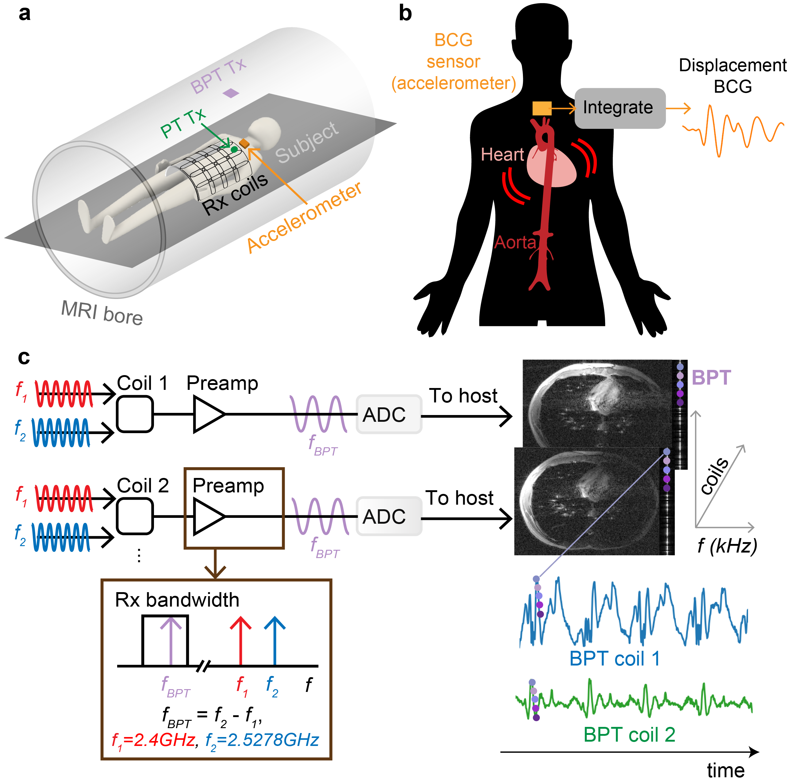
Figure 1: a) the placement of the PT transmit (Tx) antenna (green), BPT Tx antenna (purple), accelerometer (orange), and receiver (Rx) coils (black) in the MRI bore (gray). b) BCG measures the recoil of the body due to the ballistic forces of blood. It was measured with an accelerometer and integrated to obtain displacement. c) In the BPT, two tones at frequencies $$$f_1$$$ and $$$f_2$$$ are received by the coil array and mixed in the receiver chain to produce a signal with a beat frequency $$$f_{BPT} = f_2 - f_1$$$. The BPT appears as a line that is easily separated from the image.
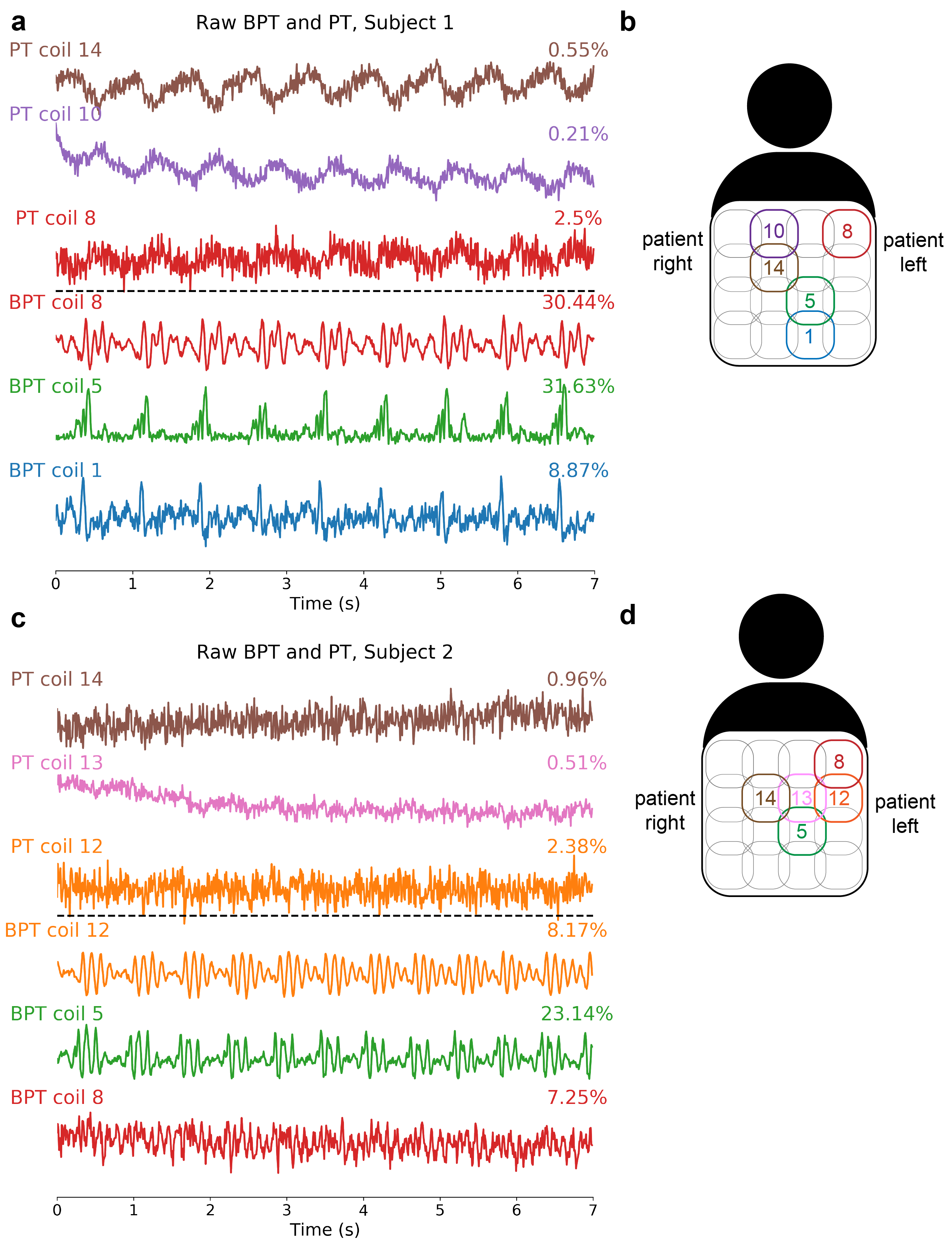
Figure 2: The raw PT and BPT signals from a breath-held scan with RF and gradients off for a) subject 1 and c) subject 2 with corresponding coil arrangements in b) and d). The data from three coils with the most energy in the cardiac frequency range were chosen. In a) and c), the percent modulation relative to the mean is displayed on the right. BPT qualitatively shows much sharper peaks and features compared to the PT. Quantitatively, BPT shows much larger modulation than the PT for both subjects.
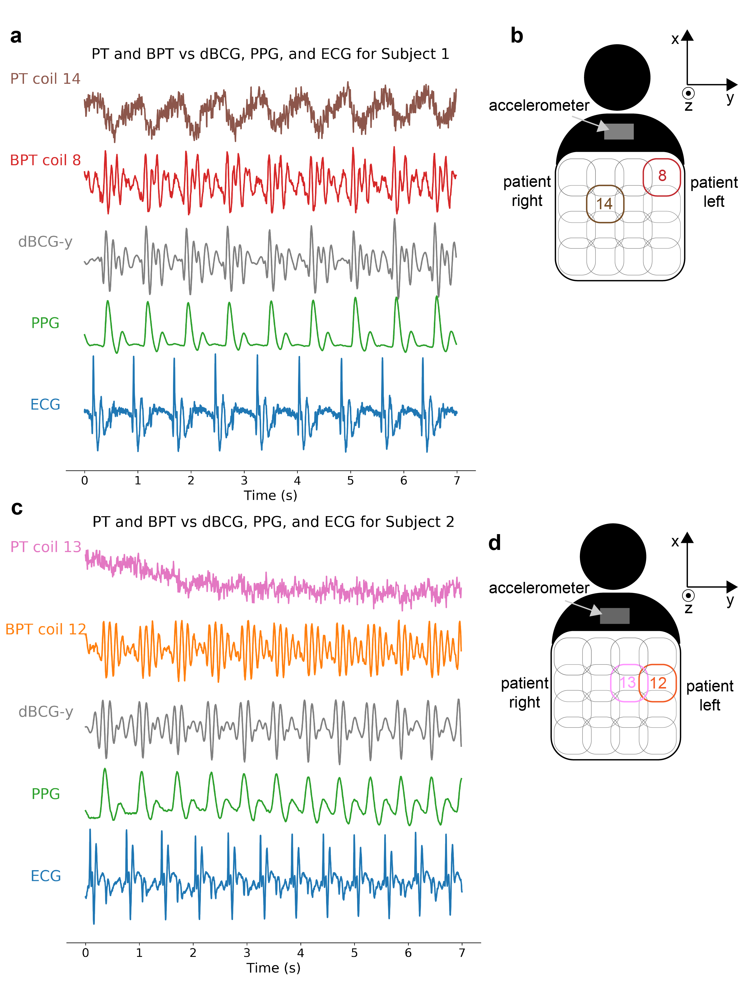
Figure 3: PT and BPT are compared to dBCG in the y-direction (dBCG-y) computed from the accelerometer, PPG, and ECG to show the relative timing for a) subject 1 and c) subject 2, with coil and accelerometer arrangements in b) and d). The first wave of the BPT and dBCG appear later than the ECG, but earlier than the PPG. The trough of the PT is similar in location to the PPG peak.
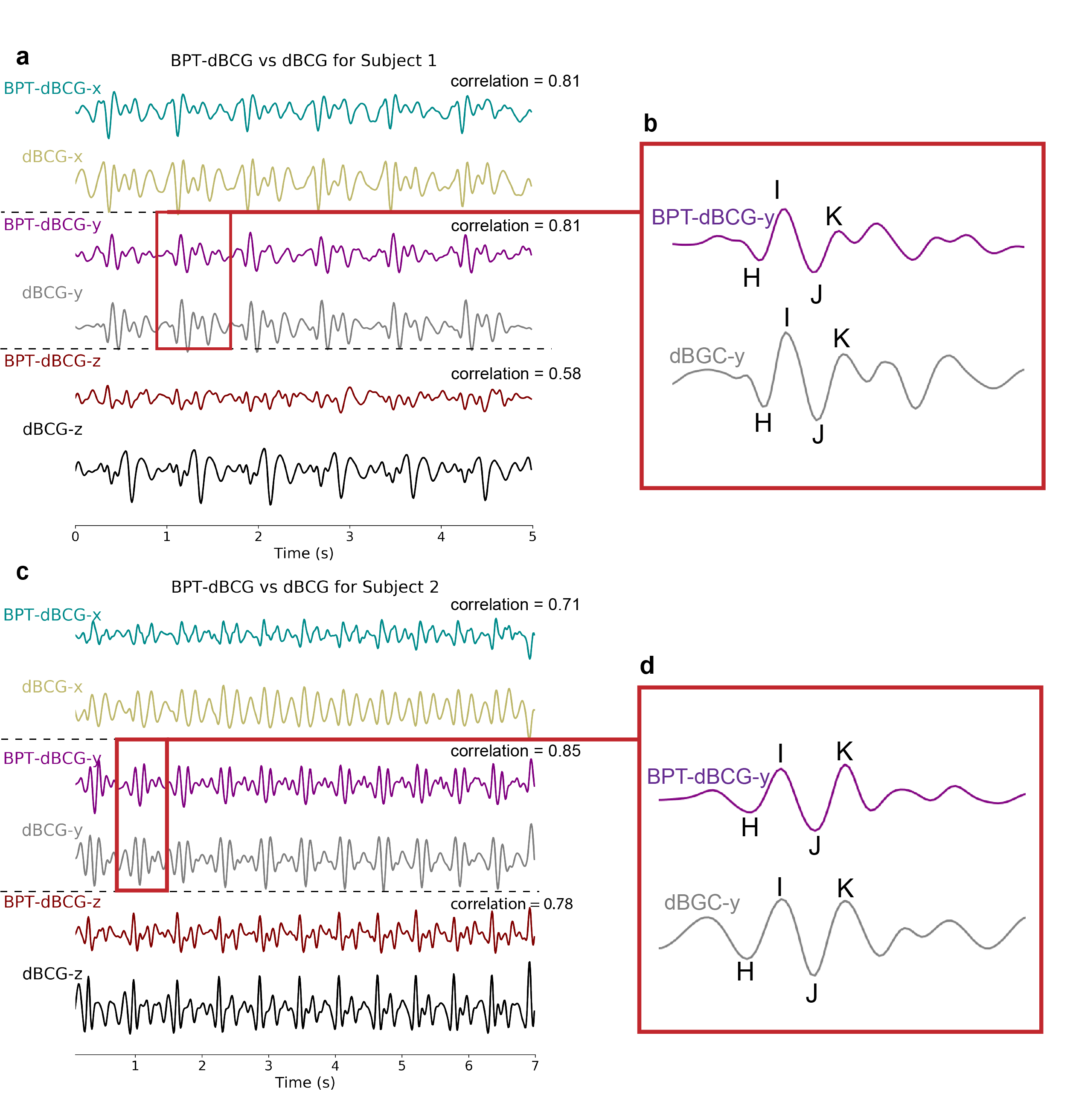
Figure 4: The BPT signals were linearly combined via a least-squares fit to the dBCG from each axis (dBCG-x, dBCG-y, and dBCG-z), then low-pass filtered with a cutoff of 15Hz for a) subject 1 and c) subject 2, with correlation coefficient displayed on the right. The correlation is greatest between BPT and dBCG-y for both subjects (0.81 and 0.85, respectively). c) and d) show a zoomed-in heartbeat and identify particular features of the dBCG, denoted by H, I, J, and K11. These features correlate with cardiac function and could be a predictor of cardiovascular risk12.
DOI: https://doi.org/10.58530/2023/0756