0755
Development of a cost-effective, fiber optic-based, MRI-compatible EEG system: a proof-of-concept study
Michael Potter1, Emily Holz1, Lindsay Demblowski1, Kyle Hunkar1, Udunna Anazodo2, Stefan Preble1, and Iris Asllani1,3
1Rochester Institute of Technology, Rochester, NY, United States, 2McGill University, Montreal, QC, Canada, 3University of Sussex, Brighton, United Kingdom
1Rochester Institute of Technology, Rochester, NY, United States, 2McGill University, Montreal, QC, Canada, 3University of Sussex, Brighton, United Kingdom
Synopsis
Keywords: Multimodal, fMRI (task based), EEG, EEG recording, MRI compatible EEG
A cost-effective, 8-channel, MRI-compatible optical EEG prototype was implemented and tested. The protoype has the potential to be especially suited for low-field MRI applications. The system uses PhotrodeTM technology, a high impedance device that can pick up signals without the need for "wet" contact with the skin. The minimum resolvable voltage of the modulator was ~ 12.5 uV, sufficient for most EEG waves.INTRODUCTION
Studies probing brain function under normal and diseased conditions using MRI have become increasingly complex thus necessitating development of multi-modal imaging methods that integrate MR- and non-MR signals toward a more comprehensive assessment of the brain's function and metabolism. Information from simultaneous acquisition of these signals, which capture different aspects –spatially and temporally– of brain activity, can be integrated to explore questions that could not be addressed by each modality alone. EEG fMRI is a prime example. The added value of concurrent acquisition of EEG and fMRI signals has been long recognized1,2. Yet, despite advancements in hardware and software, the use of EEG MRI systems remains limited. Factors such as cost and procedural complexity make routine recording of EEG signals during MRI scanning challenging. Moreover, current MRI-compatible EEG systems are made with conventional wires, which can interact with RF pulses and produce artifacts1. Here, we present results from a feasibility study aimed at implementing a low cost, fiber optics-based, universally fitting, 8-electrode MRI-compatible EEG system. The prototype is based on using lithium niobate photonic electrodes, referred to as PhotrodesTM, which harnesses the "extremely" high electrical input impedance of Mach-Zehnder Intensity electro-optic modulators to detect microvolt and millivolt physiological signals, including electrical activity in the brain3. The prototype developed here could be especially suited for low-field MRI applications.METHODS
A schematic overview of the EEG prototype that was built is described in Figure 1. The light from a continues wave laser source is sent to a PhotrodeTM placed on the head. The EEG signal received by the photrode (which is realized via dry contact) instantaneously modulates the intensity of the light from the laser source. The modulated signal is then detected and further processed by an optical receiver. The overall design and its electrical components are shown in Figures 2 and 3, respectively. To make the modulator MRI-compatible, a plastic case was 3D-printed to replace the manufacturer's original metal case. A custom laser controller printed circuit board (PCB) was constructed and its performance was compared to that of a high-end, Agilent tunable laser controller. Foam electrode testing was also performed to test whether foam interfaces on the electrode would increase the SNR of the alpha waves during the EEG recording. The MR compatibility of the system was tested on a 500 MHz NMR spectrometer.RESULTS
The minimum resolvable voltage of the modulator tested was ~ 12.5 uV, sufficient for most EEG waves. EEG data was collected from a test subject (Figure 4A). When the subject’s eyes closed, the alpha wave activity was seen in the filtered data (Figure 4B). The raw data was fed into the modulator and transmitted via optics. The modulator was able to recover the alpha wave activity (Figure 4C). There was considerable laser drift over time at high power with the Agilent laser controller (Figure 5, orange). After the laser was integrated into the custom controller PCB, the downward power drift stabilized (Figure 5, blue). The foam interface between the electrodes and the skin improved the SNR by 21%.DISCUSSION
This project served as an initial proof-of-concept that PhotrodesTM can be adapted to acquire EEG data in the presence of a magnetic field. The commercial modulators required a bias voltage (not MRI-compatible). This requirement may be circumventable in low-field applications. For mid- and high-field applications, however, a bias-free modulator will need to be custom-designed based on the path lengths in the interferometer. Another challenge encountered with the current design was the inconsistent modulator performance: the minimum resolvable voltage was not reproduced in other modulators with similar specifications. Better understanding of the sensitivity differences between modulators is needed to guide future modulator design. This may require the design and fabrication of bespoke, bias-free modulators that are pre-biased to the appropriate point on the sine-squared intensity curve. Future work is needed to investigate the effect of gradients on the performance of the system. Here, again, low-field MRI systems may prove more suitable.Acknowledgements
The authors are grateful to Prof. Andrew Webb for his crucial feedback.References
1Goldman, R.I. et al., Clin. Neurophysiol 111 (11), 2000; 2Laufs H., et al., NeuroImage 40, 2008; 3Kingsley, Stuart A. et. al. “Photrodes for Physiological Sensing.” SPIE Photonics, 2004.Figures
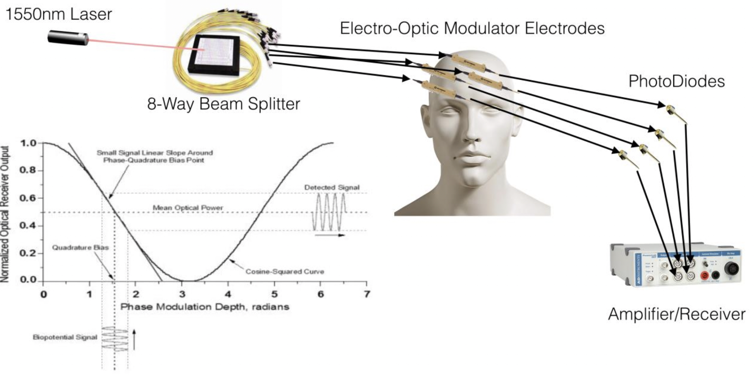
Figure 1: Schematic overview of the optic system.
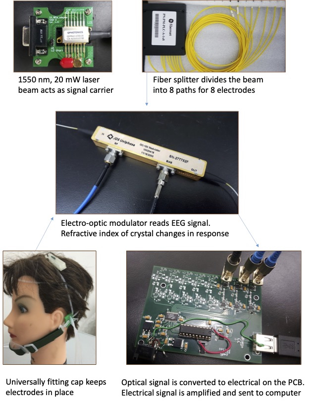
Figure 2: Design overview.
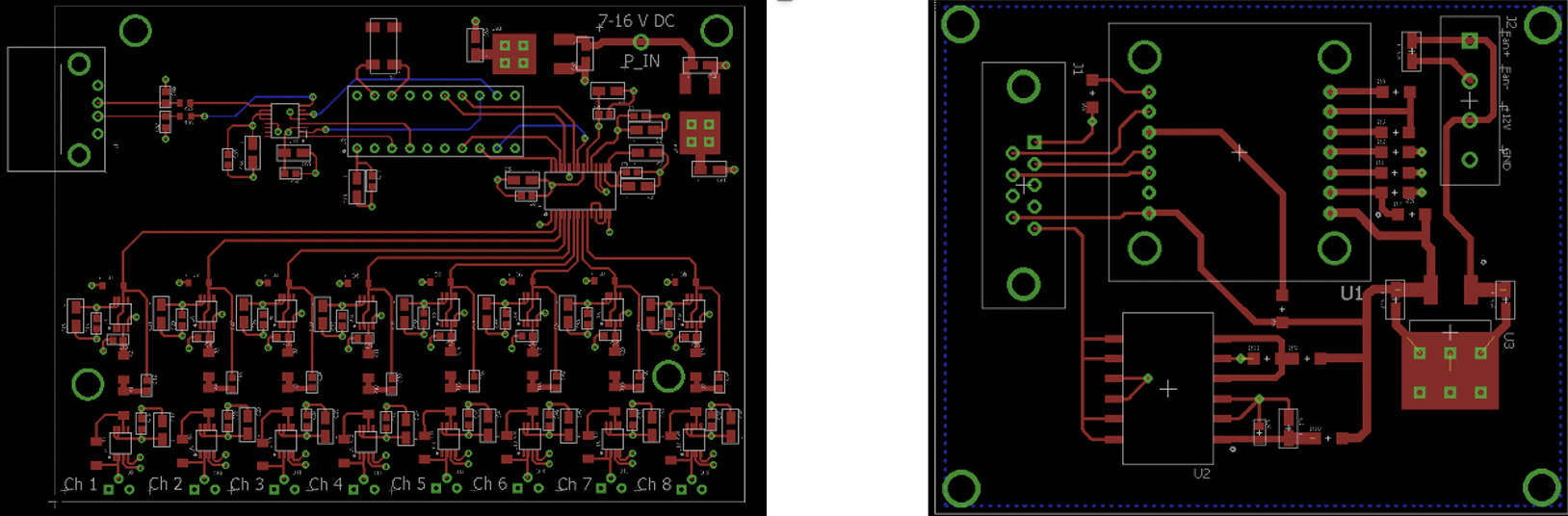
Figure 3: Electrical Components: Signal receiving board (left): 8 paths containing a photodiode, amplifier and filter. These paths converge into an ADC, microcontroller, and UART-USB converter pathway, which sends data to a computer. Laser controller board (right): includes a power controller, laser temperature controller, and power regulation circuitry.
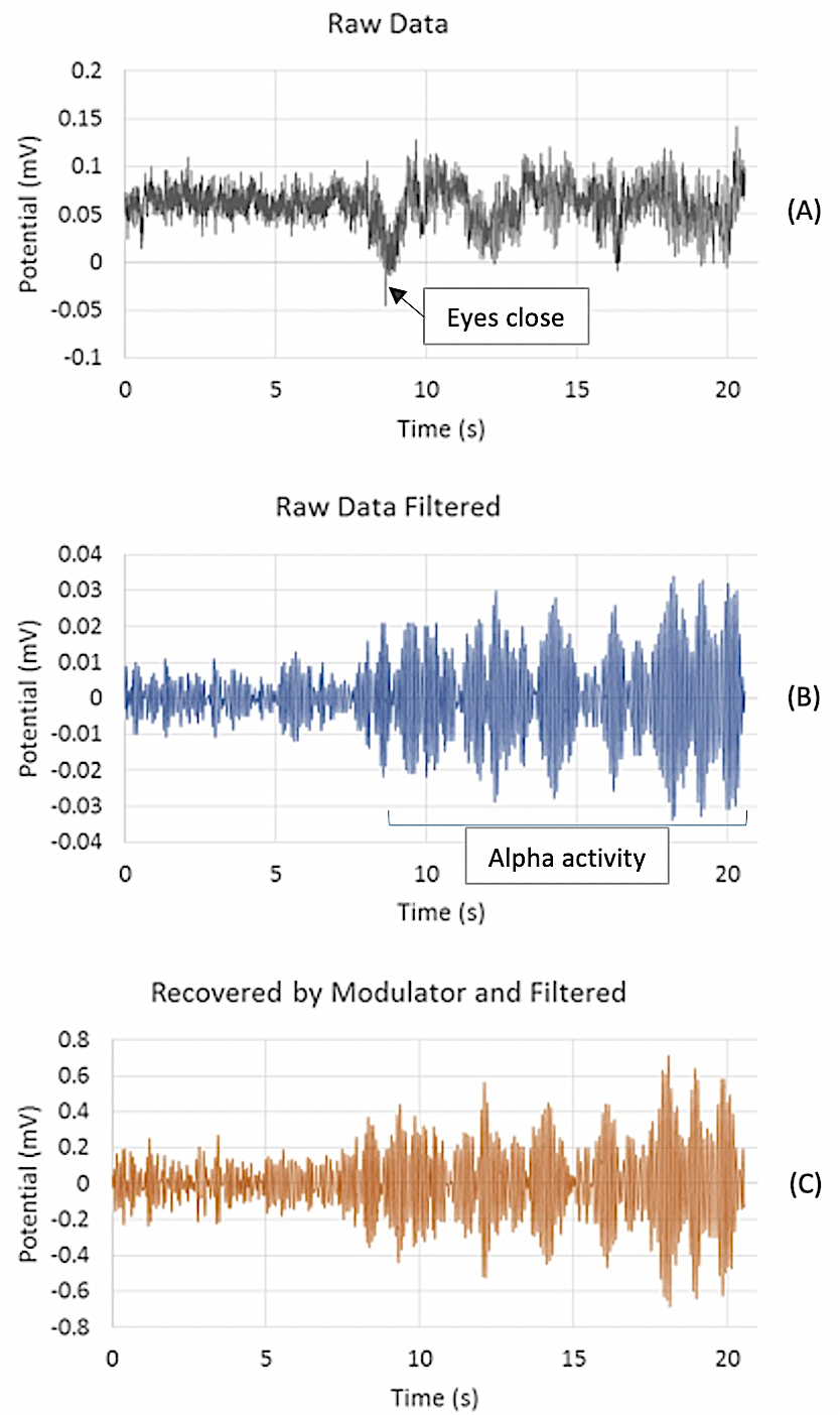
Figure 4: Alpha wave detection, (c.f. the text for details).
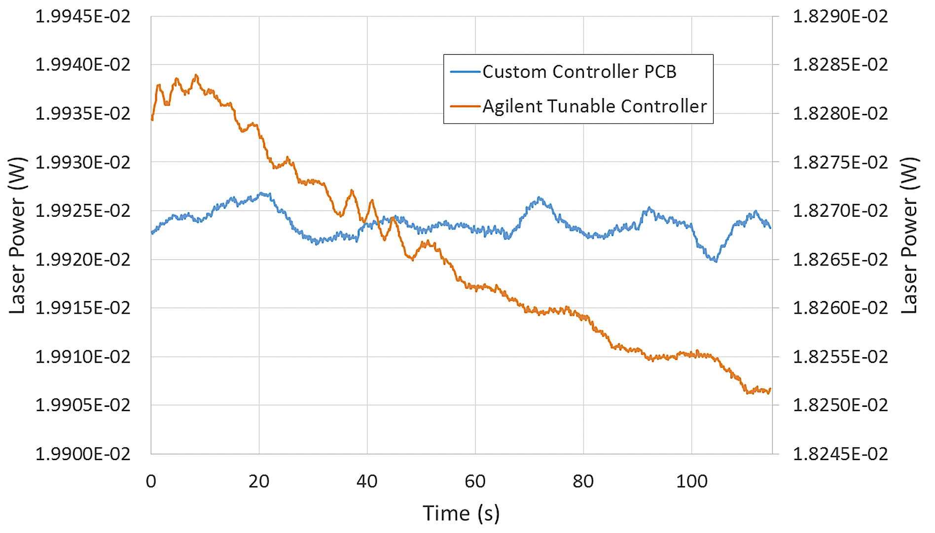
Figure 5: A considerable laser drift (orange line) was detected over time at high power with the Agilent laser controller. After the laser was integrated into the custom controller PCB, this downward power drift stabilized (blue line).
DOI: https://doi.org/10.58530/2023/0755