0753
Motion Monitoring using a Wireless Ultrasound-Based Sensor and an Integrated RF/Wireless Coil Array1Harvard Medical School, Boston, MA, United States, 2Radiology, Brigham and Women's Hospital, Boston, MA, United States, 3Medical Physics Graduate Program, Duke University, Durham, NC, United States, 4Brain Imaging and Analysis Center, Duke University, Durham, NC, United States, 5GE Healthcare, Aurora, OH, United States, 6Brain Imaging And Analysis Center, Duke University, Durham, NC, United States
Synopsis
Keywords: New Devices, Body
A wireless, battery-powered, MR-compatible ultrasound device consisting of an integrated RF/wireless coil, an organ-configuration motion sensor and its associated electronics was used to acquire OCM sensor signals on a healthy volunteer. Signals were obtained that characterized internal motion, which were wirelessly transmitted to the console room. Validation was performed against a real-time MR acquisition.Introduction
Patient physiological motion can significantly degrade MR image quality because the duration of time required to form an MR image is often too long to ‘freeze’ the underlying motion. By detecting/monitoring such underlying motion one can enable prospective (at the acquisition stage) and/or retrospective (at the reconstruction stage) motion compensation strategies that can mitigate the detrimental effects of motion on image quality. Organ-configuration motion sensors (OCM) are small, MR-compatible, ultrasound based sensors that can detect internal physiological motion1,2. These types of sensors have been used in the past to monitor breathing motion as part of wired systems that required a physical connection between sensors in the bore and MR-incompatible electronics in the console room. In the present work, a novel wireless coil technology called an integrated RF/wireless (iRFW) coil3 was combined with novel wireless OCM sensors. MR-incompatible electronics were replaced by a pulser/receiver printed circuit board (PCB), analog-to-digital converter PCB and a modified Raspberry Pi (RPi) CM4 (Fig. 1) to create a wireless, battery-powered, MR-compatible device. The device requires no wired connections and all electronics are integrated into a small form factor (about 20x10x10 cm), thereby making the device truly portable and able to travel with a patient throughout the hospital while maintaining Wi-Fi connection. Ultrasound and MRI signals tend to be complementary in nature4-8, and the present work introduces a practical approach to combine the two.Methods
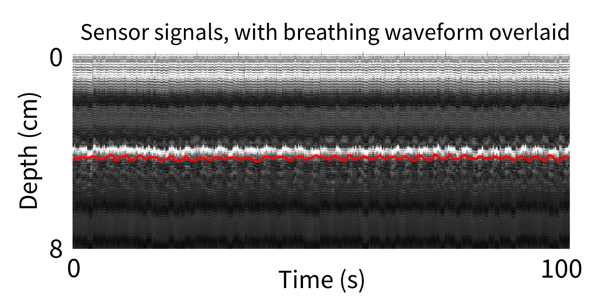
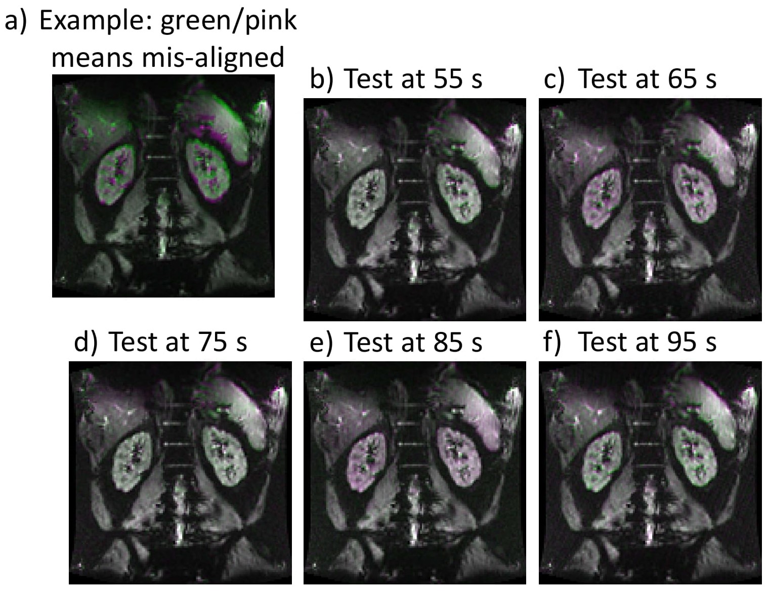
Signals were acquired, digitized, and wirelessly transmitted from the iRFW-OCM device to a laptop in the console room. Apache Kafka, an easily-scalable wireless streaming network protocol, was configured on the iRFW-OCM device and the laptop in the console room to streamline the wireless transmission of the OCM sensor signals (~1MB/s) throughout the experiment. For synchronization of the MRI and OCM sensor signals, MRI RF trigger pulses of a given sequence were measured in the machine room, time stamped on a Raspberry Pi 3 (RPi 3) and wirelessly transmitted to a laptop in the console room (Fig. 1). Both the iRFW-OCM device and Raspberry Pi 3 clocks were initialized using a network time protocol with millisecond precision managed by the laptop in the console room to align the ultrasound and trigger data timestamps for the MRI and ultrasound data time-synchronization. A time series of gradient-echo echo planar imaging (EPI) images and OCM signals were acquired on a healthy volunteer, using a 24-channel iRFW coil array and the iRFW-OCM device, respectively. The MRI images were acquired at a rate of 1 Hz, while the ultrasound data was acquired at a rate of ~60 Hz to comprise 100 MR images and ~6000 ultrasound signals. The data sets were aligned using the recorded RF trigger pulses, and a trace charactering motion (i.e., breathing waveform) in the MRI images was generated by averaging the observations of two independent human observers. A breathing waveform was generated from the OCM sensor signals and was down-sampled such that a given point would correspond to the MR image formed in that time. Synthesized MR images were generated from the second half of the OCM sensor signals for each time point by 1) identifying the closest matching time-point of OCM-generated trace from the first half of the OCM sensor signals and 2) finding the corresponding MR image at that time point. For validation, the synthesized MR images were compared with the acquired MR images.Results and Discussion
The TR specified by the scanner was 1000 ms, and the average times recorded by the RPi 3 between each transmit pulse and each TR were 99.99 +- 0.01 ms and 999.90 +- 0.12 ms, respectively. We observed no packet loss throughout the experiment. The breathing waveform generated from the MR images is shown plotted against M-mode ultrasound data (Fig. 2) and qualitatively shows a similar pattern to that of the M-mode ultrasound data. Synthesized MR images are shown overlaid with acquired MR images for various time points (Fig. 3). Misalignment leads to green and/or pink hue, as demonstrated in Fig. 3a by comparing two different time frames (i.e., different breathing states). Actual results are shown in Fig. 3b-f, where actual and synthetic frames are compared.Conclusion
In conclusion, we used the wireless, MR-compatible, battery-powered iRFW-OCM device to generate breathing waveforms and synthetic MR images. Potential future applications could include generating synthetic MR images outside of the scanner for interventional applications, which is made practical by the wireless and portable design of the iRFW-OCM device.Acknowledgements
Support from NIH grant number R01EEB030470 and T32EB025823.References
1. Madore B, Preiswerk F, Bredfeldt JS, Zong S, Cheng CC. Ultrasound-based sensors to monitor physiological motion. Med Phys 2021;48(7):3614-3622.
2. Preiswerk F, Toews M, Cheng CC, Chiou JG, Mei CS, Schaefer LF, Hoge WS, Schwartz BM, Panych LP, Madore B. Hybrid MRI-Ultrasound acquisitions, and scannerless real-time imaging. Magn Reson Med 2017;78(3):897-908.
3. Darnell D, Cuthbertson J, Robb F, Song AW, Truong TK. Integrated radio-frequency/wireless coil design for simultaneous MR image acquisition and wireless communication. Magn Reson Med 2019;81(3):2176-2183.
4. Günther M, Feinberg DA. Ultrasound-guided MRI: preliminary results using a motion phantom. Magn Reson Med 2004;52(1):27-32.
5. Arvanitis CD, Livingstone MS, McDannold N. Combined ultrasound and MR imaging to guide focused ultrasound therapies in the brain. Phys Med Biol 2013;58(14):4749-4761.
5. Bednarz BP, Jupitz S, Lee W, Mills D, Chan H, Fiorillo T, Sabitini J, Shoudy D, Patel A, Mitra J, Sarcar S, Wang B, Shepard A, Matrosic C, Holmes J, Culberson W, Bassetti M, Hill P, McMillan A, Zagzebski J, Smith LS, Foo TK. First-in-human imaging using a MR-compatible e4D ultrasound probe for motion management of radiotherapy. Phys Med 2021;88:104-110.
6. Kording F, Yamamura J, de Sousa MT, Ruprecht C, Hedstrom E, Aletras AH, Ellen Grant P, Powell AJ, Fehrs K, Adam G, Kooijman H, Schoennagel BP. Dynamic fetal cardiovascular magnetic resonance imaging using Doppler ultrasound gating. J Cardiovasc Magn Reson 2018;20(1):17.
7. Petrusca L, Cattin P, De Luca V, Preiswerk F, Celicanin Z, Auboiroux V, Viallon M, Arnold P, Santini F, Terraz S, Scheffler K, Becker CD, Salomir R. Hybrid ultrasound/magnetic resonance simultaneous acquisition and image fusion for motion monitoring in the upper abdomen. Investigative radiology 2013;48(5):333-340.
Figures
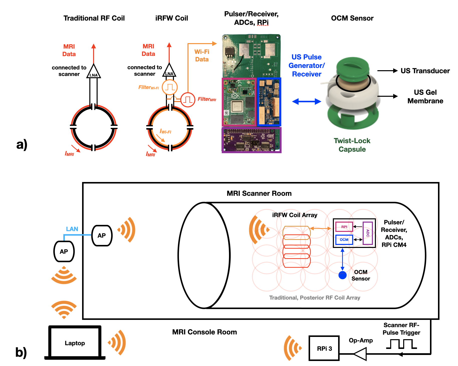
Figure 1: a) iRFW-OCM device consisting of an OCM sensor, a pulser/receiver, ADCs, an RPi CM4, and an iRFW coil array. a) iRFW-OCM device scanner setup: the iRFW-OCM device acquires, digitizes, and wirelessly transmits ultrasound data to an AP on the scanner wall, which is connected to a laptop in the console room. An RPi 3 measures scanner trigger pulses and wirelessly sends them to the console room laptop. The clocks of the iRFW-OCM device are initialized through NTP to the console room laptop and an Apache Kafka network manages the data stream (~MB/s) from the iRFW-OCM device to the laptop.