0743
Quantitative assessment of the lumbar paraspinal muscle in patients with unilateral lumbar disc herniation by IDEAL-IQ MR sequence1Sichuan Province Orthopedic Hospital, Chengdu, China, 2Chengdu Sport University, Chengdu, China, 3GE Healthcare China, Beijing, China
Synopsis
Keywords: Muscle, Fat, Quantitative imaging, Lumbar disc herniation
The development, progression, treatment and rehabilitation of lumbar disc herniation (LDH) are strongly associated with morphology and structural characteristics of lumbar paraspinal muscle. The IDEAL-IQ MR sequence allows the direct quantification of lumbar paraspinal muscle properties including fat fraction (FF) and cross-sectional area (CSA). The side-to-side differences between quantitative properties of bilateral paraspinal muscles in patients with unilateral LDH remain unclear. Therefore, this study aimed to investigate the possible unilateral changes of the paraspinal muscles in patients with unilateral LDH. Our results found no difference in FF and CSA values of paraspinal muscles between the affected and unaffected sides.Introduction
Lumbar paraspinal muscles play an important role in maintaining the upright posture and the dynamic stability of the lumber spine, and help to reduce the load on the intervertebral disc. Strong paraspinal muscles can greatly increase the stability of the lumbar spine, while the degeneration of the paraspinal muscles is closely related to the low back pain (LBP) caused by a variety of diseases. Lumbar disc herniation (LDH) is an important cause of LBP. The widely concerned unilateral LDH can often lead to nerve compression, and may cause paraspinal muscle changes. The muscle cross-sectional area (CSA) and fat infiltration are two common imaging indicators to evaluate paraspinal muscle properties. The fat infiltration of paraspinal muscles was indirectly evaluated on routine T1- or T2-weighted images in previous studies (1). The chemical shift-encoded IDEAL-IQ MR sequence (2) can provide accurate fat fraction (FF) values that directly quantify the fat infiltration of paraspinal muscles (3). The side-to-side differences between quantitative properties of bilateral paraspinal muscles in patients with unilateral LDH remain controversial (1,4). Therefore, this study aimed to investigate the possible unilateral changes of the paraspinal muscles in patients with unilateral LDH using IDEAL-IQ MR sequence.Methods
Patients: After IRB-approved written informed consent was obtained, preoperative patients with LBP were scanned on 3.0 T MRI (SIGNA Architect, GE Healthcare, USA). Finally, 208 patients diagnosed with unilateral LDH at L4-L5 and L5–S1 levels, which was confirmed by MRI and clinical symptoms, were included in this retrospective study.Imaging parameters: The MR scan included axial T2 FSE (0.5×0.8×3.0 mm3, TR=3224 ms, TE=120 ms), axial IDEAL-IQ (2.0×2.0×2.0 mm3, TR=8.0 ms, TE=3.6 ms, number of TEs=6, number of shots=2).
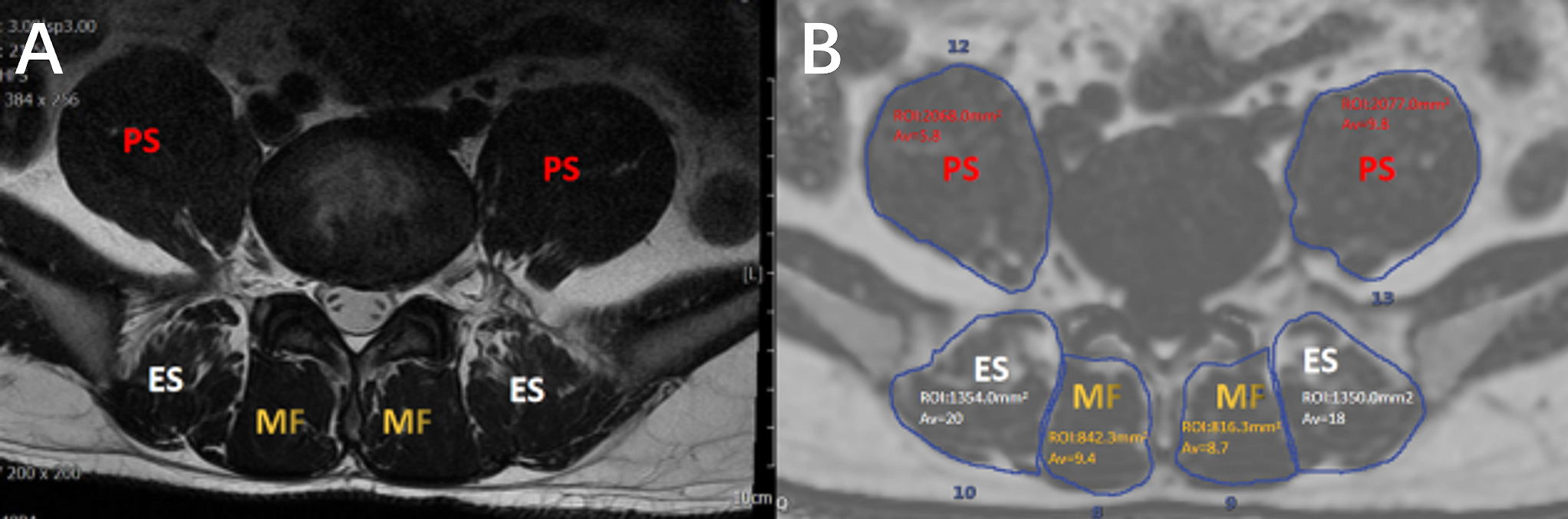
Data processing: The IDEAL-IQ images were processed in the AW4.7 workstation (GE Healthcare) to calculate the FF map. The mean CSA and FF values of the affected and unaffected paraspinal muscle were obtained on a ROI basis at the central level of L1-L2, L2-L3, L3-L4, L4-L5, and L5-S1 (Figure 1), respectively. Axial T2-weighted images were used to confirm the size and shape of herniated discs, from which the patients were separated into three groups according to the MSU classification system (5).
Statistical analysis: All analyses were performed using SPSS 22.0 software. Kolmogorov-Smirnov test was applied to assess the normal distribution of the variables. After that, the difference of paraspinal muscle properties (CSA and FF) between affected and unaffected sides in each group were tested by the independent samples t-test. Spearman’s rank correlation was used to assess the correlation between paraspinal muscle properties and disc levels or groups, respectively. The differences in age, weight, height, and BMI among different groups were tested by one-way analysis of variance (ANOVA). The p-value < 0.05 was considered statistically significant.
Results
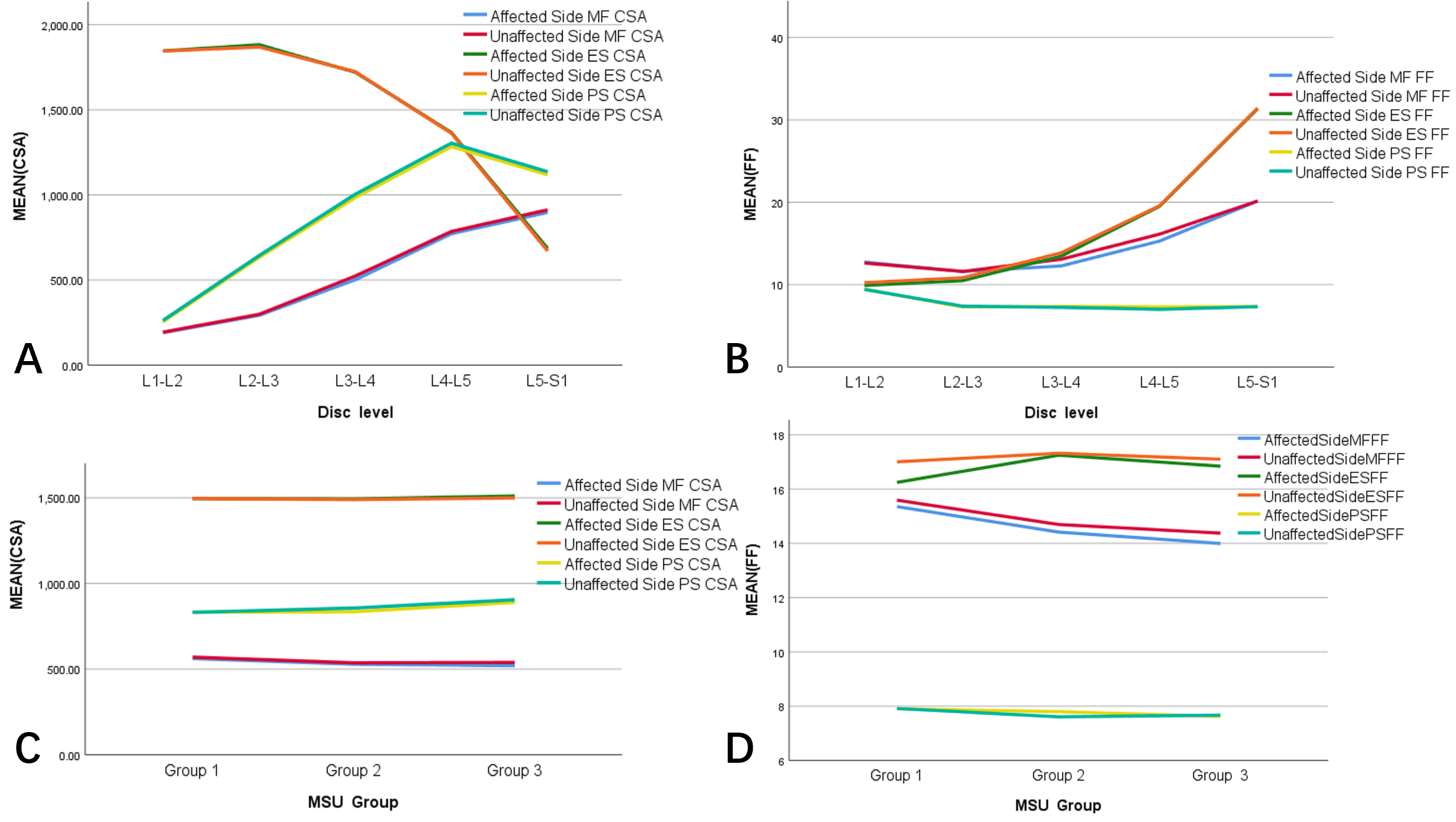
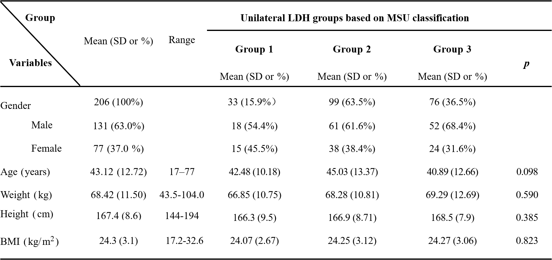
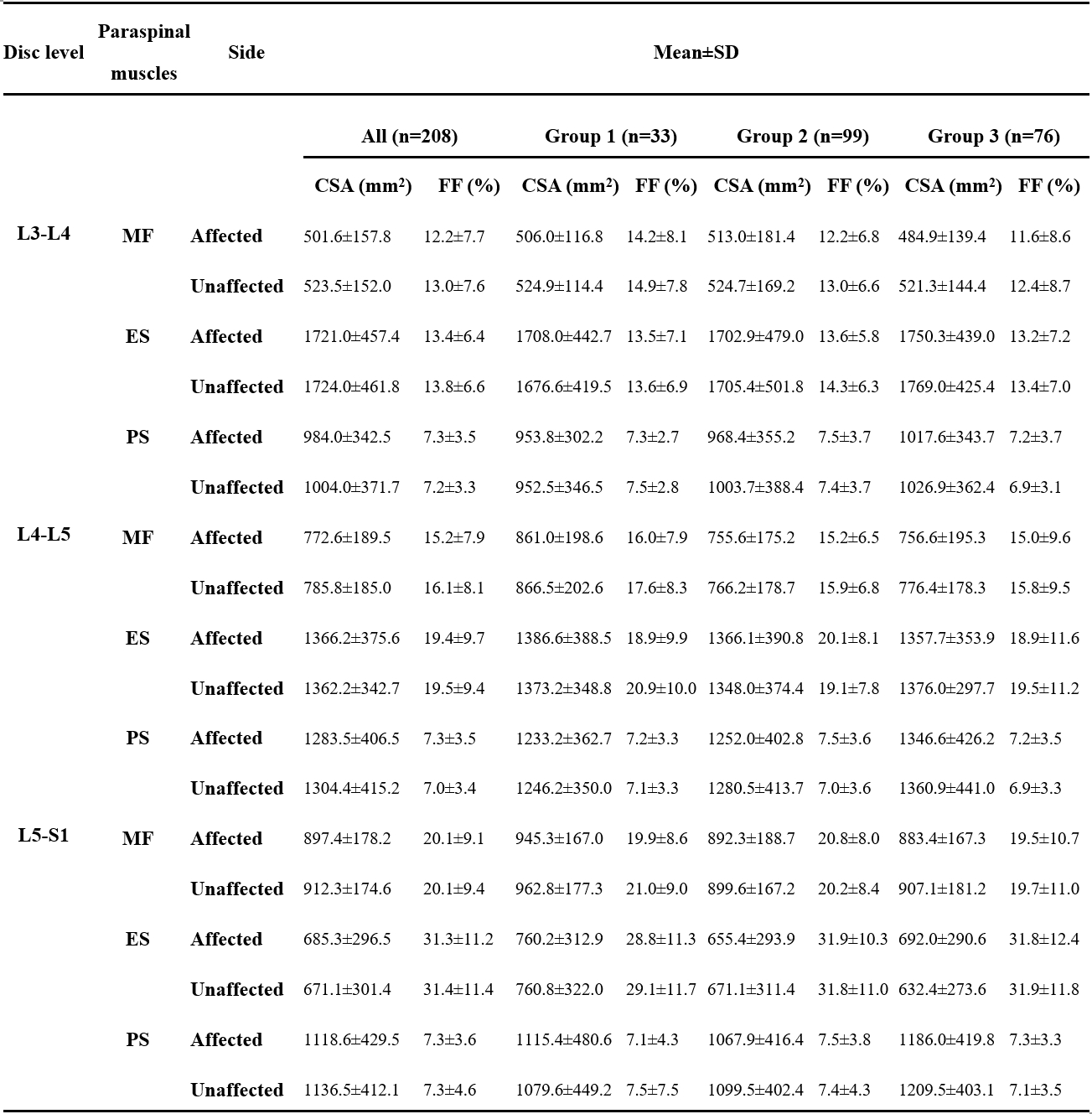
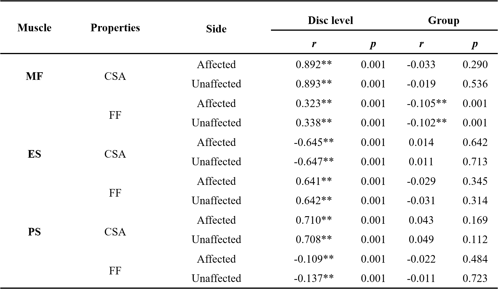
As shown in Table 1, all patients were divided into three groups based on the MSU system. No significant difference in age, weight, height, and BMI was found among three groups. As shown in Table 2, independent samples t-test found no significant side-to-side difference in CSA and FF values of three paraspinal muscles at any disc level or in any group. As shown in Table 3 and Figure 2, The disc level had a positive correlation with MF/PS CSA and MF/ES FF (affected side: r = 0.892, 0.710, 0.323, 0.641, p = 0.001), had a negative correlation with ES CSA (affected side: r = -0.645, p = 0.001), and had a very weak negative correlation with PS FF (affected side: r =-0.109, p = 0.001). MSU based group was not correlated to CSA of MF/ES/PS and FF of ES/PS, and had a very weak negative correlation with FF of MF (affected side: r = -0.105, p = 0.001).Discussion
Paraspinal muscles are vital to the support and stabilization of the spine. Previous studies have shown conflicting results on the relationship between LBP and morphology of paraspinal muscles, especially for fatty infiltration of muscles (6). The study aimed to explore whether side-to-side changes of paraspinal muscle properties occur in patients with unilateral LDH. Previous studies assessed microscopic muscle properties of paraspinal muscle and shown the presence of ipsilateral multifidus muscle changes in patients with unilateral LDH (7). Yoshihara et al suggested that this microscopic change was limited to the segment where the lumbar disk herniation is located (8). Some macroscopic studies found increased fat infiltration and atrophy in multifidus muscle of the affected side, manifested as increased fat content and reduced CSA (1,9). However, we found no side-to-side difference in CSA and FF of paraspinal muscles in patients with unilateral LDH, consistent with the results of a recent study (4). In addition, the correlations between CSA and FF of paraspinal muscles and disc level or MSU grade were identified here.Conclusion
Quantitative assessment offered by IDEAL-IQ MR sequence found no difference in FF and CSA values of paraspinal muscles between the affected and unaffected sides in patients with unilateral LDH. Larger sample sizes and a possible subgroup are needed to identify characteristics of paraspinal muscles for unilateral LDH in future studies. Quantitative imaging of paraspinal muscles holds promise to improve our understanding of unilateral LDH.Acknowledgements
No acknowledgement found.References
1. Stevens S, Agten A, Timmermans A, Vandenabeele F. Unilateral changes of the multifidus in persons with lumbar disc herniation: a systematic review and meta-analysis. Spine J. 2020;20:1573–1585.
2. Yu H, Shimakawa A, McKenzie CA, Brodsky E, Brittain JH, Reeder SB. Multiecho water-fat separation and simultaneous R2* estimation with multifrequency fat spectrum modeling. Magn. Reson. Med. Off. J. Int. Soc. Magn. Reson. Med. 2008;60:1122–1134.
3. Fischer MA, Nanz D, Shimakawa A, et al. Quantification of muscle fat in patients with low back pain: comparison of multi-echo MR imaging with single-voxel MR spectroscopy. Radiology 2013;266:555–563.
4. Xiao Y, Fortin M, Ahn J, Rivaz H, Peters TM, Battié MC. Statistical morphological analysis reveals characteristic paraspinal muscle asymmetry in unilateral lumbar disc herniation. Sci. Rep. 2021;11:15576 doi: 10.1038/s41598-021-95149-6.
5. Mysliwiec LW, Cholewicki J, Winkelpleck MD, Eis GP. MSU classification for herniated lumbar discs on MRI: toward developing objective criteria for surgical selection. Eur. Spine J. 2010;19:1087–1093.
6. Seyedhoseinpoor T, Taghipour M, Dadgoo M, et al. Alteration of lumbar muscle morphology and composition in relation to low back pain: a systematic review and meta-analysis. Spine J. 2022;22:660–676.
7. Zhao W-P, Kawaguchi Y, Matsui H, Kanamori M, Kimura T. Histochemistry and morphology of the multifidus muscle in lumbar disc herniation: comparative study between diseased and normal sides. Spine 2000;25:2191–2199.
8. Yoshihara K, Shirai Y, Nakayama Y, Uesaka S. Histochemical changes in the multifidus muscle in patients with lumbar intervertebral disc herniation. Spine 2001;26:622–626.
9. Hyun JK, Lee JY, Lee SJ, Jeon JY. Asymmetric atrophy of multifidus muscle in patients with unilateral lumbosacral radiculopathy. Spine 2007;32:E598–E602.
Figures