0729
Diagnostic performance of DCE-MRI Parameters in Distinguishing Tumor Deposits from Metastatic Lymph Nodes in Rectal Cancer
Qiaoyu Xu1, Hongliang Sun2, Yanyan Xu2, Juan Wang2, and Tongyin Zhang2
1Beijing Chao-Yang Hospital, Capital Medical University, Beijing, China, 2China-Japan Friendship Hospital, Beijing, China
1Beijing Chao-Yang Hospital, Capital Medical University, Beijing, China, 2China-Japan Friendship Hospital, Beijing, China
Synopsis
Keywords: Pelvis, Cancer
This study aimed to investigate the usefulness of diffusion-weighted imaging (DWI) derived apparent diffusion coefficient (ADC) values and dynamic contrast-enhanced magnetic resonance imaging (DCE-MRI) derived semi-quantitative parameters in distinguishing TD from MLN before surgical resection in RC patients. TD has some specific morphological features, including relatively larger size, lower short- to long-axis ratio, irregular shape and ill-defined border on T2-weighted MR images in rectal cancer. The combination of ADC values and parameters of DCE-MRI can help to improve the diagnostic efficiency of TD in rectal cancer.INTRODUCTION
Tumor deposits (TD) has been shown as an independent prognostic predictor associated with lower overall survival and lower disease-free survival in rectal cancer (RC). Moreover, there is growing evidence that TD count has specific prognostic value, even for patients with the presence of metastatic lymph nodes (MLN). Currently, MLN assessment mainly depends on morphologic characteristics of malignancy on MRI, which may overlap with TD. A growing role of diffusion-weighted imaging (DWI) and dynamic contrast-enhanced magnetic resonance imaging (DCE-MRI), as important advanced sequences of MRI over past years, have been noted. However, to the best of our knowledge, no previous reports have mentioned the role of DWI and DCE-MRI parameters in differentiating TD and LN. This study aimed to identify whether morphology, DWI and DCEparameters could help distinguish TD from MLN before surgical resection in rectal RC.METHODS
Thirty patients (totally 59 lesions, including 30 TD and 29 MLN) with pathologically proven RC and the presence of TD and/or MLN who underwent pretreatment MR examination at 3.0 T were enrolled in this study. Morphological parameters of lesions on T2-weighted imaging were compared between TD and MLN using independent-samples t-test and Fisher’s exact test. Apparent diffusion coefficient (ADC) values and semi-quantitative parameters of DCE-MRI, including relative enhancement (RE), maximum enhancement (ME), maximum relative enhancement (MRE), time to peak (TTP), wash in rates (WIR), wash out rates (WOR), brevity of enhancement (BRE), and area under the curve (AUC) were measured and automatically calculated on the lesions and tumor (Figure 1,2). These parameters were compared between TD and MLN, and between tumor with TD group and tumor without TD group by using independent-samples t-test and Mann-Whitney U test. The ratio of these parameters (parameter-ratio) of the lesions-to-tumor tissues was also compared between TD and MLN. Images were evaluated by two experienced gastrointestinal radiologists whom had been blinded to the histopathology results. Receiver operating characteristic curve analysis and binary logistic regression analysis were used to assess the diagnostic ability of single and combined metrics for distinguishing TD from MLN. Inter-observer variability was analyzed using the intraclass correlation coefficient.RESULTS
The morphological features, including size, shape, and border were significantly different between TD and MLN. TD exhibited significantly lower RE, MRE, RE-ratio, MRE-ratio, ADCmin-ratio, and ADCmean-ratio than MLN ((P =0.046, 0.036, 0.001, 0.006, 0.030, and 0.032, respectively). RE-ratio showed the highest AUC (0.749) and accuracy (77.97%) among single parameters. RC exhibit higher RE, MRE, and ADCmean in tumor with TD group than tumor without TD group. (P = 0.041, 0.027, and 0.046, respectively). The combination of the DWI parameters (ADCmin-ratio, and ADCmean-ratio), the combination of the mutual independent DCE-MRI parameters (RE-ratio and MRE-ratio), and the combination of DCE-MRI and DWI parameters (ADCmin-ratio, ADCmean-ratio, RE-ratio, and MRE-ratio) together demonstrated higher diagnostic efficiency (AUC = 0.683, 0.739, 0.825, respectively).(Figure 3).DISCUSSION
Semiquantitative parameters in DCE-MRI is derived from a time-signal intensity curve and represents contrast-related signal intensity changes, which requires less complicated software algorithms and is more convenient in post-processing. We found significantly higher RE and MRE of primary tumor tissue in the tumor with TD group compared with the tumor without TD group. These DCE-MRI parameters can reflect angiogenesis and blood perfusion, of which RE is influenced by both the gadolinium-based contrast agent diffusing into the inter-tissue spaces and in intravascular spaces, and MRE mainly reflects the RE of the lesion when the tissue enhancement reaches its peak[1]. Liu et al published that RE and MRE were associated with the positive expression of hypoxia-inducible factor 1 alpha (HIF-lα), which can regulate the angiogenesis[1]. These results reflect that the presence of TD may be associated with higher angiogenic activity and capillary permeability. In our studies, 16/23 (70%) patients with TD had positive EMVI on pathology. Meanwhile, we found that the RE and MRE of TD were lower than those of the MLN. This could be due to the different histopathological characteristics between TD and MLN. The origin and mechanism of formation of TD is still not entirely clear, and even showed more than one origin, such as vascular, lymphatic, and/or perineural routes[2]. Among all the parameters, the RE-ratio of lesion to tumor showed the highest area under the curve (0.749) and accuracy (77.97%) among single parameters in identifying TD from MLN. The inter-observer agreement of RE and MRE was good to excellent. Thus, DCE-MRI may help to establish prognostic factors, as the higher RE and MRE in RC may help predict the presence of TD, and the lower RE and MRE of separate nodes within the lymph drainage area of RC may help distinguish TD from MLN.CONCLUSION
Morphological features, DWI and DCE-MRI parameters have the potential to help predict TD and distinguish TD from MLN before surgery.Acknowledgements
No acknowledgement found.References
[1] Liu L, Hu L, Zeng Q et al (2021) Dynamic contrast-enhanced MRI of nasopharyngeal carcinoma: correlation of quantitative dynamic contrast-enhanced magnetic resonance imaging (DCE-MRI) parameters with hypoxia-inducible factor 1alpha expression and tumor grade/stage. Ann Palliat Med 10:2238-2253.
[2] Nagtegaal ID, Quirke P (2007) Colorectal tumour deposits in the mesorectum and pericolon; a critical review. Histopathology 51:141-149.
Figures
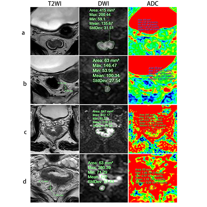
Fig. 1 Morphological features on T2WI, DWI (b= 1000 s/mm2), and ADC map of TD, MLN, and rectal cancers. A 65-year-old female RC (a) patient with the presence of TD (b) and a 59-year-old male RC (c) patient with the presence of MLN (d) but no TD. The short axis and long axis of TD and MLN were measured on T2WI. The region of interest (ROI) of lesions were manually drawn and outlined along the edges of lesions at the largest cross-sectional area of them.
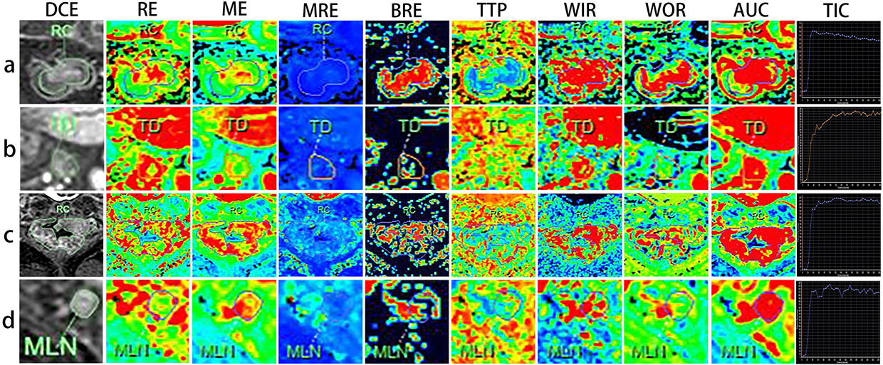
Fig. 2 DCE-MRI and semi-quantitative parameter maps of TD, MLN, and rectal cancers. A 65-year-old female RC (a) patient with the presence of TD (b) and a 59-year-old male RC (c) patient with the presence of MLN (d) but no TD. The region of interest (ROI) of lesions were manually drawn and outlined along the edges of lesions at the largest cross-sectional area of them, and then the time-signal intensity curve (TIC) of the ROI was obtained.
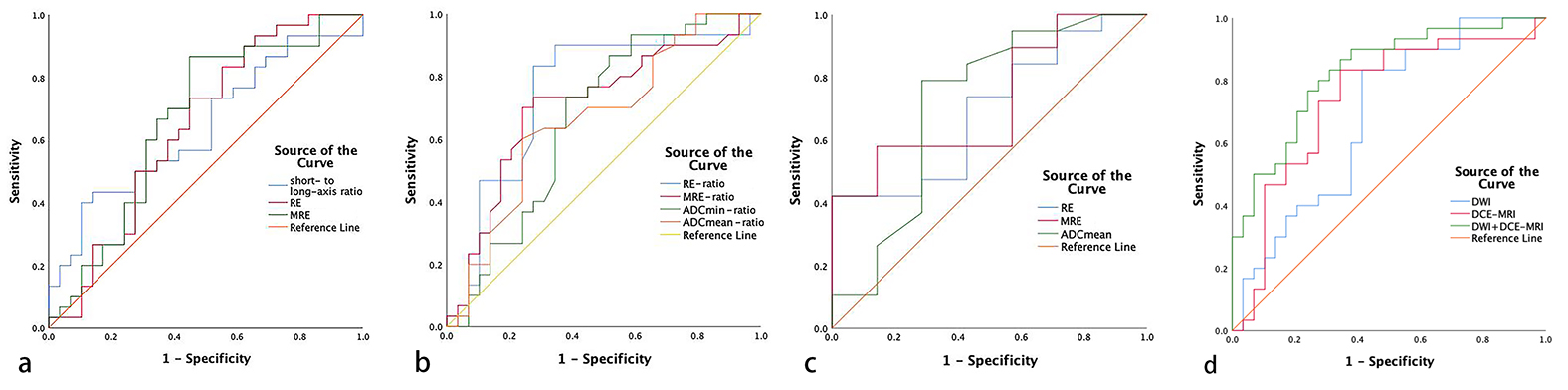
Fig. 3 ROC curve analysis of morphological parameters, ADC values, and DCE-MRI derived parameters (a) and their ratio of lesion-to-tumor (b) in distinguishing TD and MLN. (c) ROC curve analysis of ADC values and DCE-MRI derived parameters of tumor tissue between tumor with TD group and tumor without TD group. (d) ROC curve analysis of DWI parameters (ADCmin-ratio, and ADCmean-ratio), DCE-MRI parameters (RE-ratio and MRE-ratio), and the combination of DCE-MRI and DWI parameters for distinguishing TD from MLN.
DOI: https://doi.org/10.58530/2023/0729