0716
Various parameters derived from mono-exponential DWI, IVIM, SEM, DKI and CTRW in breast cancer diagnosis and the prediction of prognostic factors1Tongji Hospital, Tongji Medical College, Huazhong University of Science and Technology, Wuhan, China, 2MR Collaboration, Central Research Institute, United Imaging Healthcare, Shanghai, China
Synopsis
Keywords: Breast, Cancer
In the present study, we compared the potential of various diffusion parameters derived from five models in distinguishing breast lesions, including the mono-exponential model (Mono), intravoxel incoherent motion (IVIM) model, diffusion kurtosis imaging (DKI) model, stretched exponential model (SEM), and continuous-time random-walk (CTRW) mode. We found that diffusion-related parameters by the SEM and CTRW models are superior to ADC in discriminating breast lesions. Multiple diffusion parameters by all five diffusion models are related while the information provided is diverse.Introduction
Breast cancer is one of the most common causes of cancer-related death in women worldwide (1). Diffusion-weighted imaging (DWI) is a functional MRI technique that shows great potential in detecting and characterizing breast cancer (2). Apparent diffusion coefficient (ADC) calculated from conventional DWI with a simple mono-exponential model considers water molecule’s diffusion follows a Gaussian pattern, which may result in an inexact description of the diffusion (3). To best describe the real diffusion process, various advanced diffusion models, such as IVIM model, DKI model, SEM model, and CTRW model, have been developed. IVIM model is a bi-exponential model that might allow separating water molecular diffusion from the microcirculation while DKI model describes the non-Gaussian diffusion of water molecules caused by the complexity of tissue microstructure (4,5). Unlike DKI model, SEM and CTRW models were more powerful in revealing the tissue information regarding the structural heterogeneity. Clinical value of different models for evaluating the breast cancer has not been comprehensively quantitatively compared. Hence, this study aims to compare the diagnostic performance of mono-exponential DWI, IVIM, DKI, SEM and CTRW for diagnosing the breast cancer and predicting the prognostic factors of breast cancer.Methods
This retrospective study included 72 patients (20 benign and 52 malignant breast lesions). DWI examinations were performed on a 3.0 T scanner (uMR 790, United Imaging Healthcare) using nine b values (0 to 2000 mm2/s) and analyzed using five models: Mono (ADC), IVIM (pure diffusion coefficient (D), pseudo-diffusion coefficient (D*), and perfusion fraction (f)), DKI (mean kurtosis (MK) and mean diffusion coefficient (MD)), SEM (distributed diffusion coefficient (DDC) and diffusion heterogeneity index (α)), CTRW (spatial diffusion heterogeneity (β), temporal diffusion heterogeneity (α), and diffusion coefficient (Dm)). Comparisons between these parameters and different subgroups using Mann–Whitney U-test. Correlations between these parameters were evaluated using Spearman’s rank correlation test. The diagnostic efficiency of five models, alone and in combination, to distinguish benign from malignant breast lesions was analyzed by using receiver operating characteristics (ROC) curves and the binary logistic regression.Results
Representative MR images of patients with breast cancer and benign fibroadenoma were displayed in Figures 1 and 2. Benign lesions had significantly higher Mono-ADC, IVIM-D, IVIM-f, DKI-MD, SEM-DDC, CTRW-Dm, CTRW-α, and CTRW-β, and lower DKI-MK values than malignant lesions (Table 1), and SEM-DDC values showed the largest area under the curve (AUC) (Figure 3). As shown in Figure 4, the quantitative correlation analysis showed that there were strong correlations between Mono-ADC, IVIM-D, DKI-MD, DKI-MK, SEM-DDC, CTRW-Dm, and CTRW-α (all p < 0.01). CTRW-β correlated with all the other parameters (p < 0.05) except IVIM-f (p > 0.05). In particular, there was a strong positive correlation between Mono-ADC and CTRW-Dm (r= 0.988, p < 0.01). The IVIM-D value differed significantly between ER-positive and ER-negative tumors. All diffusion parameters except for IVIM-D*, IVIM-f, SEM-α, and CTRW-β differed significantly between PR-positive and PR-negative tumors. The DKI-MD value also differed significantly between HER2-positive and HER2-negative tumors and between Ki-67 high- and low-expression tumors.Discussion
Extensive works have been done in utilizing multiple DWI models for the differential diagnosis of breast lesions (2,4). The AUC of heterogeneity parameters (SEM-α, CTRW-α, and CTRW-β) is lower than diffusion parameters (Mono-ADC, IVIM-D, DKI-MD, SEM-DDC, and CTRW-Dm) in our study. We speculated the reason is that the difference of heterogeneity between malignant and benign lesions may be less than the difference of diffusion-related cellular density. Through logistic analysis, our results indicate that the AUC of the SEM model was higher than those of another four models. Jiang et al. (6) showed that the AUC of the SEM model was higher than that of IVIM and mono-exponential models, which is similar to this study.Conclusion
The Mono, IVIM, DKI, SEM, and CTRW models were all useful for the diagnosis of breast cancer, with the SEM model reaching the highest AUC value. Multiple parameters derived from all five models showed significant correlation with prognostic factors for breast cancer.Acknowledgements
NoneReferences
1. Demircioglu A, Grueneisen J, Ingenwerth M, et al. A rapid volume of interest-based approach of radiomics analysis of breast MRI for tumor decoding and phenotyping of breast cancer. PLoS One 2020;15:e0234871.
2. Horvat JV, Bernard-Davila B, Helbich TH, et al. Diffusion-weighted imaging (DWI) with apparent diffusion coefficient (ADC) mapping as a quantitative imaging biomarker for prediction of immunohistochemical receptor status, proliferation rate, and molecular subtypes of breast cancer. J Magn Reson Imaging 2019;50:836-846.
3. Lin M, Yu X, Chen Y, et al. Contribution of mono-exponential, bi-exponential and stretched exponential model-based diffusion-weighted MR imaging in the diagnosis and differentiation of uterine cervical carcinoma. Eur Radiol 2017;27:2400-2410.
4. Suo S, Cheng F, Cao M, et al. Multiparametric diffusion-weighted imaging in breast lesions: Association with pathologic diagnosis and prognostic factors. J Magn Reson Imaging 2017;46:740-750.
5. Karaman MM, Sui Y, Wang H, Magin RL, Li Y, Zhou XJ. Differentiating low- and high-grade pediatric brain tumors using a continuous-time random-walk diffusion model at high b-values. Magn Reson Med 2016;76:1149-1157.
6. Jiang X, Chen C, Liu J, Liu S. Application Value of Mathematical Models of Diffusion-Weighted Magnetic Resonance Imaging in Differentiating Breast Cancer Lesions. Evid Based Complement Alternat Med 2021;2021:1481271.
Figures
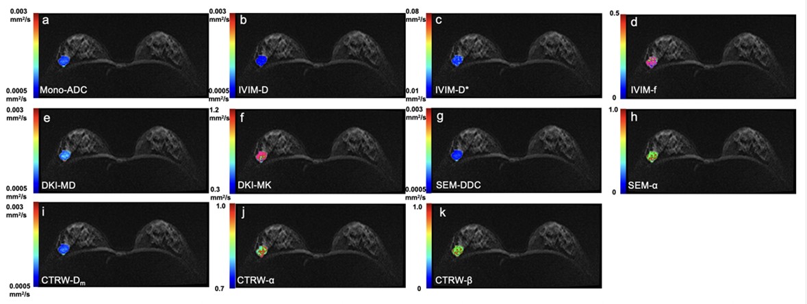
Figure 1. Parametric maps of a 26-year-old woman with invasive breast cancer in the right breast.
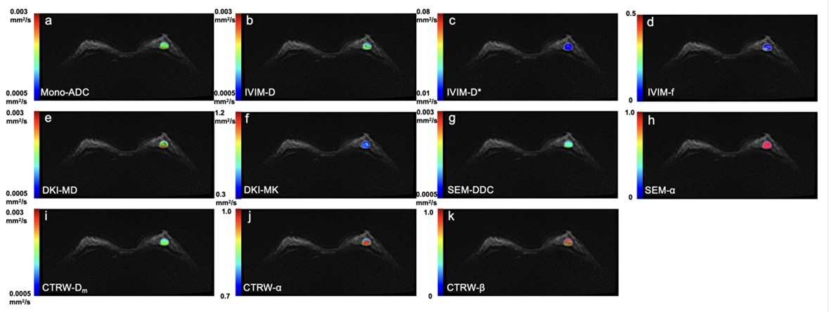
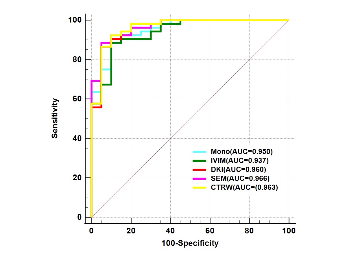
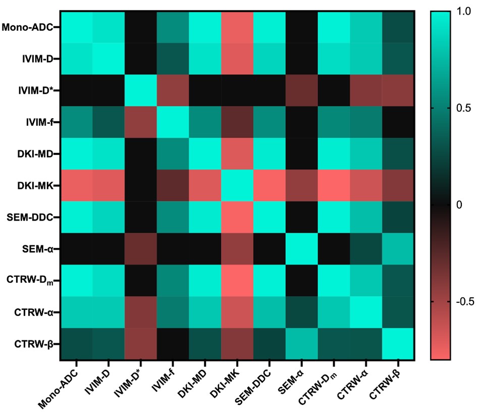
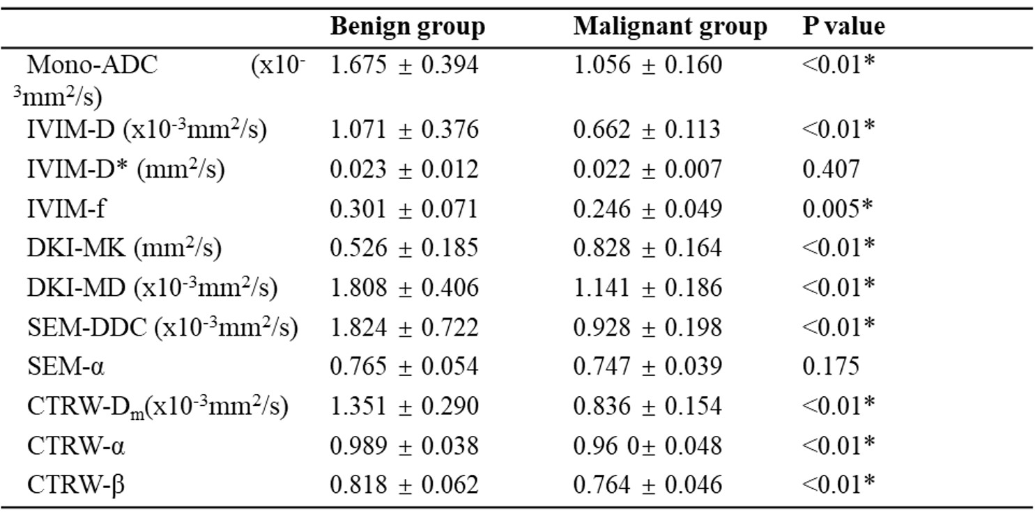
Table 1. Comparison of DWI parameters between benign (n = 20) and malignant (n = 52) breast lesions.