0713
CVT Detection with Routine Brain MRI Sequences by Multi-Modal Multi-Task Deep Learning algorithm1Beijing Chaoyang Hospital, Beijing, China, 2Infervision Medical Technology Co., Ltd, Beijing, China, Beijing, China, 3Beijing Xuanwu Hospital, Beijing, China, 4MR Scientific Marketing, Siemens Healthineers, Beijing, China, Beijing, China
Synopsis
Keywords: Stroke, Brain
A deep learning algorithm for detecting cerebral venous thrombosis using routine brain MRI achieved higher patient-level sensitivity than radiologists and reduced the number of overlooked thrombosed segments. The proposed deep learning (DL) algorithm achieved area under the receiver operating characteristic curve of 0.96 for detecting patient with cerebral venous thrombosis. The sensitivity of DL algorithm was higher than that of radiologists and obtained high specificity on patient-level.Compared to radiologists, DL algorithm found more thrombosed segments, indicating greater sensitivity and a sufficient specificity at the segment-level.Backgrounds:
Cerebral venous thrombosis (CVT) is a rare type of cerebrovascular disease1. Conventional brain MRI is usually used in diagnosing CVT2. Improving the diagnostic accuracy of CVT has always been the research that needs to be broken through3. This study aimed to develop and evaluate a novel deep learning (DL) algorithm for detecting CVT with routine brain MRI.Methods
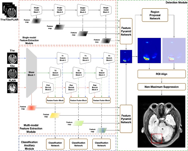
A total of 392 patients, including 294 patients with CVT (37 ± 14 years, 151 women) and 98 non-CVT patients (42 ± 15 years, 65 women), were enrolled. Of these, 100 patients (50 CVT and 50 non-CVT patients) were randomly assigned to the test set, and the rest constituted the development set. Routine brain MRI images including T1w, T2w, and FLAIR from 3.0T MR scanners (Magnetom Verio and Trio; Siemens, Germany) with 12-channel head coils of CVT patients and healthy controls from one CVT registry study were collected from April 2014 to December 2019. Different DL algorithms was constructed by multi-model feature extraction module and use global information by introducing a slice classification ancillary task4. Reference standards were defined by radiologist-adjudicated image review5. Four blinded radiologists with different levels of expertise read images and made diagnosis in the test set independently. The diagnostic performance on patient-level and segment-level, including area under the receiver-operating-characteristic curve (AUC), sensitivity, and specificity of the DL algorithm and radiologists, was evaluated, and compared in the test set.Results
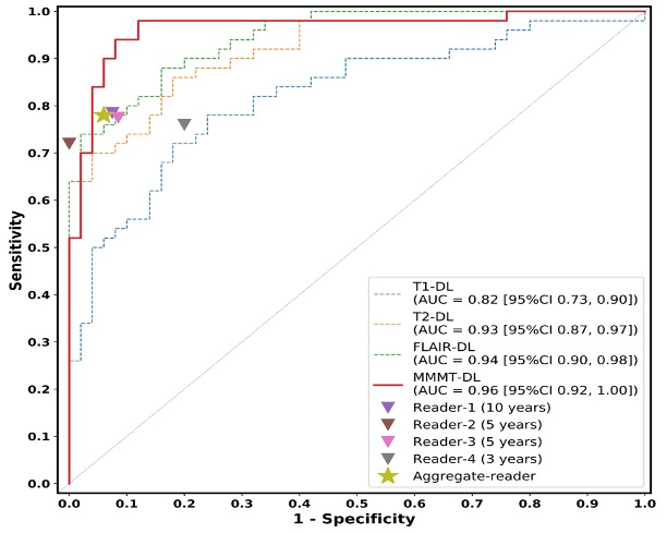
The optimal Multi-Modal Multi-Task Deep Learning (MMMT-DL) algorithm (Figure 1) achieved AUC of 0.96 with sensitivity of 94% (47/50) and specificity of 92% (46/50) on patient-level, sensitivity of 88% (129/146) and specificity of 80% (521/654) on segment-level. Compared with four radiologists, MMMT-DL had higher sensitivity on both patient-level (all p < 0.05) and segment-level (all p < 0.001) . The patient-level ROC of different DL algorithms was shown in Figure 2. The sensitivities of MMMT-DL for detecting thrombus in different segments was higher than readers. In 6 out of 8 segments, including LSS, LTS, RSS, RTS, SS, and SSS, MMMT-DL had a sensitivity of over 90%.Discussion
In this study, we developed a multi-modal multi-task deep learning algorithm tailored for CVT detecting via routine brain MRI. MMMT-DL took T1w, T2w, and FLAIR images jointly as input to exploit complementary multi-modal information and combined the image’s global and local information through a multi-task learning strategy. It achieved high sensitivity on patient-level and segment-level while not introducing excessive false-positive detection. The results of the reader study illustrated that the diagnostic performance of MMMT-DL in diagnosing CVT was as good as or better than that of radiologists.Conclusion
The CVT-detected DL algorithm improved diagnostic performance of routine brain MRI with high sensitivity and specificity.Acknowledgements
We thanks all patients and authors.References
1. Bousser MG, Ferro JM. Cerebral venous thrombosis: An update. Lancet Neurol. 2007;6:162-170
2. Sadigh G, Mullins ME, Saindane AM. Diagnostic performance of mri sequences for evaluation of dural venous sinus thrombosis. AJR. American journal of roentgenology. 2016;206:1298-1306
3. Long B, Koyfman A, Runyon MS. Cerebral venous thrombosis: A challenging neurologic diagnosis. Emergency medicine clinics of North America. 2017;35:869-878
4. Solovyev R, Wang W, Gabruseva T. Weighted boxes fusion: Ensembling boxes from different object detection models. Image Vision Comput. 2021;107:104117
5. Ferro JM, Bousser MG, Canhão P, Coutinho JM, Crassard I, Dentali F, et al. European stroke organization guideline for the diagnosis and treatment of cerebral venous thrombosis - endorsed by the european academy of neurology. European journal of neurology. 2017;24:1203-1213
Figures