0703
High Resolution TOF-MRA Using SmartSpeed-AI for the Visualization of Lenticulostriate Arteries at 3.0 T: a Preliminary Study1Department of Radiological Technology, Hokkaido University Hospital, Sappro, Japan, 2Department of Diagnostic and Interventional Radiology, Hokkaido University Hospital, Sapporo, Japan, 3Philips Japan, Ltd, Tokyo, Japan, 4Department of Diagnostic Imaging, Hokkaido University Graduate School of Medicine, Sapporo, Japan, 5Global Center for Biomedical Science and Engineering, Faculty of Medicine, Hokkaido University, Sappro, Japan
Synopsis
Keywords: Blood vessels, Image Reconstruction
SmartSpeed-AI is recently introduced as a physics driven type deep learning-based novel image reconstruction technique. We investigated the utility of SmartSpeed-AI for the better visibility of lenticulostriate artery (LSA) in high spatial resolution TOF-MRA by comparing the compressed-sensing sensitivity-encoding (compressed SENSE) algorithm. Both quantitative and qualitative assessment revealed that the visibility of LSAs were significantly higher under the SmartSpeed-AI reconstruction than compressed SENSE. SmartSpeed-AI can be helpful to provide superb image quality for the depiction of small arteries like LSA in high resolution TOF-MRA.
Purpose
Lenticulostriate Arteries (LSA) are perforating vessels originating from Middle Cerebral Artery (MCA). The occlusion or stenosis of LSA leads to small infarcts or lacunar stroke. Therefore, assessing the structure of LSA has important clinical implications. Recently, several visualization methods of LSA in the human brain using 7.0T-MRA have been reported.1,2) However, the 3.0T-MRI widely used in daily practice is inferior in signal-to-noise ratio(SNR) compared to 7.0T-MRI, making it difficult to set a high spatial resolution.Recently, novel noise-reduction technique using deep learning algorithm embedded in image reconstruction process, a physic driven type deep learning reconstruction named ‘SmartSpeed-AI’, was introduced to provide the marked improvement in image quality.3) The aim of this study was to investigate whether it is possible to observe LSA noninvasively by SmartSpeed-AI-based TOF-MRA using 3.0T MRI. For this purpose, we compared the image quality of SmartSpeed-AI-based and conventional Compressed-sensing sensitivity encoding (Compressed SENSE; Con-CS)-based TOF-MRA using MRA with full sampling data as the gold standard as reference.
Methods
MethodsSubjects
Five healthy volunteers (5 men; age range 29–34 years; mean age 30.5 ± 2.9 years) were included in this study. All subjects received the MR scanning of the head.
MR scanning and reconstruction
All TOF-MRA scanning was performed using 3-Tesla MR unit (Ingenia Elition; Philips Healthcare, Best, The Netherlands) with a 32-channel Head coil. High resolution TOF-MRA was acquired with following parameters: TR=20ms, TE=3.5ms, Slice thickness=0.4mm, FOV=200*190mm, Acquisition voxel =0.4*0.4 mm (Reconstruction voxel= 0.2*0.2 mm), flip angle =21 deg. The scanning with the reduction factor of 1 (R factor,1) was acquired as reference (scan time=38.87min). Thereafter, total three scanning datasets with the R factor of 2 , 4, and 6 was respectively obtained. We used SmartSpeed-AI for image reconstruction; this AI-based reconstruction technique consists of a specific convolutional neural network (CNN): Adaptive-CS-Net4) allows to reconstruct images acquired with Compressed SENSE based variable density undersampling patterns. This CNN is applied prior to coil combination, removing the noise from the images in order to obtain good image quality from accelerated acquisitions5). We also performed compressed SENSE-based image reconstruction using the same image dataset for the comparison to SmartSpeed AI reconstruction. Images with R factor of 2, 4, and 6 were reconstructed using SmartSpeed AI and compressed SENSE technique respectively. Finally, we obtained the seven datasets of MRA source images (R factor 1 for reference, R factor 2, 4, and 6 with CS reconstruction, R factor 2, 4, and 6 with SmartSpeed-AI reconstruction). In each image dataset, 25 mm thickness coronal-based thin MIP was further reconstructed with setting the MCA located at the center of images.
Data analysis
For the quantitative assessment, the number of visible LSAs in thin MIP images were counted. Subsequently, length of depicted LSA was manually measured; the longest diameter in visible LSA per subject was measured linearly with head-feet direction (Fig.1). For the qualitative assessment, 1) overall image quality and 2) visibility of the peripheral LSA was visually evaluated by board-certified neuroradiologist using four-point grading system (1 poor, 2 fair, 3 good, 4 excellent).
Statistical analysis
Four qualitative and quantitative variables (the number of visible LSA, the length of LSA, overall image quality, and visibility of the peripheral LSA) were respectively compared between SmartSpeed-AI and Con-CS in each R factor, using Wilcoxon signed rank test. In addition, quantitative variables (i.e., number of visible LSA, the length of LSA) obtained from SmartSpeed-AI with R factors of 2, 4, and 6 were compared to the reference image (i.e., images acquired with the R factor of 1, 38.87 min scanning), using Dunnett test. Statistical significance was set to P<0.05.
Results
In comparison between SmartSpeed-AI and Con-CS, the number of visible LSA in SmartSpeed-AI was significantly higher than Con-CS in R factor of 4 and 6 (P<0.05). Length of LSA depicted in SmartSpeed-AI tended to be longer in all R factors compared to Con-CS; significant difference was observed in R factor of 6 (P<0.05). Overall image quality in SmartSpeed-AI was significantly higher than Con-CS in R factor of 4 and 6 (P<0.05). Visibility of the peripheral LSA was significantly higher than Con-CS in all R factors (2, 4, and 6) (P<0.05). Results in quantitative and qualitative assessment were presented in Table1 and Table2, respectively.In comparison of each R factor in SmartSpeed-AI to reference images acquired with R factor 1, no significant difference was observed in the evaluation of the number of visible LSA. In the evaluation of the length of LSA, reference images with R factor of 1 was significantly higher than SmartSpeed-AI only in R factor of 6 (P<0.01).A representative case are presented in Fig. 2.
Discussion and conclusion
The present study demonstrated the better quantitative and qualitative visibility of LSA in high-resolution MRA images reconstructed by SmartSpeed-AI than that by Con-CS. We speculated the Adaptive-CS net-based deep learning reconstruction scheme in SmartSpeed-AI achieved more clear visibility of LSA with effective denoising compared to Con-CS algorithm. SmartSpeed-AI can be useful for the evaluation of disease pathology in several intracranial small vessel disease due to its clear visibility of small arteries.Acknowledgements
None
References
1. Cho ZH, Kang CK, Han JY, et al. Observation of the lenticulostriate arteries in the human brain in vivo using 7.0T MR angiography. Stroke. 2008;39(5):1604-1606.2. Grochowski C, Krukow P, Jonak K, Stępniewski A, Wawrzycki K, Maciejewski R. The assessment of lenticulostriate arteries originating from middle cerebral artery using ultra high-field magnetic resonance time-of-flight angiography. J Clin Neurosci. 2019;68:262-265.
3. Pezzotti N, Yousefi S, Elmahdy M, et al. : An Adaptive Intelligence Algorithm for Undersampled Knee MRI Reconstruction. IEEE Access, 2020; vol 8: 204825-204838.
4. Pezzotti N, de Weerdt E, Yousefi S, et al. Adaptive-CS-Net: FastMRI with Adaptive Intelligence. arxiv. 2019;(NeurIPS).
5. Philips SmartSpeed. No compromise Image quality and speed at your fingertips. Hans Peeters PhD, Hayley Chung PhD, Giuseppe Valvano PhD, Deniz Yakisikli MSc., Jeroen van Gemert PhD, Elwin de Weerdt PhD and Kim van de Ven PhD. https://www.philips.com/c-dam/b2bhc/master/landing-pages/mri-innovations/philips-smart-speed-brochure-lr.pdf
Figures
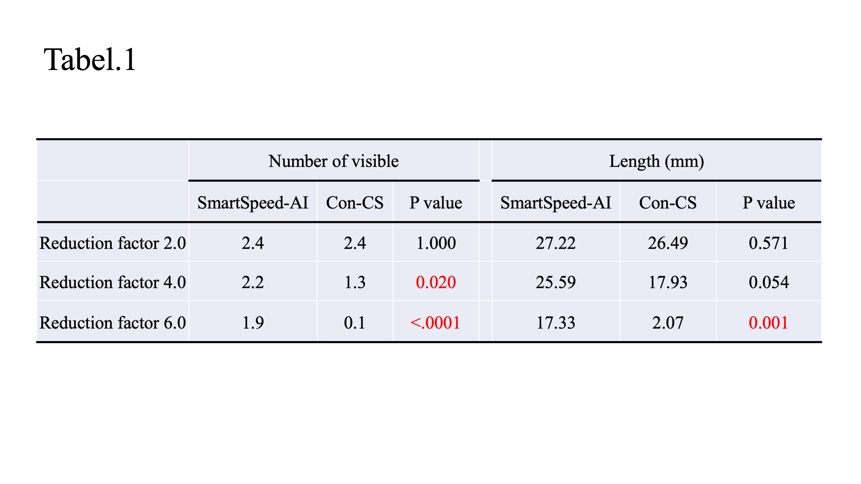
Qualitative assessment (the number of visible LSA and Length of LSA) in comparison between SmartSpeed-AI and compressed SENSE
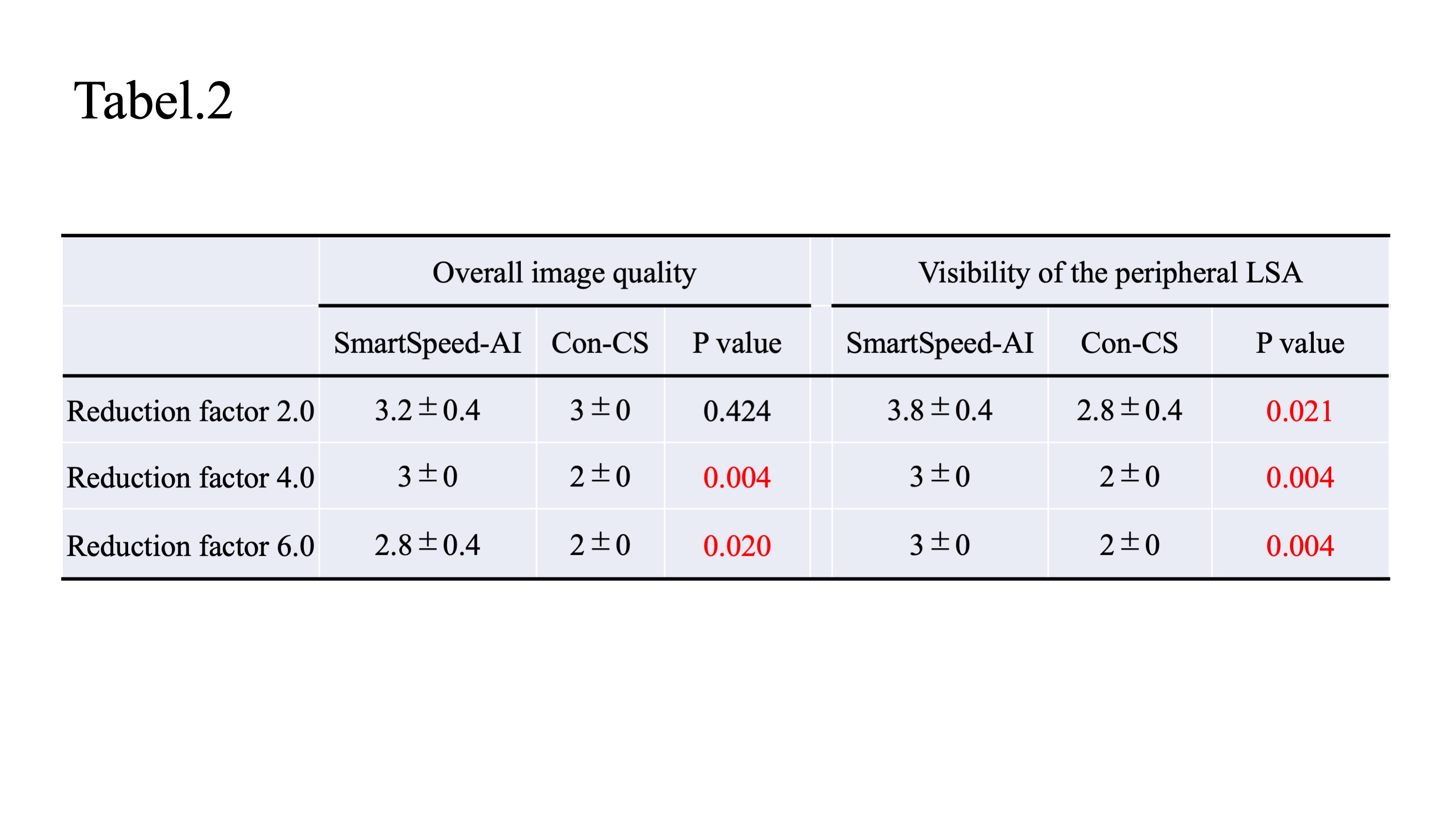
Qualitative assessment (overall image quality and visibility of the peripheral LSA) in comparison between SmartSpeed-AI and compressed SENSE
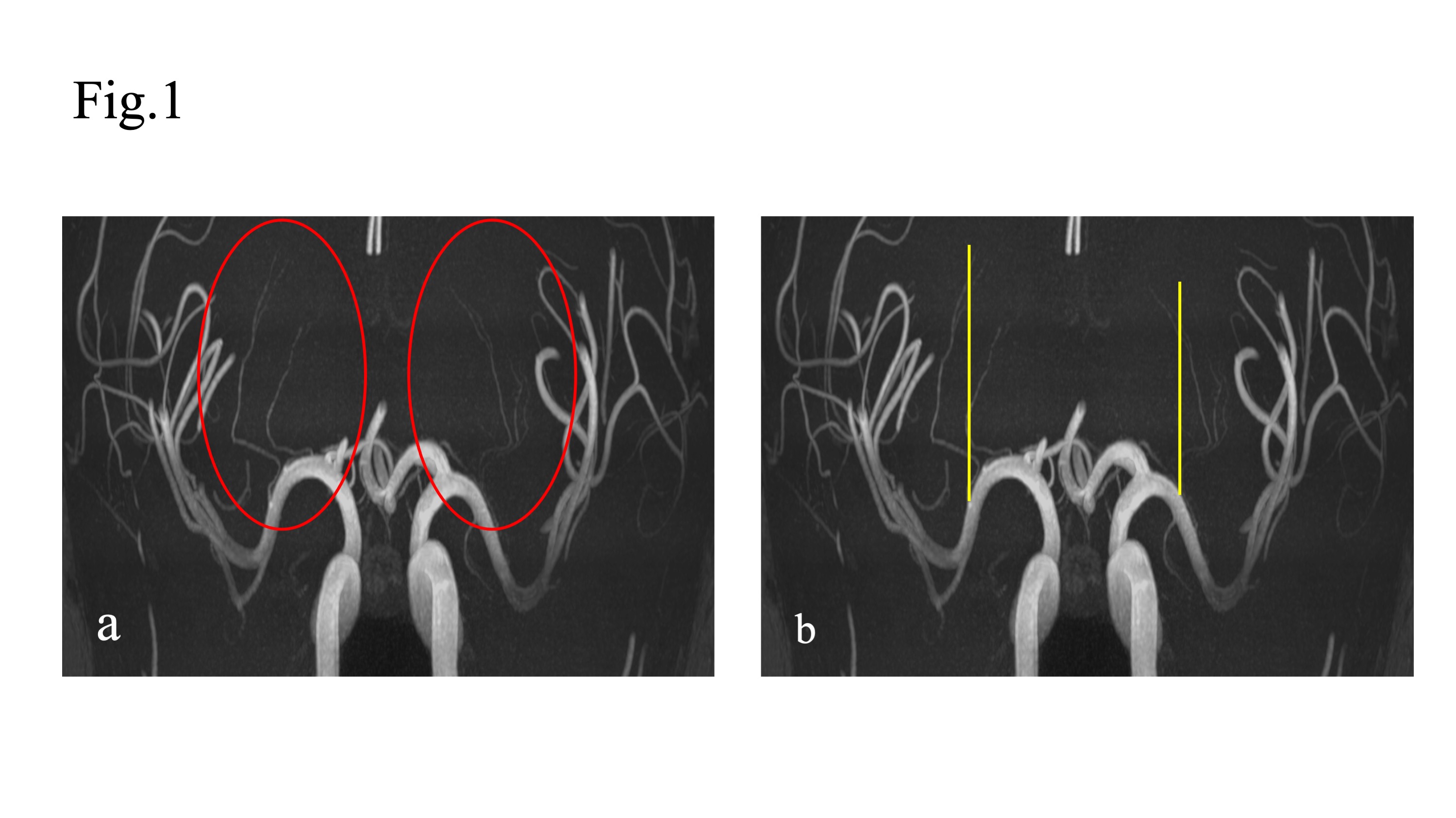
Measurement of the length of LSA.
On thin MIP TOF-MRA images, the length of depicted LSA was measured manually. From all of depicted LSAs (a; red oval line), the longest diameter in visible LSA was linearly measured with head-feet direction (b; yellow line).
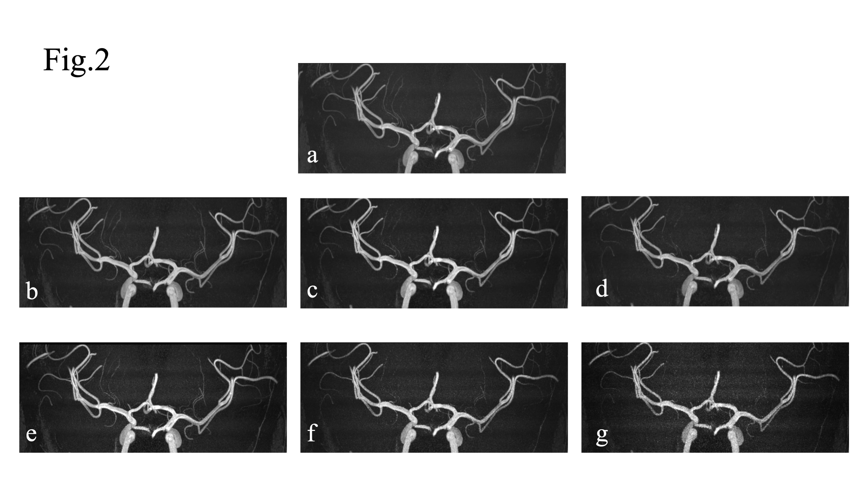
Representative case of thin MIP images reconstructed by SmartSpeed AI and Con-CS.
Reference image (R factor 1), images reconstructed by SmartSpeed-AI (b; R factor 2, c; R factor 4, and d; R factor 6), and images reconstructed by compressed SENSE (e; R factor 2, f; R factor 4, and g; R factor 6) were respectively presented.