0702
Deep Learning Based Algorithm to Identify Large Vessel Stenosis and Occlusion on Contrast Agent-free Magnetic Resonance Imaging
Di Wu1, Mengzhou Sun2, Yi Li3, Xiaoyun Liang3, Feng Huang3, and Wenzhen Zhu1
1Radiology, Tongji Hospital, Tongji Medical College, Huazhong University of Science and Technology, Wuhan, China, 2Neusoft Medical Systems Co. Ltd, Shenyang, Liaoning, China, Beijing, China, 3Neusoft Medical Systems Co. Ltd, Shenyang, Liaoning, China, Shanghai, China
1Radiology, Tongji Hospital, Tongji Medical College, Huazhong University of Science and Technology, Wuhan, China, 2Neusoft Medical Systems Co. Ltd, Shenyang, Liaoning, China, Beijing, China, 3Neusoft Medical Systems Co. Ltd, Shenyang, Liaoning, China, Shanghai, China
Synopsis
Keywords: Stroke, Machine Learning/Artificial Intelligence
Large vessel occlusion detection based on clinical scales is of low sensitivity and that based on CTA needs contrast agent exposure. This study aims to develop a deep learning (DL) algorithm for detecting intracranial large vessel steno-occlusion on contrast agent-free MR techniques including DWI and ASL. The accuracy of the DL algorithm was 88.2% with a sensitivity of 88.0%, comparable to CTA-based DL algorithms with sensitivity ranging from 67% to 94%. The MR-based DL algorithm is feasible to accurately detect intracranial large vessel steno-occlusion without intervention, radiation exposure and contrast agent, which could optimize stroke workflow and guide clinical decision-making.Introduction
It is essential for patients with acute anterior circulation ischemic stroke caused by large vessel occlusion (LVO) to be triaged and transferred to endovascular thrombectomy-capable center 1. Many prehospital stroke scales and in-hospital artificial intelligence (AI) software based on CT angiography (CTA) for LVO detection are designed to meet the demand 2-4. However, they either have relatively low sensitivity or need radiation and contrast agent exposure. To our knowledge, there have been no studies describing AI for LVO detections from MR images like diffusion-weighted imaging (DWI) and arterial spin labeling (ASL)-based perfusion image which are sensitive to ischemic core and perfusion deficit 5, 6. Therefore, we aim to develop a deep learning (DL) algorithm to deeply mine various features of DWI and ASL, so as to provide a non-invasive, contrast agent-free and image-based triage scheme for large vessel stenosis and occlusion recognition.Materials and methods
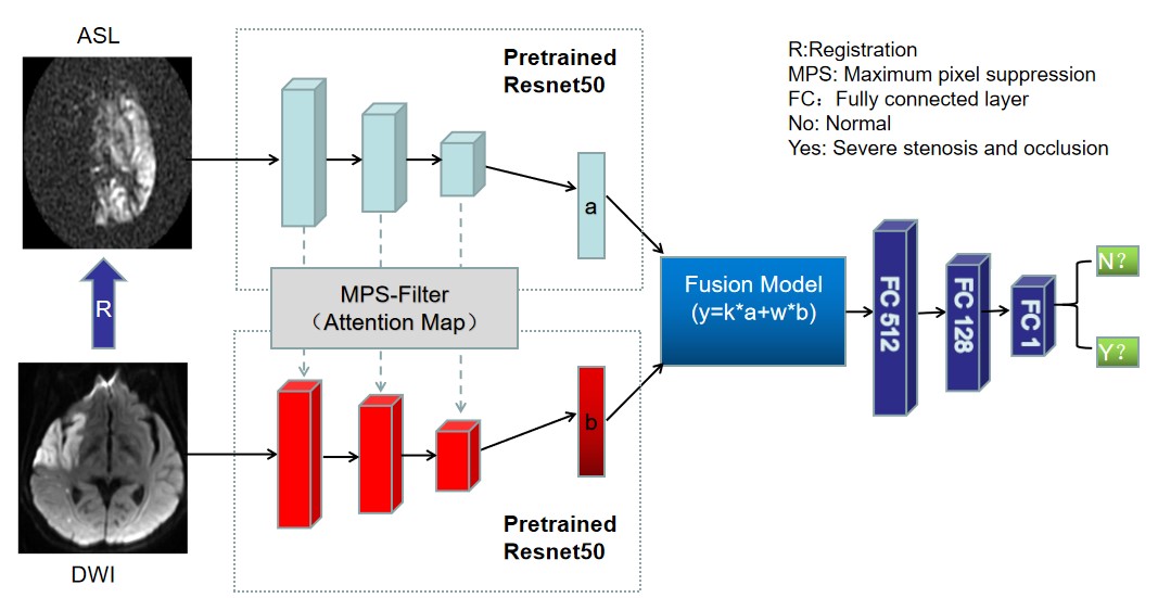
Subjects: Three hundred and eighty patients (256 males, 67.4%; mean age: 55 years) who were suspected with ischemic stroke and acquired routine DWI, ASL, time-of-flight MR angiography (TOF-MRA) and CTA scans were included in this study. Neuroradiologist analysis: CTA or TOF-MRA readings by neuroradiologists were recognized as the clinical reference standards for large vessel steno-occlusion assessment. Percent stenosis was measured using WASID method 7. Greater than 60.0% diameter reduction of the intracranial internal carotid artery (ICA) and M1 segment of middle cerebral artery (MCA) was regarded as severe stenosis. LVO was defined as 100% stenosis of the intracranial ICA and MCA (M1 and M2 segment). Development of the algorithm: DWI and ASL images were used to develop the algorithm that was called two-stream adaptive suppression network (Figure 1). Convolutional block attention module was added into pre-training ResNet50 8, 9, extracting the low- and high- dimensional information of the original data (such as shape feature, texture feature, etc.). An adaptive fusion module was used to aggregate feature map between convolution layers and fully connected layers 10. The algorithm iterates a total of 40 epochs, with batch size set to 4, the learning rate set to 0.00001, and a total of 304 training sets and 76 test sets. Statistical analyses were carried out using IBM SPSS Statistics 26 (Armonk, NY, USA).Results
Three hundred and twenty-one patients (84.5%) showed hyperintensity on DWI while 166 patients (43.7%) had severe large artery stenosis (25, 6.6%) and LVO (141, 37.1%) (Table 1, Figure 2). Interobserver agreement between neuroradiologist and DL algorithm was good (kappa = 0.740). The accuracy for the identification of severe stenosis and occlusion was 88.2% with the DL algorithm (sensitivity: 88.0%; specificity: 88.2%; positive predictive value: 78.6%; negative predictive value: 93.8%) which only missed 3 of the 25 diseased arteries in the test sets (Figure 3).Discussion
In this study, we proposed a DL algorithm for detection of severe stenosis and occlusion of intracranial large arteries using contrast agent-free MR techniques for the first time. The DL algorithm has three advantages: first, the attention module suppresses the interference information in the image, which improves the accuracy of the feature extraction. The adaptive fusion module aggregates affinity features and difference features of DWI and ASL, which can effectively reduce the misjudgment caused by a single linear combination of the two image features. These two modules facilitate the comparable performance of the DL algorithm to that of CTA-based algorithms with sensitivity ranging from 67% to 94% 2; second, it is free from radiation exposure, exogenous contrast agent allergy, and renal fibrosis; third, the ischemic lesion, perfusion deficit, and collateral status can be clearly delineated by DWI-derived ADC maps and ASL-derived CBF maps, providing comprehensive information of the brain function 5, 11, 12. Limitations were single site data and inclusion of the severe stenosis of intracranial ICA and M1 segment of MCA instead of LVO alone due to the restricted sample size. In conclusion, contrast agent-free MR techniques, namely DWI and ASL, are of feasibility of intracranial large artery steno-occlusion detection using artificial intelligence which could help optimize stroke workflow and guide clinical decision-making.Acknowledgements
No acknowledgement found.References
1. Goyal M, Menon BK, van Zwam WH, et al. Endovascular thrombectomy after large-vessel ischaemic stroke: a meta-analysis of individual patient data from five randomised trials. Lancet 2016; 387: 1723-1731. DOI: 10.1016/S0140-6736(16)00163-X.2. Murray NM, Unberath M, Hager GD, et al. Artificial intelligence to diagnose ischemic stroke and identify large vessel occlusions: a systematic review. Journal of neurointerventional surgery 2020; 12: 156-+. DOI: 10.1136/neurintsurg-2019-015135.
3. Duvekot MHC, Venema E, Rozeman AD, et al. Comparison of eight prehospital stroke scales to detect intracranial large-vessel occlusion in suspected stroke (PRESTO): a prospective observational study. Lancet Neurology 2021; 20: 213-221. DOI: 10.1016/S1474-4422(20)30439-7.
4. Smith EE, Kent DM, Bulsara KR, et al. Accuracy of Prediction Instruments for Diagnosing Large Vessel Occlusion in Individuals With Suspected Stroke: A Systematic Review for the 2018 Guidelines for the Early Management of Patients With Acute Ischemic Stroke. Stroke 2018; 49: e111-e122. 2018/01/26. DOI: 10.1161/str.0000000000000160.
5. Song K, Guan M, Li W, et al. Acute ischemic stroke patients with diffusion-weighted imaging-Alberta Stroke Program Early Computed Tomography Score ≤ 5 can benefit from endovascular treatment: a single-center experience and literature review. Neuroradiology 2019; 61: 451-459. 2019/02/07. DOI: 10.1007/s00234-019-02177-1.
6. Wang K, Shou Q, Ma SJ, et al. Deep Learning Detection of Penumbral Tissue on Arterial Spin Labeling in Stroke. Stroke 2020; 51: 489-497. 2019/12/31. DOI: 10.1161/strokeaha.119.027457.
7. Samuels OB, Joseph GJ, Lynn MJ, et al. A standardized method for measuring intracranial arterial stenosis. AJNR American journal of neuroradiology 2000; 21: 643-646.
8. Li M, Wang YW, Zhang FC, et al. Deformable medical image registration based on unsupervised generative adversarial network integrating dual attention mechanisms. 2021 14th International Congress on Image and Signal Processing, Biomedical Engineering and Informatics (Cisp-Bmei 2021) 2021. DOI: 10.1109/Cisp-Bmei53629.2021.9624229.
9. Sotoudeh-Paima S, Jodeiri A, Hajizadeh F, et al. Multi-scale convolutional neural network for automated AMD classification using retinal OCT images. Comput Biol Med 2022; 144.
10. Mihailescu RC. A weakly-supervised deep domain adaptation method for multi-modal sensor data. 2021 Ieee Global Conference on Artificial Intelligence and Internet of Things (Gcaiot) 2021: 45-50. DOI: 10.1109/GCAIoT53516.2021.9693050.
11. Nam KW, Kim CK, Ko SB, et al. Regional Arterial Spin Labeling Perfusion Defect Is Associated With Early Ischemic Recurrence in Patients With a Transient Ischemic Attack. Stroke 2020; 51: 186-192. 2019/11/14. DOI: 10.1161/strokeaha.119.026556.
12. Zhao MY, Václavů L, Petersen ET, et al. Quantification of cerebral perfusion and cerebrovascular reserve using Turbo-QUASAR arterial spin labeling MRI. Magnetic resonance in medicine 2020; 83: 731-748. 2019/09/13. DOI: 10.1002/mrm.27956.
Figures
Figure
1. The overall architecture of the DL algorithm which is called two-stream
adaptive suppression network.
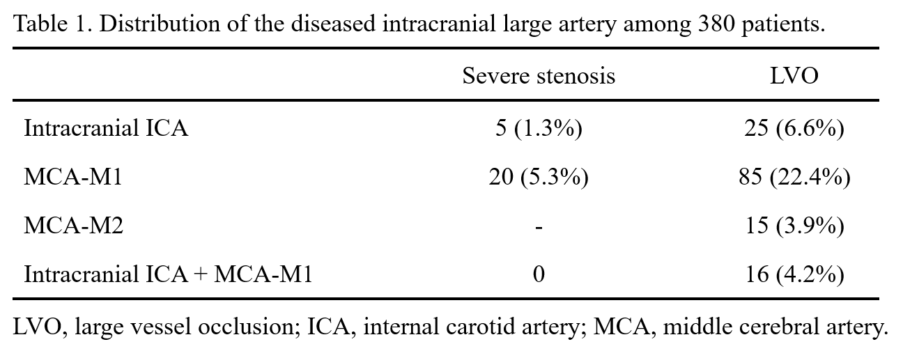
Table 1. Distribution
of the diseased intracranial large artery among 380 patients.
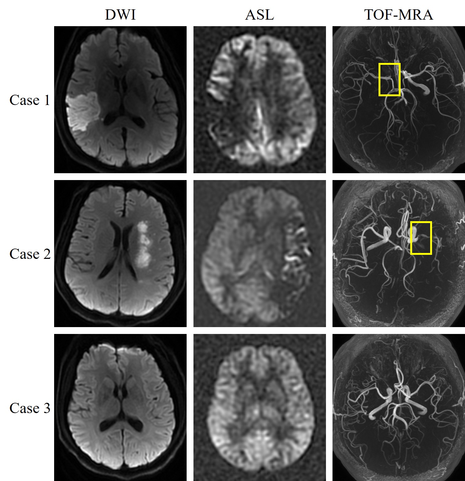
Figure 2. Three representative cases from the test sets. Case 1, a 42-year-old male diagnosed with acute ischemic stroke in the right temporal lobe. TOF-MRA showed right intracranial internal carotid artery occlusion. Case 2, a 47-year-old male diagnosed with acute ischemic stroke in the left corona radiata. TOF-MRA showed severe stenosis (71.65%) of the left middle cerebral artery (M1 segment). Case 3, a 52-year-old male diagnosed with a transient ischemic attack. TOF-MRA showed normal vascular structure. The DL algorithm predicted the three cases accurately.
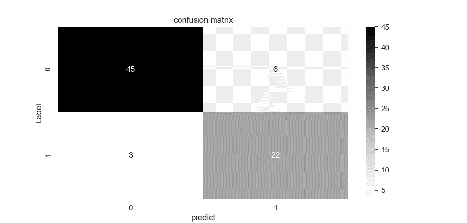
Figure 3.
Prediction results of two-stream adaptive suppression model. 1 refers to
subjects who suffer from severe stenosis and occlusion; 0 refers to normal
subjects.
DOI: https://doi.org/10.58530/2023/0702