0690
High-SNR whole-brain microstructure diffusion MRI using Romer-EPTI1Athinoula A. Martinos Center for Biomedical Imaging, MGH, Charlestown, MA, United States, 2Department of Radiology, Harvard Medical School, Charlestown, MA, United States, 3Harvard-MIT Health Sciences and Technology, MIT, Cambridge, MA, United States
Synopsis
Keywords: Diffusion/other diffusion imaging techniques, Microstructure
Diffusion MRI is a widely-used non-invasive imaging method for studying tissue microstructure but often suffers from low signal-to-noise ratios when using high b values. Here, we present a diffusion MRI acquisition technique that can achieve significantly higher SNR efficiency while providing distortion-free high-quality images. It achieves high robustness to phase variations and motion and provides an efficient acquisition technique for microstructural diffusion imaging. Ultra-high b-value and time-dependent experiments were performed to evaluate the improved SNR efficiency of Romer-EPTI.Introduction
Diffusion MRI is a widely-used non-invasive imaging method for studying tissue microstructure1-4 but often suffers from low signal-to-noise ratios when quantifying subtle microstructure features. For example, strong diffusion-weighting (high or ultra-high b values) is often used, which leads to signal decay and low image SNR even at standard or moderate spatial resolution. In addition, advanced diffusion models have shown great promise to provide more specific information about tissue diffusion and microstructure features1-4. However, the diffusion model fitting could be sensitive to the data’s SNR level, and a large number of diffusion encoding directions or multiple diffusion times are usually needed. This can result in a lengthy scan that compromises its practicality and clinical feasibility.Here, we present a Romer-EPTI (ROtating-view Motion-robust supEr Resolution Echo Planar Time-resolved Imaging) technique for microstructure diffusion MRI that can achieve significantly higher SNR efficiency and therefore reduced acquisition time, while providing distortion-free high-quality images. Importantly, it provides high robustness to phase variations, which is especially critical in high b-value diffusion scans where large phase variations exist between different excitations. Romer-EPTI integrates the efficient ky-t encoding5 with a rotating-view thick-slice acquisition that spatially encodes the readout-slice dimensions6-8 and resolves high isotropic resolution voxels from the multiple thick-slice volumes. While the ky-t EPTI encoding enables continuous readout with minimal dead time, minimal TE, and optimal readout length that significantly improve the image SNR, the rotating-view thick-slice acquisition achieves an additional SNR gain of >2x. We have shown preliminary results of this technique to provide significant SNR gain that enables mesoscale diffusion MRI at 500um isotropic resolution using a multi-shot ky-t EPTI encoding9. However, for diffusion MRI with high b-values, the large shot-to-shot phase variations could be detrimental to the image quality and diffusion metrics when using multi-shot acquisition or multi-slab encoding techniques. Therefore, in this work, we developed Romer-EPTI with single-shot ky-t encoding to achieve high robustness to phase variations and motion, and provide an efficient and practical acquisition technique for microstructural diffusion imaging. We then demonstrate its significant SNR gain in high b-value diffusion and time-dependent diffusion applications1-4.
Methods
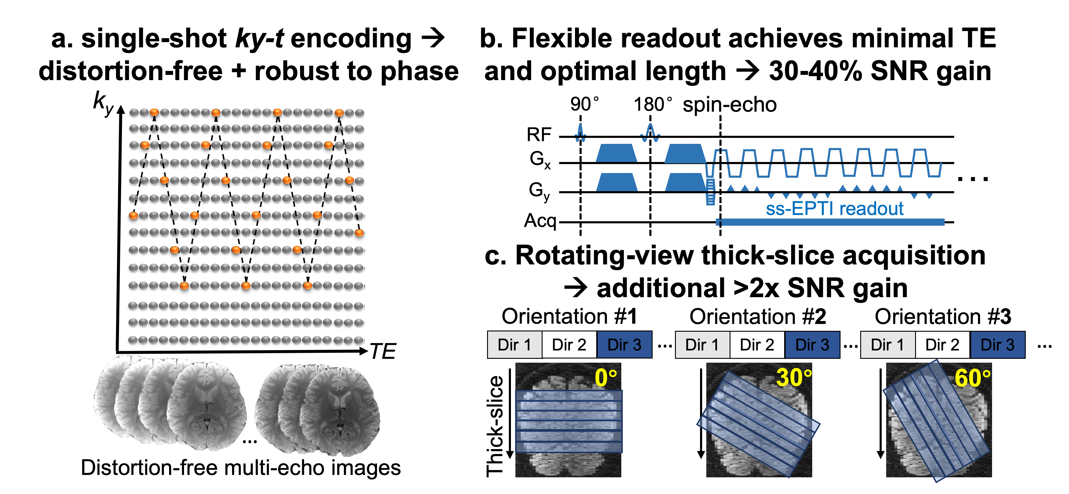
Romer-EPTI acquisition: Fig. 1a shows the single-shot EPTI encoding5,10 in the ky-t space. The complementary encoding along ky takes better advantage of the spatiotemporal correlation in the k-t space, while the zig-zag trajectory that continuously traverses through the k-t space reduces dead time and achieves stronger spatiotemporal correlation for improved reconstruction. The distortion-free images are then obtained from the reconstructed full k-t space. The continuous readout with minimal dead time using the ky-t encoding is applied, which is able to achieve minimal TE and optimal readout length for SNR improvement. Thick-slice volumes are acquired at different slice orientations (rotated around the anterior-posterior axis, PE) as shown in Fig.1c. For example, thick-slice volumes with 6 orientations (a 30° increment) were acquired to provide a super-resolution factor of 4 for 2-mm isotropic resolution data (2x SNR efficiency gain). It acquires different diffusion directions first for each slice orientation to avoid spin-history artifacts. A subspace reconstruction is employed to recover distortion-free images from the full k-t space, which is combined with a motion-robust super-resolution reconstruction to recover isotropic resolution9. All data were acquired using Romer-EPTI at 3T Siemens Prisma with a 32-channel coil. The application of Romer-EPTI for ultra-high b-value (b = 5000 s/mm2) imaging and time-dependent dMRI were preliminarily evaluated in this work with large whole-brain coverage.Results
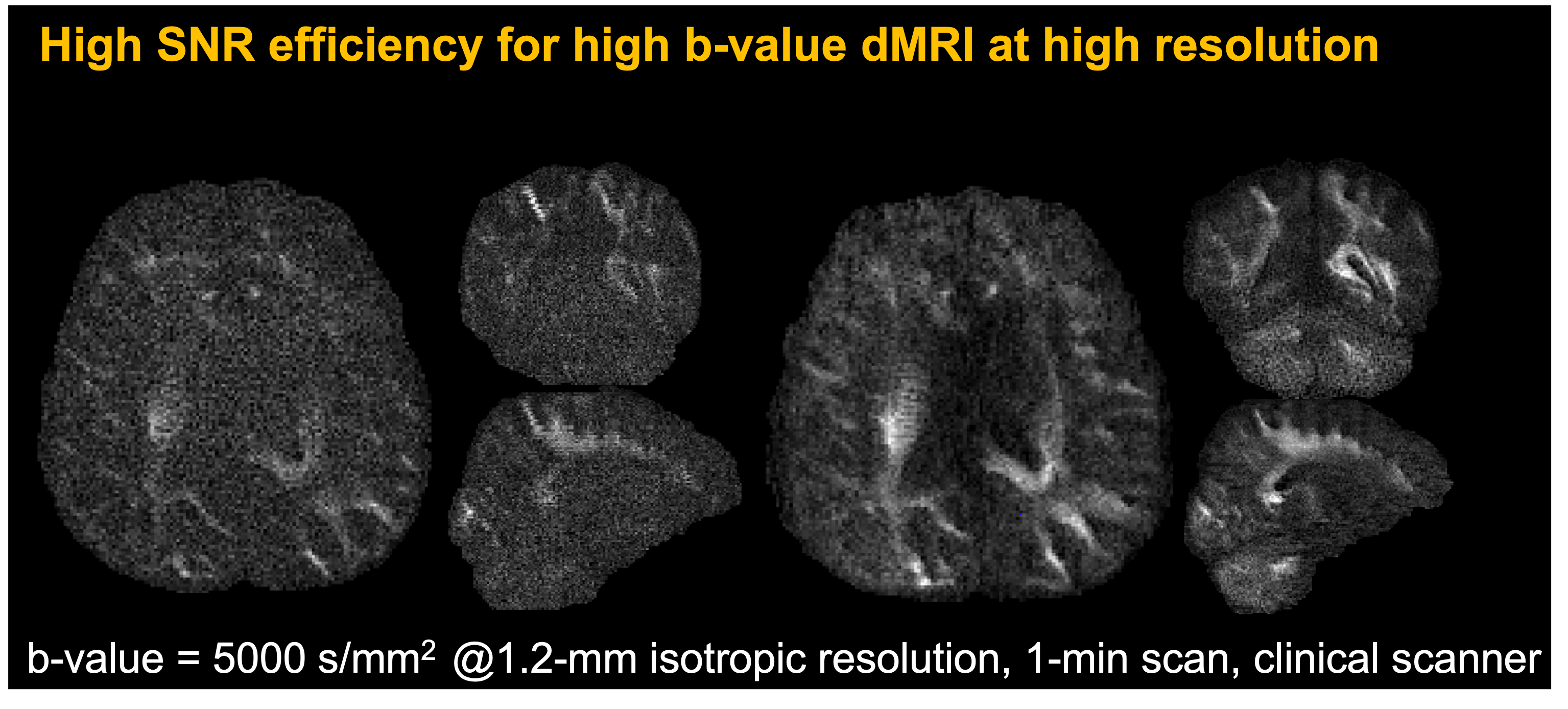
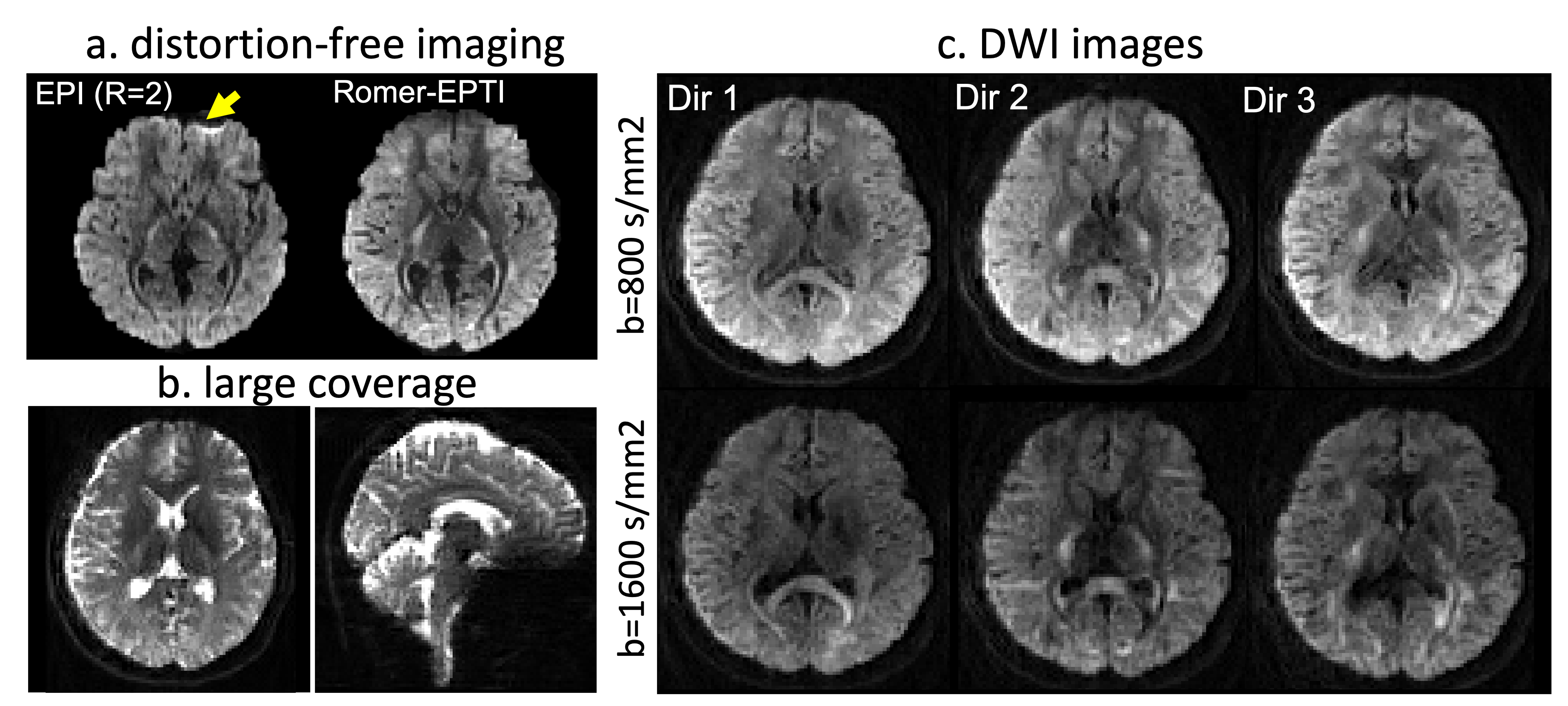
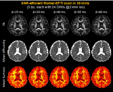
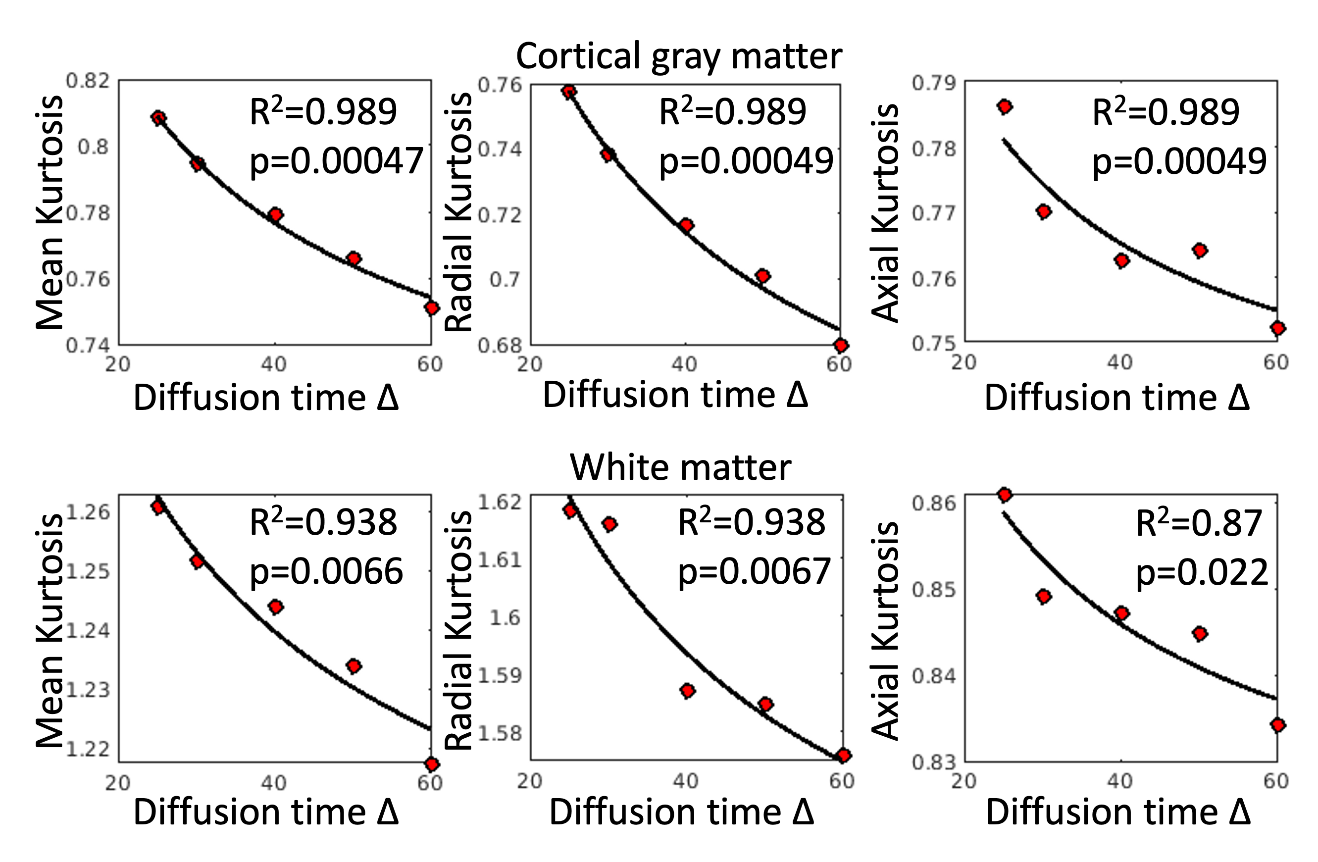
Fig. 2 shows the significantly higher SNR of Romer-EPTI for high b-value diffusion (b=5000 s/mm2) at a high spatial resolution of 1.2-mm, compared to the conventional EPI data with matching scan time. Fig. 3 shows the examples of diffusion images provided by Romer-EPTI. It eliminates geometric distortions that exist in conventional EPI, and achieves large whole-brain coverage. Fig. 4 shows the diffusion metrics estimated from Romer-EPTI data acquired with different diffusion times in a short scan time (each about 6 mins, and in a total of 30 mins). The preliminary kurtosis measurements show strong time-dependency (p value <0.05) in cortical gray matter and in white matter.Conclusion
We present an efficient diffusion MRI technique that can achieve significantly higher SNR efficiency and robustness to phase variations while providing distortion-free high-quality images for diffusion imaging applications such as high b-value and time-dependent diffusion, future work will explore its benefit in quantifying diffusion microstructure features.Acknowledgements
This work was supported by the NIH NIBIB (R01-EB019437, P41EB030006), NIH OD and NIDCR (DP5OD031854) and the instrumentation Grants (S10OD023637, S10-RR023401, S10-RR023043, and S10-RR019307).References
1 Lee, H.-H., Papaioannou, A., Novikov, D. S. & Fieremans, E. In vivo observation and biophysical interpretation of time-dependent diffusion in human cortical gray matter. Neuroimage 222, 117054 (2020).
2 Fieremans, E. et al. In vivo observation and biophysical interpretation of time-dependent diffusion in human white matter. Neuroimage 129, 414-427 (2016).
3 Burcaw, L. M., Fieremans, E. & Novikov, D. S. Mesoscopic structure of neuronal tracts from time-dependent diffusion. NeuroImage 114, 18-37 (2015).
4 Novikov, D. S., Jensen, J. H., Helpern, J. A. & Fieremans, E. Revealing mesoscopic structural universality with diffusion. Proceedings of the National Academy of Sciences 111, 5088-5093 (2014).
5 Wang, F. et al. Echo planar time‐resolved imaging (EPTI). Magnetic resonance in medicine (2019).
6 Plenge, E. et al. Super-resolution methods in MRI: can they improve the trade-off between resolution, signal-to-noise ratio, and acquisition time? Magnetic resonance in medicine 68, 1983-1993, doi:10.1002/mrm.24187 (2012).
7 Shilling, R. Z. et al. A super-resolution framework for 3-D high-resolution and high-contrast imaging using 2-D multislice MRI. IEEE Trans Med Imaging 28, 633-644, doi:10.1109/TMI.2008.2007348 (2009).
8 Vis, G., Nilsson, M., Westin, C. F. & Szczepankiewicz, F. Accuracy and precision in super-resolution MRI: Enabling spherical tensor diffusion encoding at ultra-high b-values and high resolution. NeuroImage 245, 118673, doi:10.1016/j.neuroimage.2021.118673 (2021).
9 Dong, Z., Polimeni, J. R., Wald, L. L. & Wang, F. SuperRes-EPTI: in-vivo mesoscale distortion-free dMRI at 500μm-isotropic resolution using short-TE EPTI with rotating-view super resolution In Proceedings of the 30th Annual Meeting of ISMRM 2022.
10 Wang, F. et al. Improving fMRI acquisition using single-shot EPTI with distortion-free high-SNR high-CNR multi-echo imagingProc Intl Soc Mag Reson Med. 0330.
Figures