0652
Prominent role of cerebellar communication in schizophrenia reflected by resting state fMRI connectivity1Institute of Neuroscience and Medicine - 4 (INM- 4), Forschungszentrum Jülich, Juelich, Germany, 2Department of Psychiatry, Psychotherapy and Psychosomatics, Uniklinik RWTH Aachen, Aachen, Germany, 3Department of Nuclear Medicine, Uniklinik RWTH Aachen, Aachen, Germany, 4Department of Neurology, Uniklinik RWTH Aachen, Aachen, Germany, 5Institute of Neuroscience and Medicine 11 (INM - 11), Forschungszentrum Jülich, Juelich, Germany
Synopsis
Keywords: Psychiatric Disorders, fMRI (resting state), Schizophrenia
Schizophrenia is a complex neuropsychiatric disorder, the pathophysiology of which is unclear. Several studies have shown the involvement of altered brain communication. Several new voxel-level connectivity measures such as radial similarity, radial correlation, inter-hemispheric connectivity and local correlation have enabled a deeper understanding of alterations in communication. Our results reveal the prominent role of the cerebellum in schizophrenia by investigating voxel-level connectivity in resting state fMRI data.Introduction
Schizophrenia is a complex disorder characterized by abnormal interpretation of reality in people. It often presents itself as a combination of symptoms such as hallucinations, delusional perceptions, disordered thinking, and social impairment. Several studies have tried to explain the pathophysiology, but they remain to be inconclusive1. Alterations in brain communication is by far one of the most widely accepted hypotheses2. Recently several new connectivity measures investigating voxel-level communication through fMRI have come to light. These measures such as intrinsic connectivity, global correlation, local correlation, inter-hemispheric connectivity, radial similarity and so on, enable a deeper investigation of alterations in communication, allowing us to further understand the complex mechanisms of schizophrenia. In this study, several such methods have been employed to resting state fMRI data and have reflected the prominent role of the cerebellum in schizophrenia.Methods
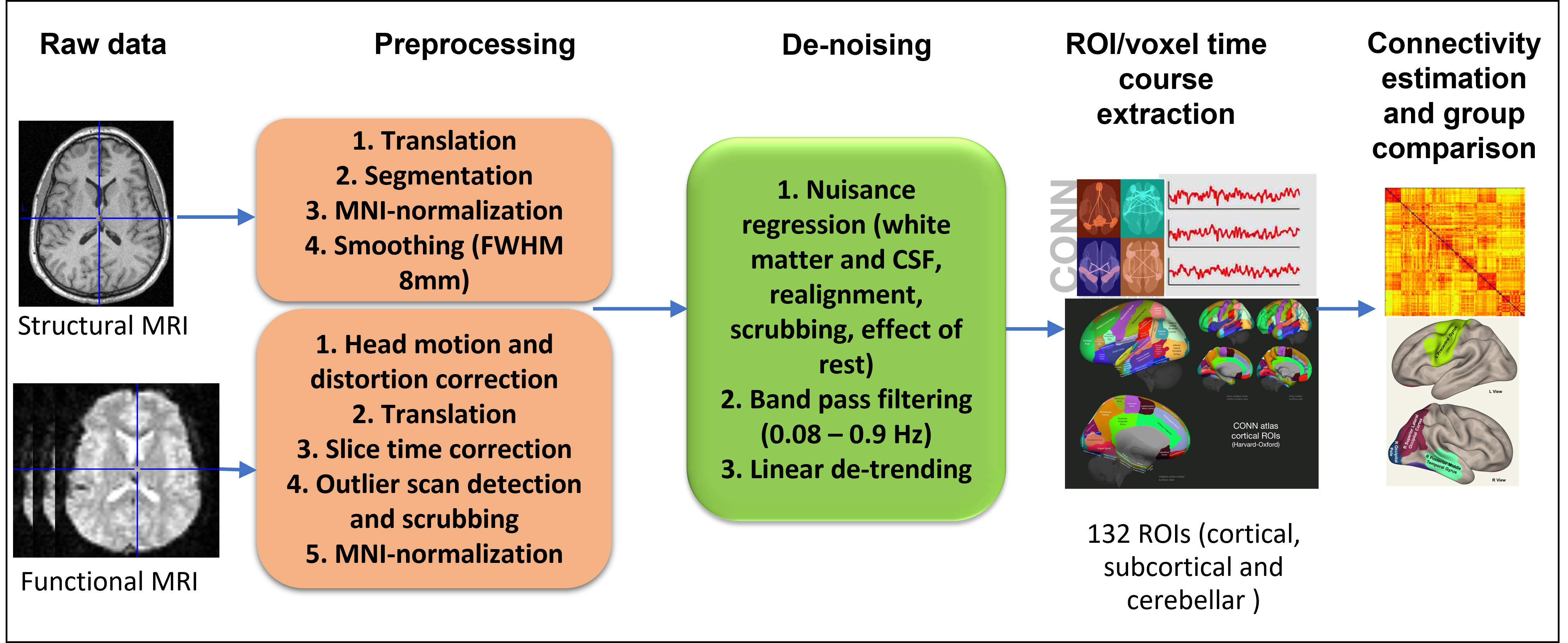
Resting state fMRI data of 28 male Schizophrenia patients (age: 38 ± 10) and 41 group-level age-matched male healthy controls (age: 36 ± 11) recorded simultaneously with EEG and PET on a 3T BrainPET MR Scanner (Trio, Siemens, Germany) were used for this study. The acquisition protocol and sequence parameters can be found in another study3. The raw data were preprocessed and denoised using the steps shown in figure 1. After denoising the first level analyses were performed at the ROI and the voxel level. All analyses were performed in CONN v2021a4. For the ROI level analysis, average timeseries of 132 ROIs covering the entire brain (Harvard-Oxford cortical and subcortical and AAL cerebellar) were extracted, and ROI-ROI bivariate correlation was performed. For the voxel level, the following network measures were computed5:1. Intrinsic Connectivity (ICC) – It is a measure of voxel centrality given by the root mean square of correlation between this voxel and all other voxels of the brain.
2. Global Correlation (GCOR) – It is also a measure of voxel centrality given by the average of correlation between this voxel and all other voxels in the brain.
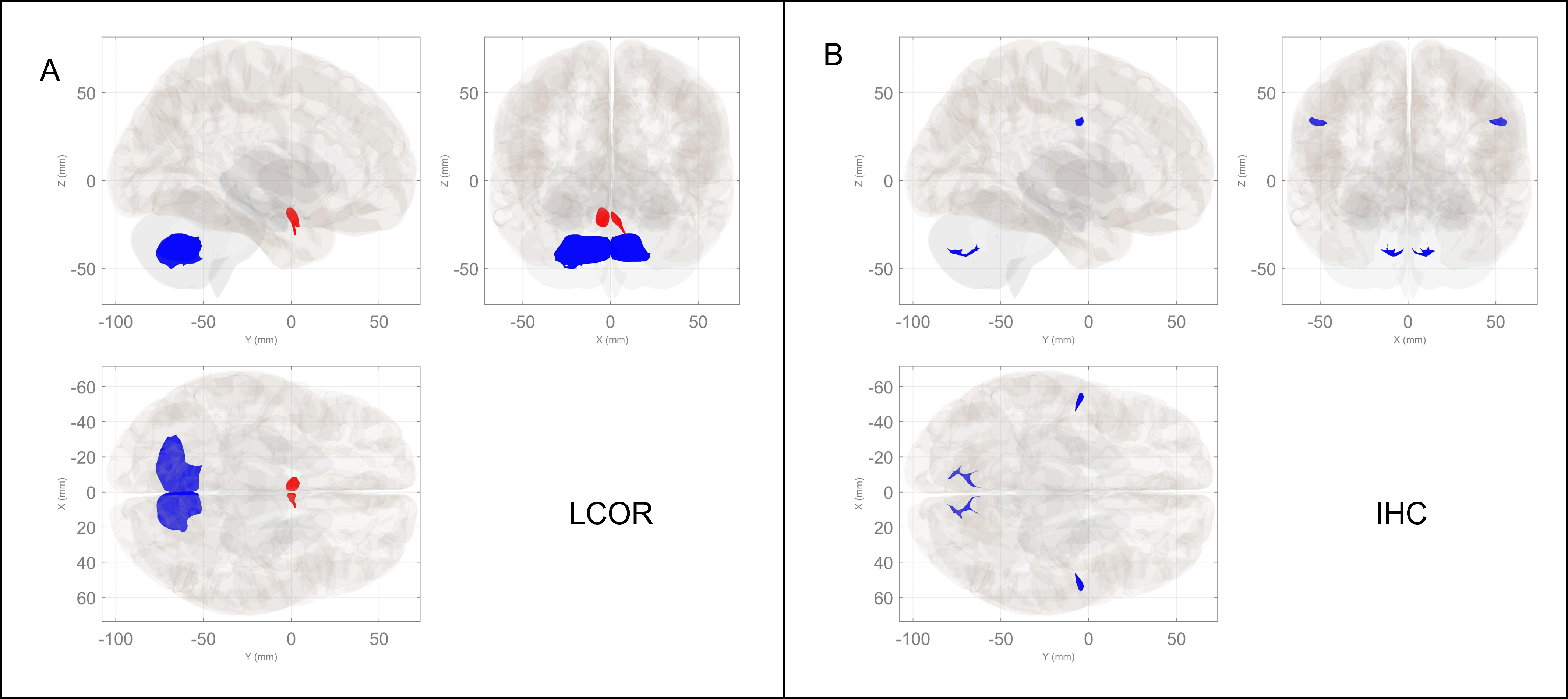
3. Local Correlation (LCOR) – It is a measure of local coherence given by the average correlation between this voxel and its neighbours.
4. Interhemispheric Connectivity (IHC) – It characterizes connectivity between hemispheres given by the correlation between this voxel and its counterpart (same anatomical location) in the other hemisphere.
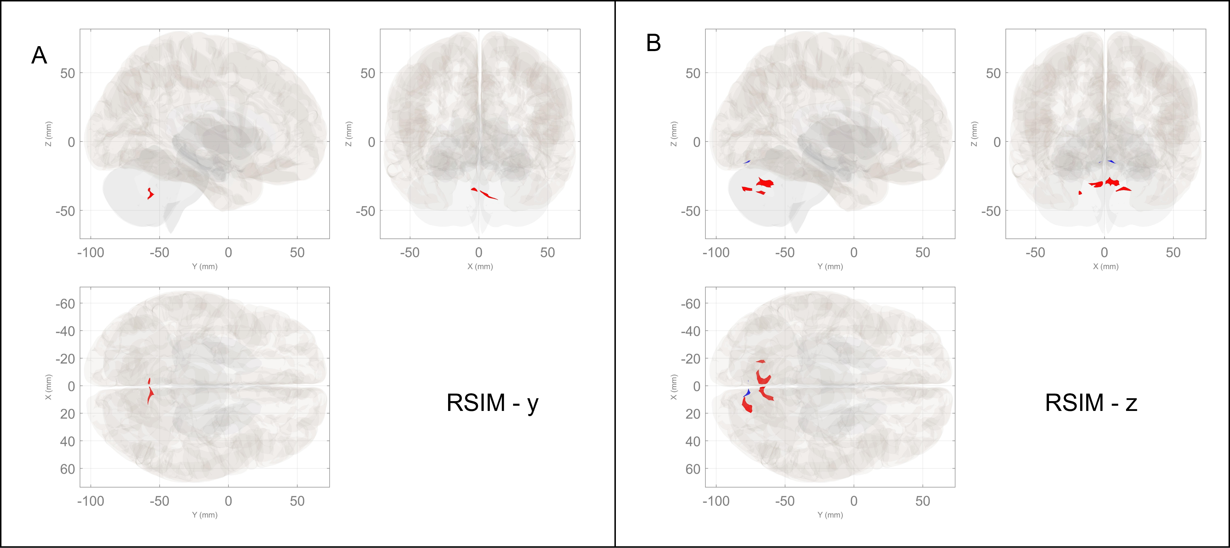
5. Radial Similarity (RSIM) – It is given by the strength of the spatial gradient of connectivity between this voxel and the entire brain in the x, y and z directions.
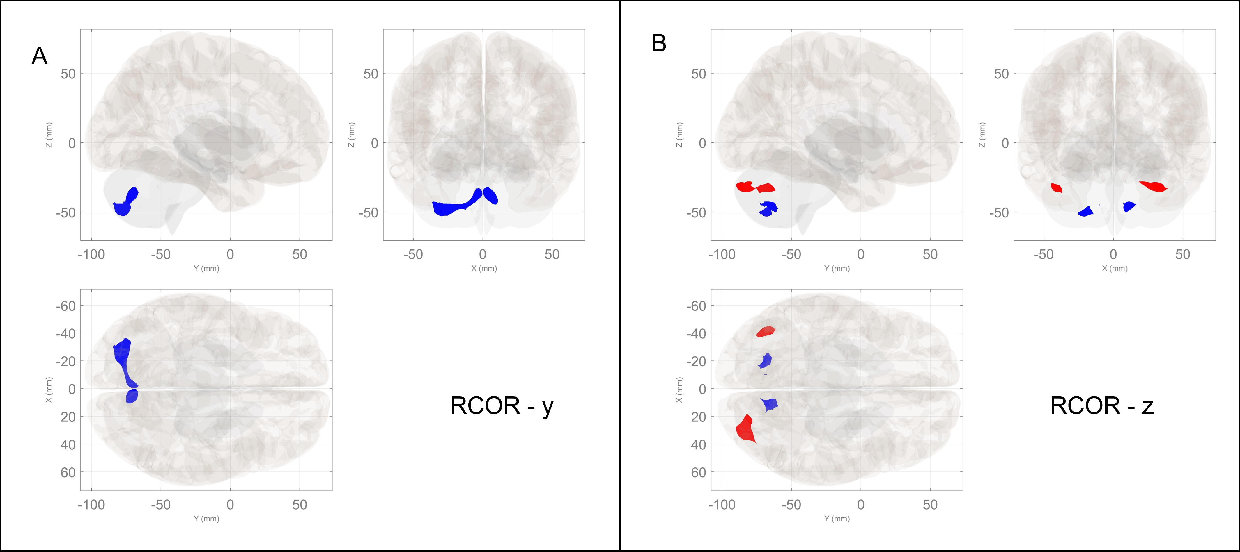
6. Radial Correlation (RCOR) – It is the spatial gradient of local connectivity between this voxel and its neighbours in the x, y and z directions.
7. Fractional Amplitude of Low Frequency Fluctuations (fALFF) – It is the root mean square ratio of BOLD signal fluctuations in the frequency band of interest compared to the entire frequency band.
Following this, second level group analyses were performed. For ROI analyses spatial pairwise clustering6 was used with a cluster threshold of p<0.05 cluster-level p-FDR corrected and connection threshold of p<0.01 p-uncorrected. For the voxel analyses, Gaussian Random Field Theory7 was used with a cluster threshold of p<0.05 cluster-size p-FDR corrected; and voxel threshold of p<0.001 p-uncorrected.
Results
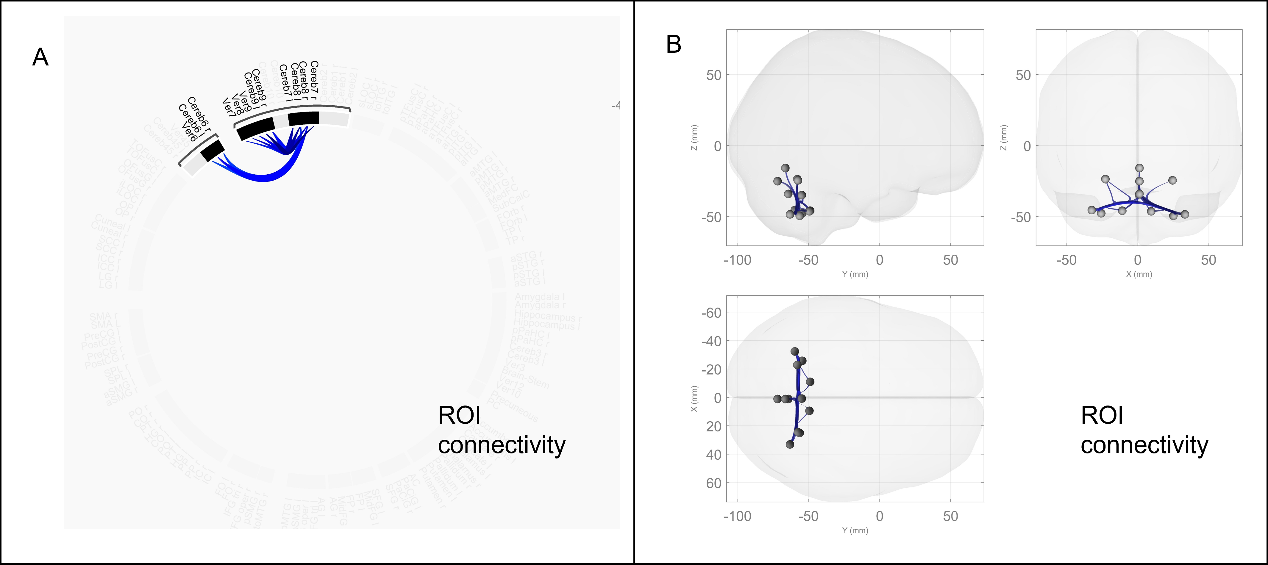
The ROI level analyses revealed reduced connectivity between several regions of the cerebellum as shown in figure 2. The voxel level network measures RCOR, RSIM, LCOR and IHC revealed distinct clusters in the cerebellum as shown by figures 3,4 and 5. The RSIM in the y and z directions was higher in patients for two cerebellar clusters. The RCOR in the y direction was lower in patients but in the z direction it was higher for one cluster and lower for the other. These two clusters corresponded to two different cerebellar parcellations which are known to have different functions8. The LCOR and IHC were also reduced in clusters of the cerebellum in patients. The other measures ICC, GCOR and fALFF did not reveal any significant differences between the two groups.Discussion
The cerebellum has been traditionally known for its role in movement and balance. More recently, however, it’s involvement in higher order functions has come to light8. It is now known that the cerebellum is fundamentally involved in motor, default-mode (task negative) and attentional/executive (task positive) processing8. The reduced local correlation and reduced interhemispheric connectivity reflect disrupted local subnetwork performance as well as disrupted information mirroring. The increased radial similarity in the coronal (y) and axial (z) directions indicates faster global information spread to the cortex through these regions. This might be indicative of lack of inhibition. Similarly, the reduced radial correlation in the coronal and increased and reduced in the axial directions indicate the slower or faster local information spread in these directions through the respective parcellations. All these findings put together emphasize the role of cerebellar communication in the complex pathophysiology of schizophrenia and pave the way for a new understanding of the disease.Conclusion
Schizophrenia is a complex neuropsychiatric disorder, the pathophysiological mechanisms of which are still unclear. The application of novel voxel-level network measures to resting state fMRI data has revealed the prominent role played by cerebellar communication in the complex pathophysiology of schizophrenia, paving the way to a new understanding of the disease.Acknowledgements
No acknowledgement found.References
1. Keshavan, M. S. et al. Neuroimaging in schizophrenia. Neuroimaging Clin. N. Am. 30, 73 (2020).
2. Friston, K., Brown, H. R., Siemerkus, J. & Stephan, K. E. The dysconnection hypothesis (2016). Schizophr. Res. 176, 83–94 (2016).
3. Rajkumar, R. et al. Excitatory–inhibitory balance within EEG microstates and resting-state fMRI networks: assessed via simultaneous trimodal PET–MR–EEG imaging. Transl. Psychiatry 11, 1–15 (2021).
4. Whitfield-Gabrieli, S. & Nieto-Castanon, A. Conn: A Functional Connectivity Toolbox for Correlated and Anticorrelated Brain Networks. Brain Connect. 2, 125–141 (2012).
5. Alfonso Nieto-Castanon. Handbook of functional connectivity Magnetic Resonance Imaging methods in CONN. (2020).
6. Zalesky, A., Fornito, A. & Bullmore, E. On the use of correlation as a measure of network connectivity. Neuroimage 60, 2096–2106 (2012).
7. Worsley, K. J. et al. A unified statistical approach for determining significant signals in images of cerebral activation. Hum. Brain Mapp. 4, 58–73 (1996).
8. Guell, X. & Schmahmann, J. Cerebellar Functional Anatomy: a Didactic Summary Based on Human fMRI Evidence. Cerebellum 19, 1–5 (2020).
Figures