0635
Local $$$B_1^+$$$ shimming improves visualization of the bone-metal interface in patients with orthopedic hardware
Iman Khodarahmi1, Mahesh B Keerthivasan2, and Jan Fritz1
1NYU Langone School of Medicine, New York, NY, United States, 2Siemens Medical Solutions USA Inc., Malvern, PA, United States
1NYU Langone School of Medicine, New York, NY, United States, 2Siemens Medical Solutions USA Inc., Malvern, PA, United States
Synopsis
Keywords: Bone, Bone
$$$B_1^+$$$ field inhomogeneity is a source of metal artifacts in patients with orthopedic hardware. Local $$$B_1^+$$$ shimming can potentially decrease these artifacts and improve visualization of the bone-metal interface. Our proposed turbo-spin echo-based $$$B_1^+$$$ mapping technique enables accurate estimation of the $$$B_1^+$$$ field near the metal hardware. After optimization for in-vivo applications, the technique was successfully employed on a clinical 3.0 T parallel-transmit system aiming at $$$B_1^+$$$ shimming near the orthopedic hardware. Our results demonstrate significant improvement in visualization of the bone-metal interface compared to standard 1.5 and 3.0 T acquisitions.Introduction
Susceptibility artifacts are a well-known source of image artifacts in MRI of patients with metallic orthopedic hardware. The other much less investigated source of artifacts is the perturbation in the transmit $$$B_1^+$$$ field, which is caused by metal-induced electromagnetic fields 1-2. Such $$$B_1^+$$$ inhomogeneities may cause signal alterations at the bone-metal interface and be misinterpreted as an abnormality and obscure the underlying pathology. $$$B_1^+$$$ shimming localized to the area surrounding the metal hardware using a multi-channel transmit system can potentially provide a more homogeneous $$$B_1^+$$$ field and improve visualization of the bone-metal interface. A successful $$$B_1^+$$$ shim requires (1) obtaining reliable $$$B_1^+$$$ field maps in the presence of metal and (2) a subject-specific combination of the individual $$$B_1^+$$$ field maps from each transmit channel. This work aims to develop a turbo-spin echo (TSE)-based $$$B_1^+$$$ mapping technique for in-vivo applications and analyze the effect of local $$$B_1^+$$$ shimming on the image quality of patients with metallic orthopedic hardware using a clinical dual-transmit system.Methods
$$$\it B_1^+$$$ mapping: We have recently developed and validated a TSE-based method of $$$B_1^+$$$ mapping which relies on the acquisition of multiple images with various excitation-refocusing (EX-Ref) flip angles (FA) 3. Briefly, with a $$$B_1^+$$$ amplitude scale factor of b1 (b1 = actual $$$B_1^+$$$ / nominal $$$B_1^+$$$), the signal intensity of a TSE sequence can be expressed as: $$$S(b_{1}) = f(b_{1}.\theta,b_{1}.\phi,\psi)$$$, where θ and φ represent excitation and refocusing flip angles, respectively, ψ other imaging parameters, and f(.) is the signal Bloch model. For different sets of Ex-Ref FA, b1 can be obtained by solving the following optimization problem: $$\widehat{b_{1}} = \min_{b_1}\sum_i^n\parallel f(b_{1}.\theta_{i},b_{1}.\phi_{i},\psi)-\widehat{S}(\theta_{i},\phi_{i},\psi) \parallel_{2}$$ with $$$\widehat{S}(\theta_{i},\phi_{i},\psi)$$$ being the pixel signal obtained by the ith set of Ex-Ref FA.$$$\it B_1^+$$$ mapping optimization for in-vivo imaging: Monte-Carlo simulations were performed to study the sensitivity of $$$B_1^+$$$ relative error to the number and choice of Ex-Ref FA pairs. The TSE signal was simulated for a wide range of excitation (30-120°) and refocusing (60-180°) FA sets using Bloch equations. Gaussian noise was added to the simulated signal for each set and the mean $$$B_1^+$$$ map was estimated from one thousand realizations.
$$$\it B_1^+$$$ shimming: The composite $$$B_1^+$$$ field can be obtained by complex linear combination of the $$$B_1^+$$$ field maps of the two transmit channels using the $$$B_1^+$$$ amplitudes of the previous step and the relative $$$B_1^+$$$ phase distribution of the TSE images. For a given region-of-interest (ROI) near the metal, the shimming was performed by adjusting the relative amplitude, and phases of the two transmit elements, aiming at minimizing the difference between the $$$B_1^+$$$ in the ROI and that of the background.
MRI Experiments: After obtaining institutional review board approval and informed consent, a volunteer with a femoral intramedullary nail was imaged on a parallel-transmit 3T clinical system with the following parameters: TR/TE = 2000/29 ms, voxel size = 0.6 x 0.6 x 3.0 mm3, and turbo factor = 13. Standard and $$$B_1^+$$$ shimmed intermediate-weighted axial images were acquired, focusing on visualization of the bone-metal interface. For comparison purposes, $$$B_1^+$$$ maps were also obtained using a TurboFLASH (TFL) sequence equipped with a preceding RF pulse for magnetization preparation 4.
Results and Discussion
Monte-Carlo simulation results on the optimal number and choice of the Ex-Ref FA pairs are summarized in Figure 1. The lowest mean relative error in $$$B_1^+$$$ estimations were 27%, 19%, and 18% in 2,3, and 4 Ex-Ref FA sets, respectively. Considering the increased imaging time and specific absorption rate (SAR) values, these results suggest that acquiring more than three Ex-Ref FA sets will not result in improved $$$B_1^+$$$ estimation. In addition, the error in $$$B_1^+$$$ estimation is more pronounced at low $$$B_1^+$$$ values, as the overall low signal at low $$$B_1^+$$$ values reaches the noise floor. Using the optimal three Ex-Ref FA sets, the $$$B_1^+$$$ field was estimated in a volunteer and compared with those of the TFL method (Figure 2). As seen on magnified subplots, the $$$B_1^+$$$ field near the metal is better resolved with our technique. The effect of $$$B_1^+$$$ shimming on the visualization of the bone-metal interface is shown in Figure 3. $$$B_1^+$$$ shimming in a bone-metal containing ROI provides the optimal shim parameters, which were used for subsequent imaging. Compared with the standard 1.5 and 3.0 T images, the visibility of the bone-metal interface is significantly improved after $$$B_1^+$$$ shimming.Conclusion
The proposed $$$B_1^+$$$ mapping technique provides promising results at the bone-metal interface invisible to other mapping sequences. Our initial results suggest that patient-specific local $$$B_1^+$$$ shimming is clinically feasible and can reduce $$$B_1^+$$$-related artifacts surrounding the metal.Acknowledgements
No acknowledgement found.References
- Khodarahmi I, Nittka M, Fritz J. Leaps in Technology: Advanced MR Imaging after Total Hip Arthroplasty. Semin Musculoskelet Radiol. 2017;21(5):604-15.
- Bachschmidt TJ, Kohler M, Nistler J, Geppert C, Jakob PM, Nittka M. Polarized Multichannel Transmit MRI to Reduce Shading near Metal Implants. Magn Reson Med. 2016;75(1):217–226.
- Khodarahmi I, Keerthivasan MB, Fritz J. Turbo-spin echo based B1+ mapping in the presence of metallic hardware. 31st Annual Meeting of the International Society for Magnetic Resonance in Medicine: International Society for Magnetic Resonance in Medicine; 2022.
- Chung S, Kim D, Breton E, and Axel L. Rapid B1+ mapping using a preconditioning RF pulse with turboflash readout. Magn Reson Med. 2010;64(2):439-446.
Figures
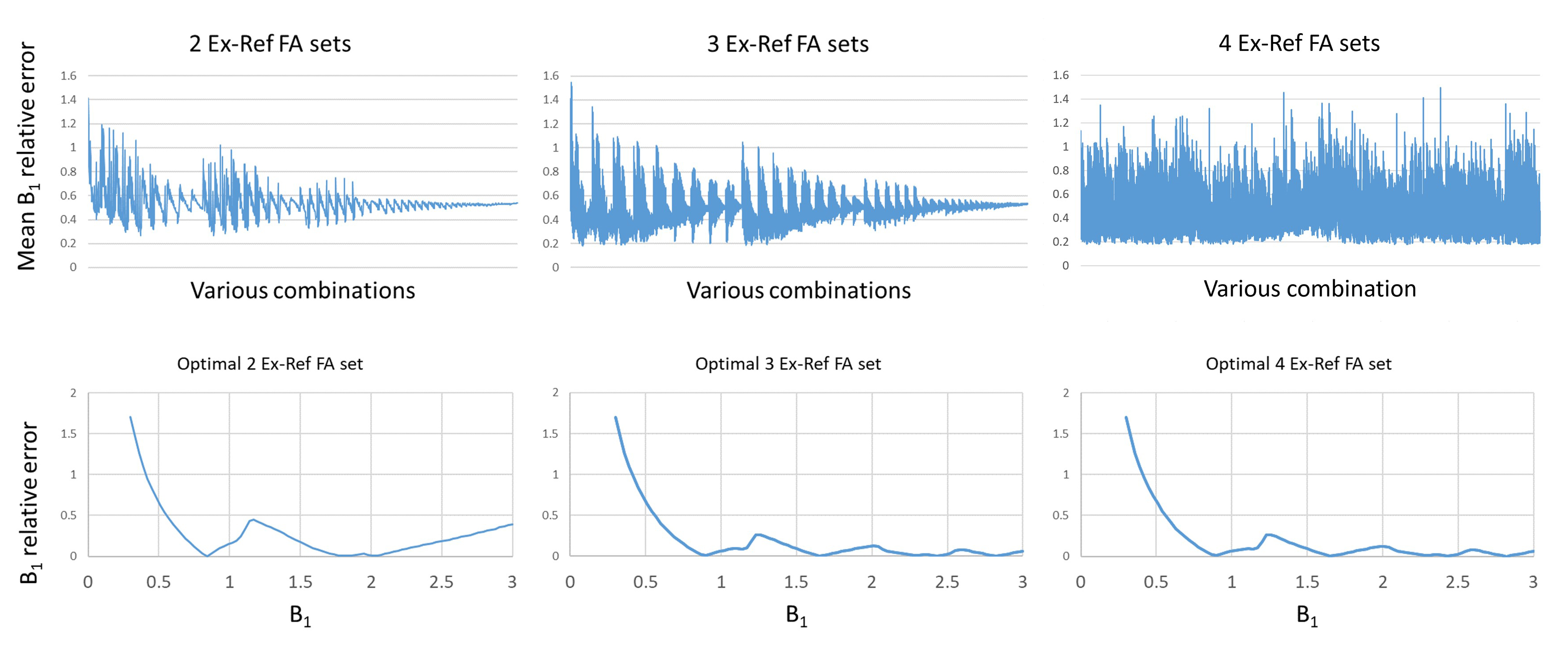
Figure 1. Top row: Mean relative error in $$$B_1^+$$$ estimation at
different excitation-refocusing flip angle (EX-Ref FA) sets. The minimum
relative error was 27%, 19% and 18% in the 2, 3 and 4 EX-Ref FA sets,
respectively. Bottom row: In all cases, the estimation errors were higher at
low $$$B_1^+$$$ values.
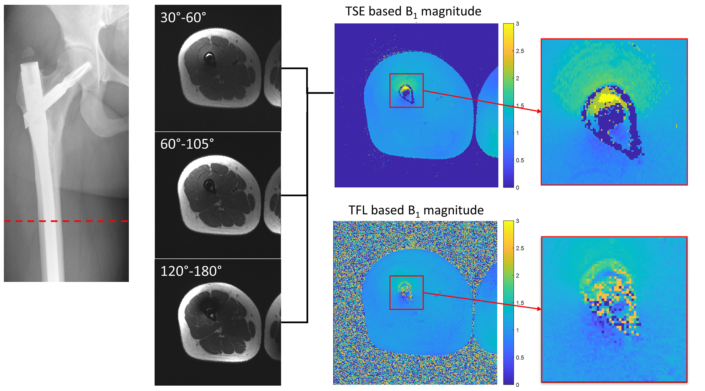
Figure 2. $$$B_1^+$$$ field maps of a patient with a femoral
intramedullary nail. Three acquisitions with optimal choice of excitation and
refocusing flip angles were obtained to encode $$$B_1^+$$$ variations. The resultant turbo spin echo
(TSE) based $$$B_1^+$$$ map
better resolves $$$B_1^+$$$ field
variations in the medullary canal than the turboFLASH (TFL) technique.
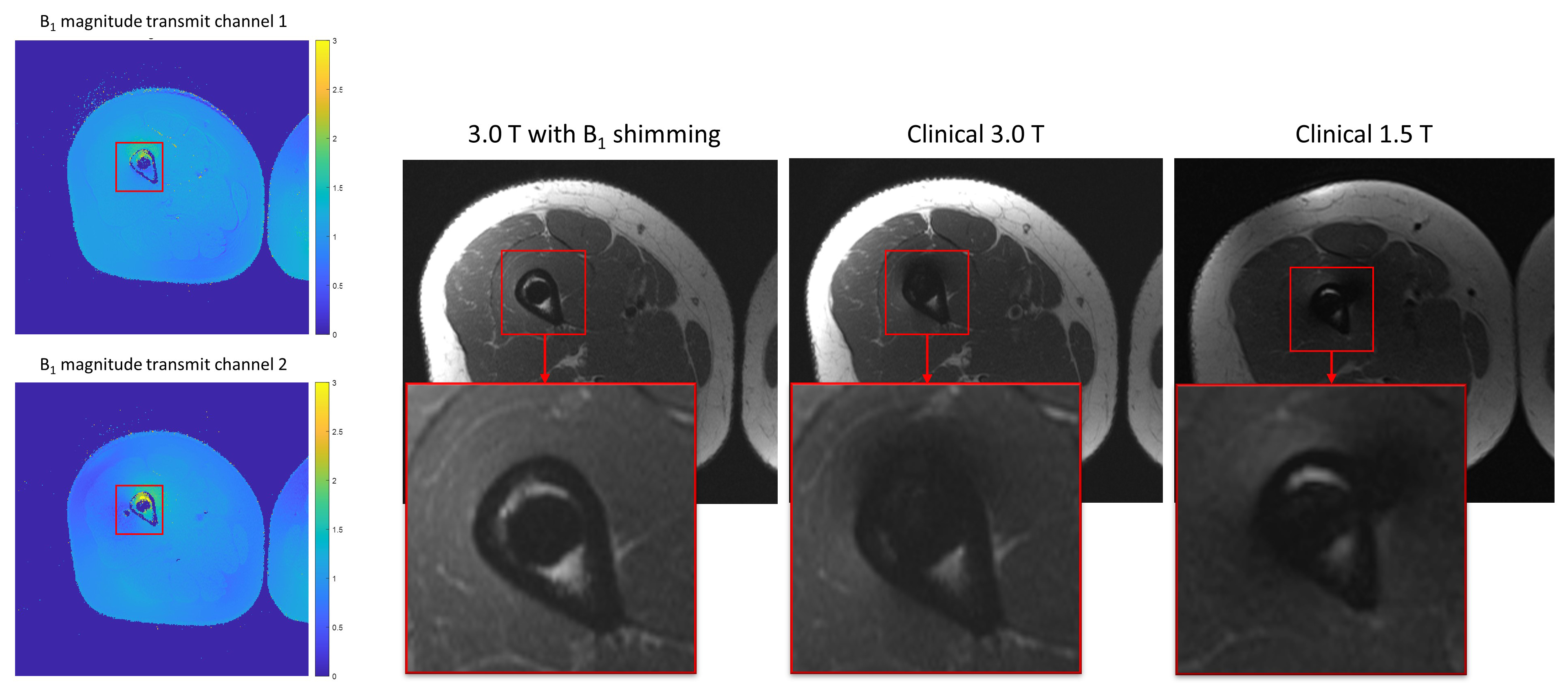
Figure 3. Local $$$B_1^+$$$ shimming. $$$B_1^+$$$ field maps of each of
the two transmit channels were obtained and combined. The shim parameters were
determined by the combination that generated the lowest $$$B_1^+$$$ variation compared to
the background. Compared with the clinical 3.0 and 1.5 T, visibility of the
bone-metal interface was significantly improved after $$$B_1^+$$$ shimming.
DOI: https://doi.org/10.58530/2023/0635