0632
Volume isotropic 3D bone Imaging with broadband IR-prepared FLORET UTE and Fibonacci interleaved trajectory ordering1Philips Japan, Tokyo, Japan, 2Department of Radiology, Faculty of Medicine, University of Miyazaki, Miyazaki, Japan, 3Division of Radiology, Miyazaki University Hospital, Miyazaki, Japan, 4Philips Healthcare, Rochester, MN, United States, 5Department of Radiology, Mayo Clinic College of Medicine and Science, Rochester, MN, United States, 6Philips Australia & New Zealand, North Ryde, Australia, 7Philips Healthcare, Best, Netherlands
Synopsis
Keywords: Bone, Bone
MR bone imaging has gained more attention for detecting and assessing bone pathology. In this study, we proposed a new technique consisting of broadband inversion recovery preparation with FLORET UTE with Fibonacci interleaved trajectory ordering (FLORET BoneVIEW) to obtain 3D isotropic bone images within a clinically feasible scan time. FLORET BoneVIEW has great potential to help a more accurate assessment of bone pathology as an alternative to CT bone imaging.PURPOSE
MR bone imaging, using ultrashort echo-time (UTE) or zero echo-time (ZTE) sequences, has gained more attention for detecting and assessing bone pathology as an alternative to CT imaging1-4. An important challenge when applying MR bone imaging clinically to various anatomies is to achieve sufficiently robust image quality. Bone-weighted imaging suffers from insufficient background suppression due to B0 inhomogeneities. Recently, a new technique based on a broadband IR prepared segmented (multispoke) radial stack-of-stars UTE sequence (3D BoneVIEW)5,6 demonstrated great potential for the assessment of ossification of the posterior longitudinal ligament (OPLL) in the cervical spine5 and low back pain in the lumbar spine6. One of the limitations of 3D BoneVIEW is its long acquisition time due to radial stack-of-stars acquisition; making it difficult to obtain 3D isotropic volume images.FLORET (Fermat looped, orthogonally encoded trajectories), on the other hand, is an efficient center-out 3D sphere spiral trajectory7 and can be combined with UTE8. The 3D FLORET UTE with Fibonacci interleaved trajectory ordering minimizes eddy current artifacts while improving temporal stability and robustness to motion artifacts, resulting in high-quality lung UTE images9.
In this study, we propose a new technique consisting of broadband IR preparation with FLORET UTE with Fibonacci interleaved trajectory ordering (FLORET BoneVIEW) to obtain 3D isotropic bone images within a clinically feasible scan time. The purpose of this study was to evaluate the feasibility of FLORET BoneVIEW in various anatomies.
METHODS
The original 3D BoneVIEW5,6 is based on broadband adiabatic IR-prepared UTE 3D stack-of-stars radial sequence. The broadband offset independent trapezoid (OIT) inversion pulses selectively invert long-T2 species and fat simultaneously10. To suppress the background signals sufficiently, we used a long-duration OIT inversion pulse (≥20ms). By using a long pulse duration, long T2 species are inverted whereas short T2 species are saturated. Bone signal is saturated because the T2 of bone is significantly shorter than the duration of the RF pulse. The TFE shot interval and inversion delay (TI) are chosen to ensure optimal nulling of the signal from both muscle and fat. This provides high-contrast morphological imaging of bone. In this study, we replaced the radial multispoke stack-of-stars sequence with 3D FLORET UTE.The 3D FLORET UTE with Fibonacci ordering sequence consists of a 2D Fermat’s spiral k-space trajectory projected onto the surface of many 3D cones. Each cone consists of a single Fermat’s spiral interleave. Sufficient cones are acquired to sample k-space over the volume of a 3D hub fully. A hub may be an entire sphere or may have conical notches at each end, as defined by the maximum cone angle used to fill the hub. In this study, three hubs with angles of 36° were applied to fill these conical gaps. In contrast with a radial trajectory, 3D FLORET samples k-space more efficiently, thus allowing for full k-space coverage in a fraction of the time. Furthermore, Fibonacci ordering provides minimal B0 artifacts and increased robustness to motion artifacts with an interleaved sampling9.
A total of seven patients were examined with a 3.0T whole-body clinical system (Ingenia CX, Philips Healthcare). The local IRB approved the study, and written informed consent was obtained from all subjects.
FLORET BoneVIEW images were compared with conventional bone imaging, including Fast field echo resembling a CT using restricted echo-spacing (FRACTURE11,12), single-echo UTE with logarithmic intensity rescaling and bias field correction1,13 (UTE), and radial stack-of-stars BoneVIEW5,6 images for image quality, especially for the overall SNR, bone-to-background contrast.
The following imaging parameters were used for FLORET BoneVIEW; 3D spiral FLORET UTE-TFE, Sagittal or Axial acquisition, voxel size=1.5*1.5*1.5mm3, TFE shot interval=250ms, flip angle=30°, turbo factor=7, TR/TE=4.6/0.10ms, TI=72ms, spiral acquisition window=1.6~1.8ms, and total acquisition time=5 to 7minutes (depending on the anatomy).
RESULTS and DISCUSSION
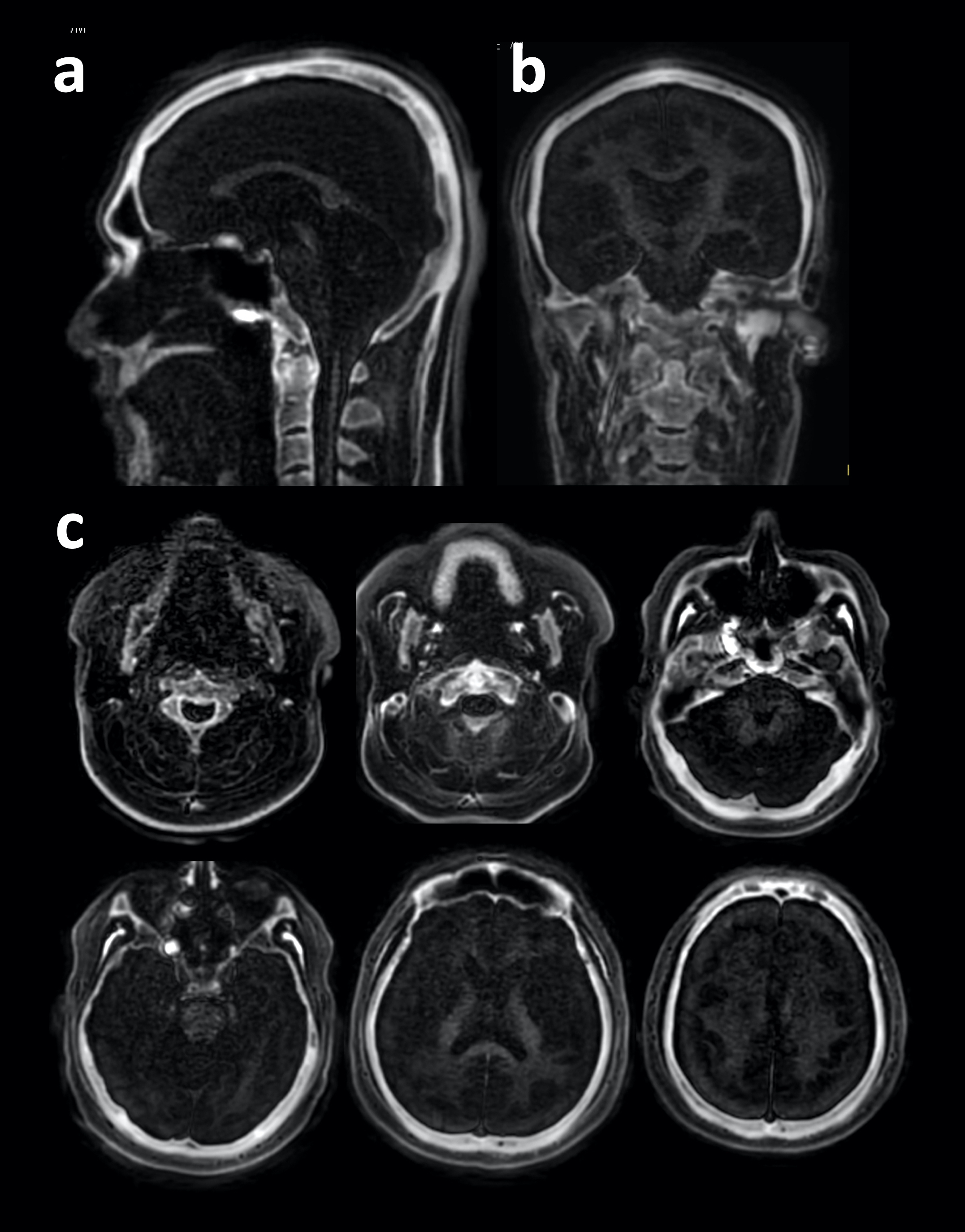
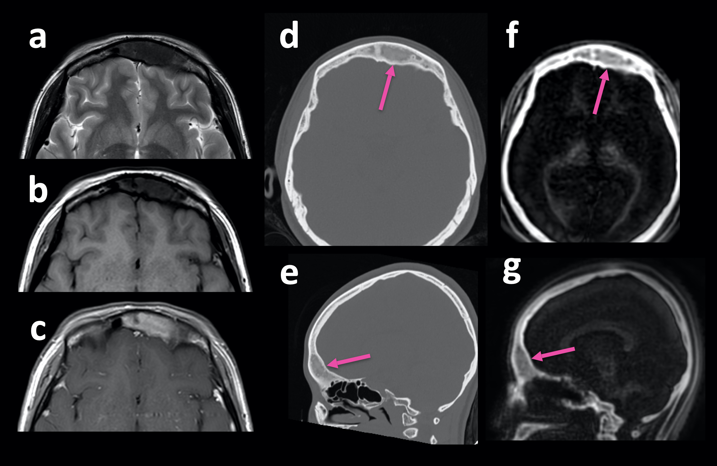
Representative sagittal, coronal and axial FLORET BoneVIEW skull images obtained from the 3D isotropic volume data are shown in Figure 2. FLORET BoneVIEW depicted the cortical bone structure of the skull while suppressing background signals sufficiently.Figure 3 shows a representative clinical case in a patient with fibrous dysplasia in comparison with the CT scan. BoneVIEW clearly showed the existence of dysplasia inside the skull as well as CT scan. Although further clinical investigation is needed, this technique can provide comparable information with CT imaging.
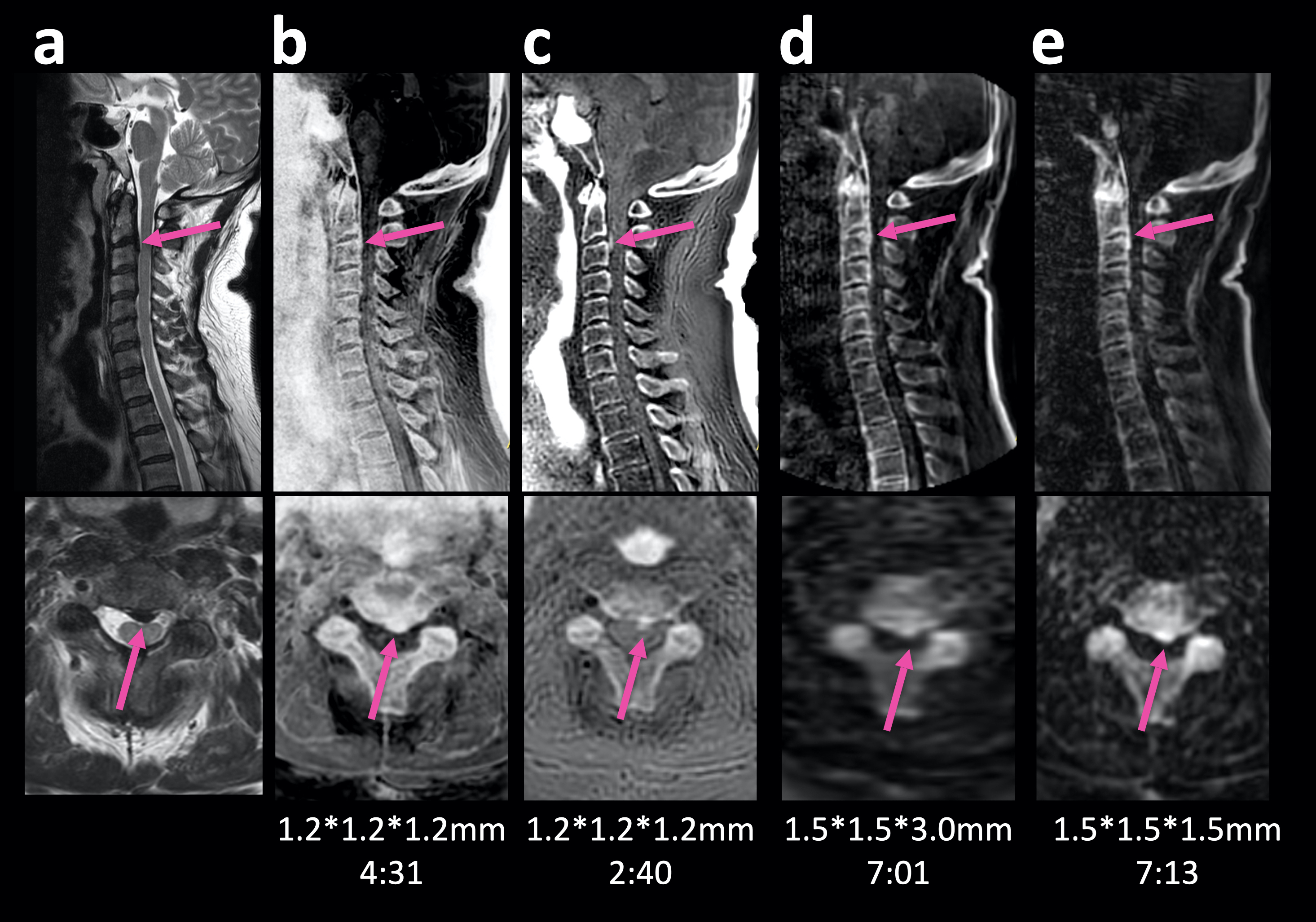
Figure 4 demonstrates a representative clinical case in a patient with OPLL compared to conventional MR bone imaging methods. FLORET BoneVIEW clearly showed the existence of ectopic hyperostosis both in sagittal source and axial MPR images thanks to isotropic volume acquisition.
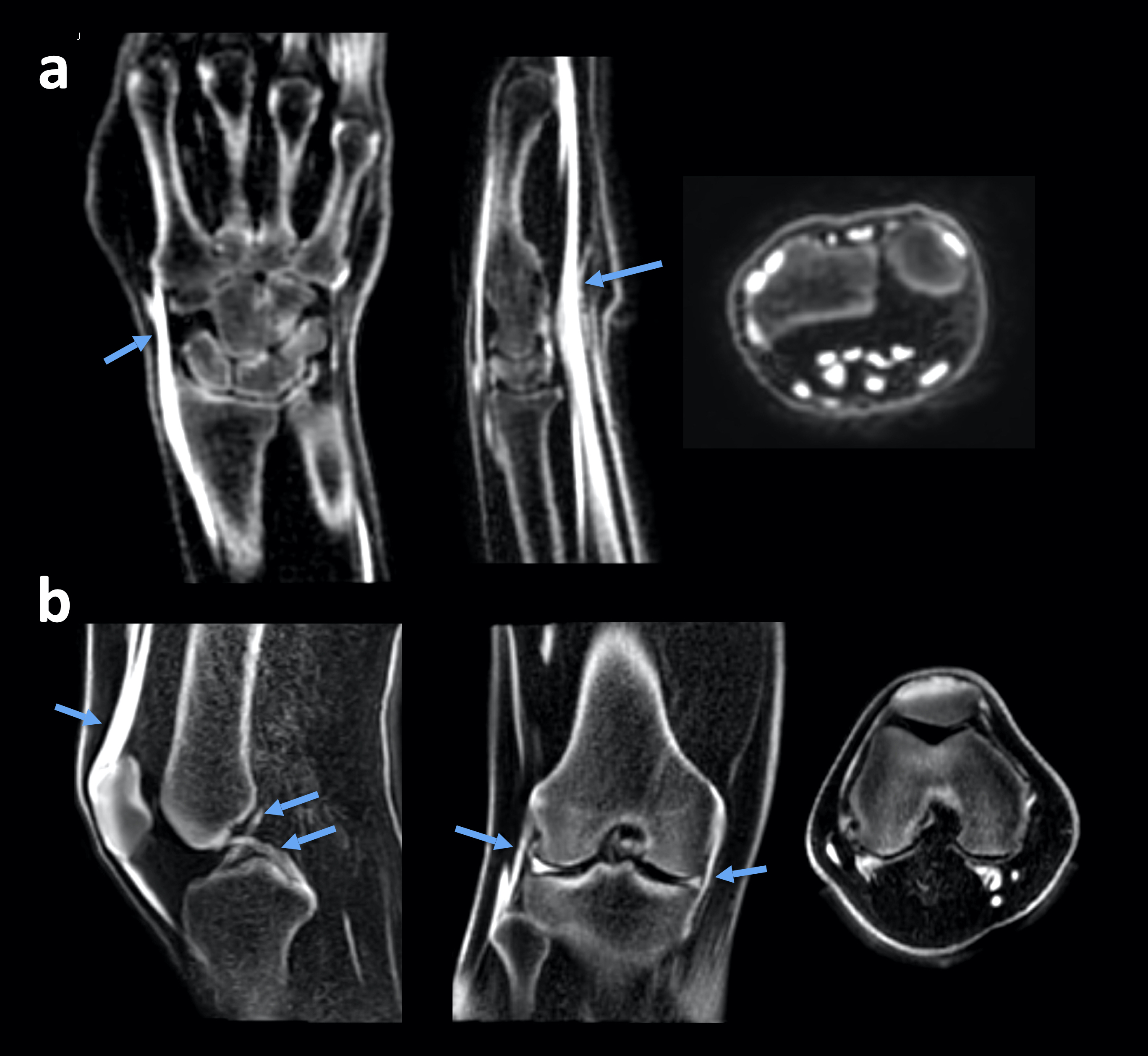
Figure 5 demonstrates representative three-orthogonal MPR bone images obtained by volume isotropic datasets in the extremities, including the wrist and knee joint. FLORET BoneVIEW showed ligaments and tendons with high intensity as well as bones. It may also be useful for assessment of ligaments and tendons.
CONCLUSION
We have demonstrated the feasibility of a new scheme for 3D volumetric isotropic MR bone imaging by using broadband IR-prepared FLORET UTE with Fibonacci interleaved trajectory ordering. This sequence has great potential to help a more accurate assessment of bone pathology as an alternative to CT bone imaging.Acknowledgements
No acknowledgement found.References
1. Delso G, et al. Clinical evaluation of zero-echo-time MR imaging for the segmentation of the skull. J Nucl Med. 2015;56:417-22.
2. Wiesinger F, et al. Zero TE MR bone imaging in the head. Magn Reson Med. 2016;75:107-14.
3. Nazaran A, et al. Three-dimensional adiabatic inversion recovery prepared ultrashort echo time cones (3D IR-UTE-Cones) imaging of cortical bone in the hip. Magn Reson Imaging. 2017;44:60-64.
4. Gersing AS, et al. Evaluation of MR-derived CT-like images and simulated radiographs compared to conventional radiography in patients with benign and malignant bone tumors. Eur Radiol. 2018 Jun 12. doi: 10.1007/s00330-018-5450-y.
5. Yoneyama M, et al. 3D broadband IR-prepared UTE bone imaging for assessment of ossification of the posterior longitudinal ligament (OPLL) in the cervical spine. Proc Intl Soc Mag Reson Med. 2019;27:2873.
6. Yoneyama M, et al. Robust motion-compensated lumbar spine bone imaging using 3D UTE with broadband inversion recovery pulse and k-space weighted navigator gating. Proc Intl Soc Mag Reson Med. 2019;27:0133.
7. Pipe JG, et al. A new design and rationale for 3D orthogonally oversampled k-space trajectories. Magn Reson Med. 2011;66(5):1303-1311. doi:https://doi.org/10.1002/mrm.22918.
8. Robison RK, et al. Three-dimensional ultrashort echo-time imaging using a FLORET trajectory. Magn Reson Med. 2017;78(3):1038-1049. doi:https://doi.org/10.1002/mrm.26500.
9. Krishnamoorthy G, et al. High-quality Lung imaging with FLORET UTE and Fibonacci interleaved trajectory ordering. Proc Intl Soc Mag Reson Med. 2022;30:3497.
10. Larson PE, et al. Using adiabatic inversion pulses for long-T2 suppression in ultrashort echo time (UTE) imaging. Magn Reson Med. 2007;58:952-61.
11. Johnson B, et al. Fast field echo resembling a CT using restricted echo-spacing (FRACTURE): a novel MRI technique with superior bone contrast. Skeletal Radiol. 2021 Aug;50(8):1705-1713.
12. Gascho D,et al. FRACTURE MRI: Optimized 3D multi-echo in-phase sequence for bone damage assessment in craniocerebral gunshot injuries. Diagn Interv Imaging. 2020 Sep;101(9):611-615.
13. Tustison NJ, et al. N4ITK: improved N3 bias correction. IEEE Trans Med Imaging. 2010 Jun;29(6):1310-20.
Figures
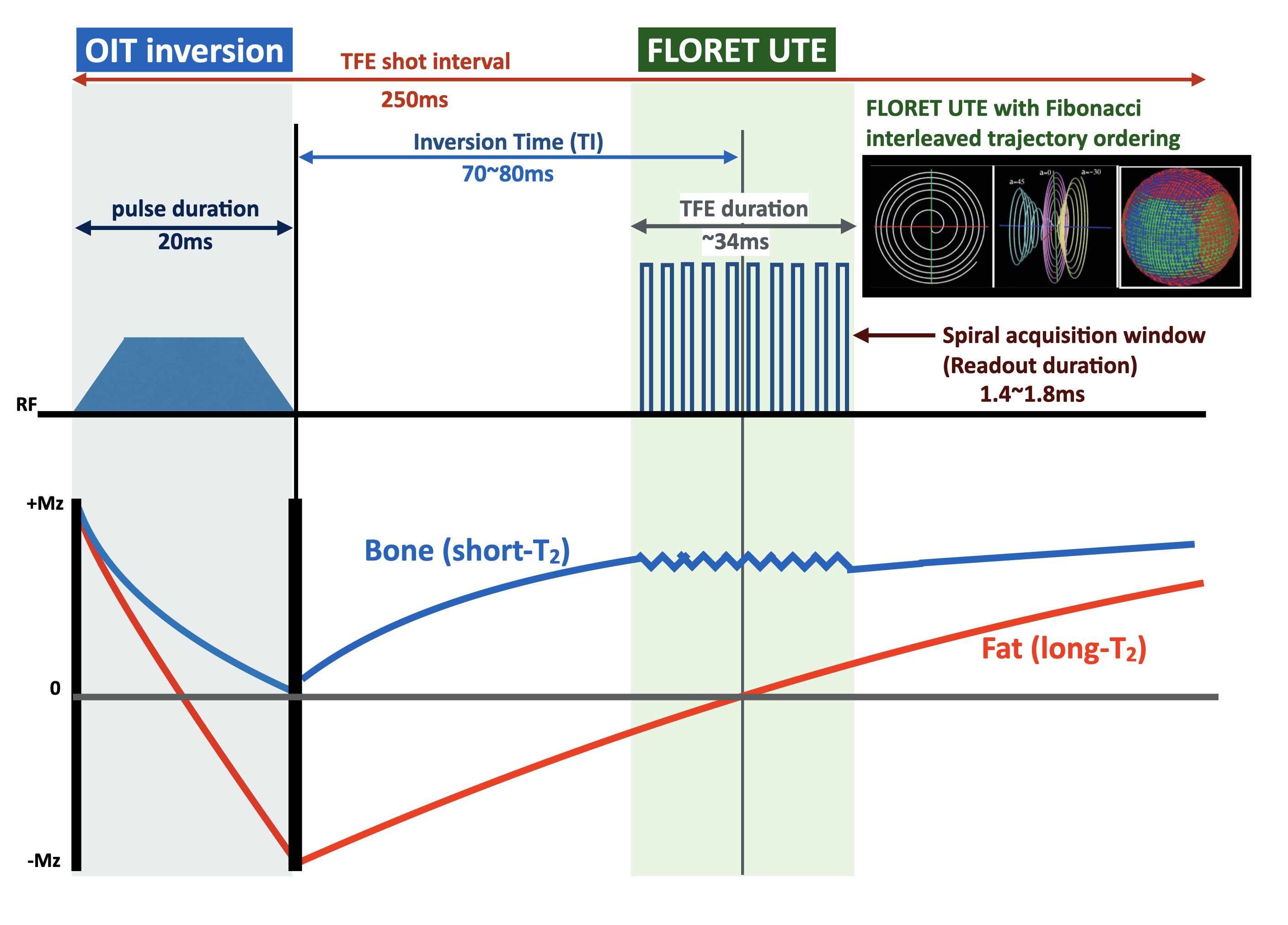
Figure 1. Scheme overview of FLORET BoneVIEW sequence.
BoneVIEW is based on broadband adiabatic IR-prepared UTE 3D stack-of-stars radial sequence. The OIT inversion pulses selectively invert long-T2 species and fat simultaneously. To suppress the background signals sufficiently, we used a long-duration OIT inversion pulse, then background signals are inverted whereas bone signals are saturated. TFE shot interval and TI are chosen for optimal nulling of the background signals. We replaced the radial stack-of-stars sequence with 3D FLORET UTE with Fibonacci ordering.