0631
Application of fast, oblique 2D-UTE to musculoskeletal imaging with a library of predistorted slice select gradient waveforms
Kevin D Harkins1,2,3, Nicholson S Chadwick1, and Mark D Does2,3
1Radiology and Radiological Sciences, Vanderbilt University Medical Center, Nashville, TN, United States, 2Institute of Imaging Science, Vanderbilt University Medical Center, Nashville, TN, United States, 3Biomedical Engineering, Vanderbilt University, Nashville, TN, United States
1Radiology and Radiological Sciences, Vanderbilt University Medical Center, Nashville, TN, United States, 2Institute of Imaging Science, Vanderbilt University Medical Center, Nashville, TN, United States, 3Biomedical Engineering, Vanderbilt University, Nashville, TN, United States
Synopsis
Keywords: MSK, Pulse Sequence Design
The adoption of 2D UTE for general musculoskeletal imaging has been slowed by technical limitations—especially gradient errors that cause slice profile distortions. In this work, a library of predistorted gradient waveforms were used to interpolate slice select gradient waveforms for oblique oriented half-pulse 2D UTE. Example multi-echo and multi-slice acquired 2D UTE images are shown in the tibia, ankle, & knee. Positive contrast in collagen rich tissues like bone and tendon could provide a new source of information to potentially diagnose MSK injuries.Introduction
Ultrashort echo time (UTE) imaging provides the ability to image signals with T2 relaxation time-constants too short to be imaged with conventional MRI imaging sequences. Several tissues are known to contain such ultrashort T2s—especially collagen rich tissues like bone, tendon, and cartilage1. However, adoption of UTE imaging has been slowed by technical limitations, especially those related to gradient trajectories for readout and excitation. For instance, half-pulse excited 2D UTE2 is sensitive to imperfections in the slice select gradient waveform, where imperfections cause slice profile distortions.Several methods have been proposed to reduce or suppress the impact of gradient infidelity on 2D slice selection3-6. One such method is gradient predistortion7, which is an iterative procedure to update the applied gradient waveform until the output waveform matches the ideal. The benefit of this method is that it converges even for nonlinear gradient systems, and it has previously been applied to UTE for bound and pore water imaging8,9. However, the method as applied was limited to pure transverse slices on gradient isocenter, and was not implemented on an oblique axis. Instead, oblique imaging would require predistortion of the slice select gradient waveform projections on each of the three physical gradient axes independently.
This abstract uses a library of predistorted gradient waveforms to interpolate obliquely applied slice select gradients. Using such a library, we applied oblique multi-slice, multi-echo 2D UTE imaging in the tibia, ankle and knee.
Methods
The iterative predistortion procedure is highlighted in Figure 1, where an applied gradient waveform is iteratively updated until the observed gradient waveform matches the ideal waveform. For this study, the ideal gradient waveform is a minimum duration bipolar slice select gradient designed for half-pulse excitation subject to maximum gradient amplitude, slew rate, and acceleration rate constraints. A library of predistorted gradient waveforms was created9, consisting of six linear spaced amplitudes on each of the physical X, Y and Z gradient axes. For half-pulse 2D UTE imaging, X, Y and Z slice select waveforms were interpolated from the library based upon slice orientation.Two healthy controls were imaged after IRB approval and informed consent was obtained. UTE signals acquired from the two half pulse passes were corrected for bulk phase shift, and 2D UTE images were reconstructed from measured trajectories by gridding and Fourier transforming density density compensated acquired signals. Two echoes were acquired, an ultrashort echo (=0.1 ms), and the first in phase image plus the ultrashort echo time image (2.3+0.1=2.4 ms), such that fat and water is equal amounts out of phase in both images. Ultrashort echo time signals were visualized by subtracting the magnitude two echo images, and subtracted signal fraction images were calculated by dividing the magnitude subtracted images by the UTE image. Since the same readout and image reconstruction was used for both echo time images, no external scaling factor was required to re-scale the images to each other.
Results and Discussion
Example 2D UTE images in the tibia are shown in Figure 2. Subtracted signal fraction images show positive contrast in the tibia, fibula, and connective tissue between muscle groups. There is the potential for fast 2D UTE to improve outcomes for diseases affecting collagen in the cortical bone in the calf. For instance, stress fractures are often assessed using a Fredericson score10, which is based the presence of fluid outside the tibia, inside the marrow, or inside the cortical bone itself, where the presence of fluid in the cortical bone of the tibia is the highest grade. Still, Fredericson scoring is poorly correlated with time to resolution.Example 2D UTE images acquired in the ankle are shown in Figure 3, highlighting positive contrast in the Achilles tendon, extensor tendon, flexor tendon, and plantar facia. These structures are typically dark in standard anatomical imaging, and injury diagnosis depends either on the presence of fluid within the tissue, or thickening of the dark band representing the tissue. Instead, UTE imaging provides positive contrast in these tissues, providing a new source of information to potentially diagnose injuries in the ankle.
Finally, Figure 4 demonstrates 2D UTE imaging in the knee of a healthy control. Subtraction and subtracted signal fraction images show positive contrast in the patella, patella tendon, and quadriceps tendon, in addition to the trabecular bone within the tibia and femur.
Conclusions
The application of fast 2D UTE imaging has the potential to provide novel contrast for the assessment of injury and disease in the musculoskeletal system.Acknowledgements
This work was supported by NIH R01EB014308References
1. Afsahi, A. M., Ma, Y., Jang, H., Jerban, S., Chung, C. B., Chang, E. Y., & Du, J. (2021). Ultrashort Echo Time Magnetic Resonance Imaging Techniques: Met and Unmet Needs in Musculoskeletal Imaging. Journal of Magnetic Resonance Imaging, 1–16. https://doi.org/10.1002/jmri.280322. Pauly, J. M. (2012). Selective Excitation for Ultrashort Echo Time Imaging. In Encyclopedia of Magnetic Resonance (Vol. 1, pp. 1–7). John Wiley & Sons, Ltd. https://doi.org/10.1002/9780470034590.emrstm1271
3. Josan, S., Kaye, E., Pauly, J. M., Daniel, B. L., & Pauly, K. B. (2009). Improved half RF slice selectivity in the presence of eddy currents with out-of-slice saturation. Magnetic Resonance in Medicine, 61(5), 1090–1095. https://doi.org/10.1002/mrm.21914
4. Josan, S., Pauly, J. M., Daniel, B. L., & Pauly, K. B. (2009). Double half RF pulses for reduced sensitivity to eddy currents in UTE imaging. Magnetic Resonance in Medicine, 61(5), 1083–1089. https://doi.org/10.1002/mrm.21879
5. Stumpf, K., Kaye, E., Paul, J., Wundrak, S., Pauly, J. M., & Rasche, V. (2020). Two‐dimensional UTE overview imaging for dental application. Magnetic Resonance in Medicine, 84(5), 2616–2624. https://doi.org/10.1002/mrm.28312
6. Harkins, K. D., Horch, R. A., & Does, M. D. (2014). Simple and robust saturation-based slice selection for ultrashort echo time MRI. Magnetic Resonance in Medicine, 00, 1–8. https://doi.org/10.1002/mrm.25361
7. Harkins, K. D., Does, M. D., & Grissom, W. A. (2014). Iterative method for predistortion of MRI gradient waveforms. IEEE Transactions on Medical Imaging, 33(8), 1641–1647. https://doi.org/10.1109/TMI.2014.2320987
8. Manhard, M. K., Harkins, K. D., Gochberg, D. F., Nyman, J. S., & Does, M. D. (2017). 30-Second bound and pore water concentration mapping of cortical bone using 2D UTE with optimized half-pulses. Magnetic Resonance in Medicine, 77(3), 945–950. https://doi.org/10.1002/mrm.26605
9. Harkins, K. D., Ketsiri, T., Nyman, J. S., & Does, M. D. (2022). Fast bound and pore water mapping of cortical bone with arbitrary slice oriented two-dimensional ultra-short echo time. Magnetic Resonance in Medicine, July, 1–7. https://doi.org/10.1002/mrm.29484
10. Kijowski, R., Choi, J., Shinki, K., Del Rio, A. M., & De Smet, A. (2012). Validation of MRI classification system for tibial stress injuries. American Journal of Roentgenology, 198(4), 878–884. https://doi.org/10.2214/AJR.11.6826
Figures
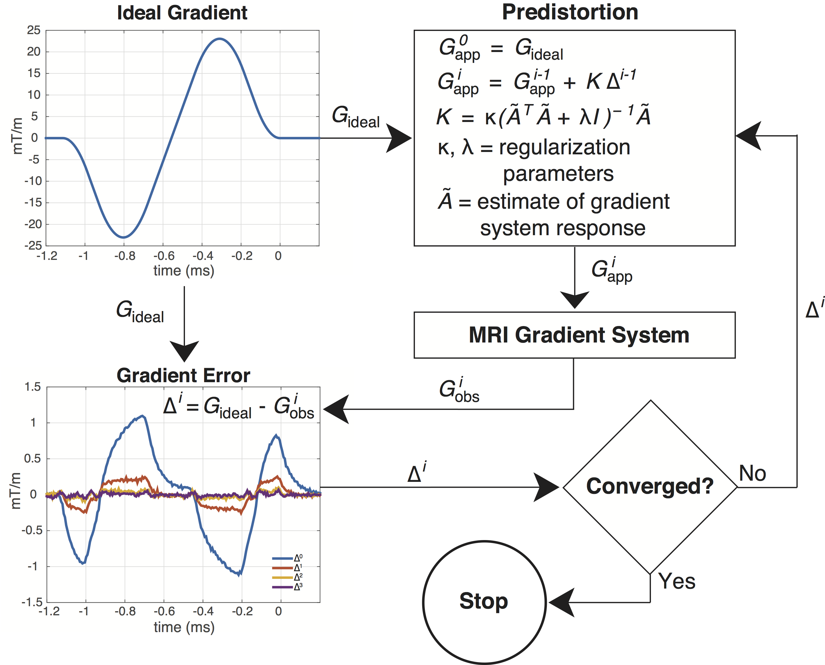
Figure 1: Iterative predistortion updates the gradient waveform applied to the system until the measured waveform matches the ideal waveform. In this case, iterative predisotrtion is used to improve the fidelity of a slice select waveform for half-pulse excited 2D UTE.

Figure 2: Four example slices of a multi-slice, multi-echo 2D UTE acquisition in the calf of a healthy control. Subtraction and subtracted signal fraction images highlight ultrashort echo time echo signals in the tibia, fibula, and connective tissues between muscle groups.
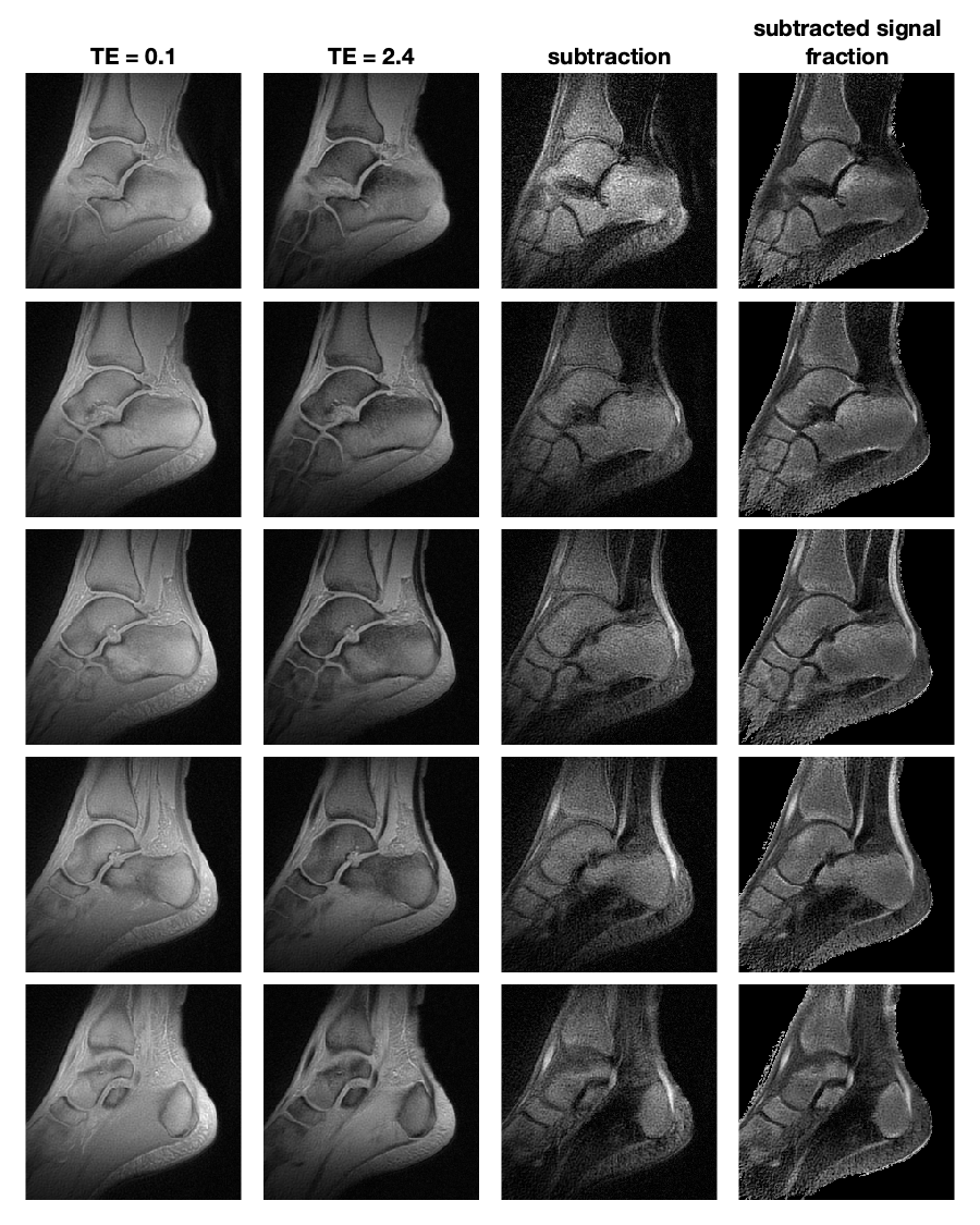
Figure 3: Five example slices of a multi-slice, multi-echo 2D UTE acquisition in the ankle of a healthy control. Subtraction and subtracted signal fraction images provide positive contrast for collagen rich tissues like the Achilles tendon, extensor tendon, flexor tendons, and trabecular bone.
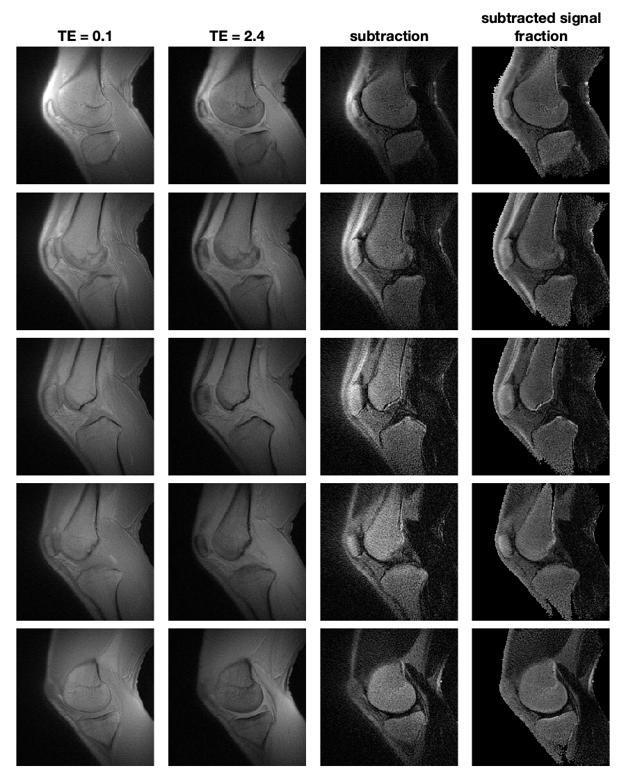
Figure 4: Five example slices of a multi-slice, multi-echo 2D UTE acquisition in the knee of a healthy control.
DOI: https://doi.org/10.58530/2023/0631