0613
All-inclusive Safety Models Limit Imaging Performance at 7T1CUBRIC, School of Psychology, Cardiff University, Cardiff, United Kingdom
Synopsis
Keywords: Safety, High-Field MRI, RF Pulse Design & Fields; Parallel Transmit & Multiband
Safety models on scanners are patient-position unaware and may include multiple patient positions to ensure safety. This may lead to overestimation of the specific absorption rate (SAR) and limit scanning performance. This study investigates the effect of an all-inclusive safety model on SAR estimation for parallel-transmit and quadrature-excitation at 7T. Results show that more than 4-fold SAR overestimation can be commonly observed. RF shimming suffered the most, with 11-fold overestimation at the worst-case and more than 4-fold overestimation in 37% of cases. RF shimming also offered the lowest peak local SAR and may be unnecessarily penalized by overconservative safety models.Background
Ultrahigh field MRI (UHF-MRI) scanning performance is often limited in practice due to safety concerns. Scanners and/or transmit coils are shipped with (computational-modelling based) safety models that are used for real-time safety calculations. These safety models may range from a single virtual body model simulated at a single position (less representative, under-conservative) to multiple models at all possible body positions within the coil (more representative but over-conservative). Recent studies have demonstrated that the former can lead to considerable underestimation of peak local SAR in the presence of positional mismatches or patient motion 1-6. This study investigates the latter; i.e., the effect of using all head positions within an 8-channel transmit coil on SAR estimations.Methods
The virtual body model Ella was simulated at 161 positions inside a generic 8-channel parallel-transmit (pTx) array (radius: 115 mm) using Sim4Life (Zurich MedTech,Zurich,CH). Up to 20 mm displacements and degrees-of-rotations along/around each axis were included (anterior-posterior displacement within ±10 mm) including off-axis displacements (axial/coronal planes). Electromagnetic fields were exported to Matlab (Mathworks,Natick,MA,USA). Realistic small-tip angle 7 pulses were designed (target: homogeneous 30-degrees flip-angle) using an adaptation of the spatial-domain method Matching Pursuit-guided-Conjugate Gradient 8-10 with Tikhonov regularization (β=0.5) to balance flip-angle error and channel-by-channel RF power. Details omitted here followed previous literature 3. Slice-selective single-channel (quadrature-excitation) 1-spoke; and parallel-transmit 1-/2-/3-/4-/5-pulses (RF shimming: 1-spoke) were designed for seven axial slices at five different head positions (centre; combinations of ±10mm right-left/anterior-posterior displacements, indicated in Figure 3). 10-gram averaged Q-matrices 11,12 averaged over cubical volumes 13 were used for local SAR calculations.Body models at all positions were combined to act as an all-inclusive safety model. Local SAR values calculated using the Q-matrices at the actual head positions were compared with those calculated using the safety model. The effect of including each head position in the safety model was also investigated.
Results and Discussion
Using all head positions in the safety model causes considerable overestimation of peak local SAR. In the worst-case, the estimated peak local SAR was 110 W/kg when the actual peak local SAR was 10 W/kg. Figure 1 shows that a majority of the head positions included in the safety model yield relatively lower SAR overestimations. Nevertheless, the overall estimate of the safety model can often exceed 4-fold overestimation: 20%, 37%, 3%, 9%, 14%, 11% of the pulses for quadrature, RF shimming and 2-/3-/4-/5-spokes pulses, respectively.SAR overestimation was highest for the superiormost slice, for which SAR was consistently overestimated by at least 86%. Nevertheless, overestimations of more than 4-fold were observed for all slices (Figure 1b).
RF shimming (1-spokes) was worst affected from SAR overestimation, with its SAR being overestimated by at least 80% across cases. RF shimming is inherently self-correcting when coils are distributed around the slice of interest 3,6,14. I.e., when the head is not centred, coil elements closer to the head are turned down and farther elements turned up, to achieve the target flip-angle. This also keeps local SAR somewhat balanced, albeit indirectly (Figure 2a). When the safety model contains head positions closer to the turned-up coils, local SAR estimations increase considerably. This behaviour is not similar for more spokes (they target complementarily inhomogeneous flip-angle distributions, turning coils up/down non-trivial) or other slice orientations (coils provide less degrees-of-freedom) 3,6. Therefore, axial RF shimming suffers from an all-inclusive safety model more than other modes of operation.
Figure 2 shows maximum-intensity projections (MIPs) of three-dimensional local SAR distributions along the superior-inferior direction. The overestimation exceeded 10 W/kg in a volume of 166 cm3 (panel c). An example 2-spokes pulse shows that an all-inclusive safety model may misestimate the location of the peak local SAR (here, estimated: Right-Anterior instead of actual: Left-Middle). This could mislead SAR-hopping schemes 15. Panels d-e also show that peak local SAR estimations are not dominated by the head position closest to coil elements (here, left-posterior) or always observed in the same region.
Figure 3 shows how including a head position in the safety model affects SAR overestimation. As the distance between the actual head position and the head position included in safety calculations increases, SAR overestimation increases. Figure 3e shows that several head positions led to SAR overestimations of more than 3-fold when they were included in the safety model.
Figure 4 plots SAR overestimation with respect to the actual peak local SAR values. RF shimming yields comparatively the lowest peak local SAR across all pTx pulse types, and also suffers from the highest overestimation factors. The (lower-SAR) benefit of RF shimming would increase further if pulses were time-normalized (i.e.,same duration) 16.
Figure 5 compares the actual SAR with SAR estimated using the safety model. The increased dynamic range of estimation values would limit scanning performance due to the implied limitations on scan parameters (such as flip-angle or TR), especially for RF shimming.
Future work will expand this investigation to more body and coil models.
Conclusion
Using all possible head positions within a coil structure may cause substantial overestimation of peak local SAR, and thereby affect scanning performance considerably. Having better safety models that are aware of patient position can help improve scanning performance, especially for RF shimming.Acknowledgements
This project was supported in part by the Wellcome Trust [204824/Z/16/Z], by the Welsh Government [Wales Data Nation Accelerator project].
References
1. Kopanoglu E, Deniz CM, Erturk MA, Wise RG. Specific absorption rate implications of within-scan patient head motion for ultra-high field MRI. Magn Reson Med 2020;84(5):2724-2738.
2. Ajanovic A, Hajnal J, Malik S. Positional Sensitivity of Specific Absorption Rate in Head at 7T. In: Proceedings of the 28th Annual Meeting of ISMRM; 2020; online. p 4251.
3. Kopanoglu E. Patient specific parallel transmit pulses are patient position dependent while safety models are fixed: safety implications. In: Proceedings of the 29th Annual Meeting of ISMRM; 2021; online. p 2299.
4. Malik SJ, Hand JW, Satnarine R, Price AN, Hajnal JV. Specific absorption rate and temperature in neonate models resulting from exposure to a 7T head coil. Magn Reson Med 2021;86(3):1299-1313.
5. Ajanovic A, Hajnal J, Tomi-Tricot R, Malik S. Motion and Pose Variability of SAR Estimation with Parallel Transmission at 7T. In: Proceedings of the 29th Annual Meeting of ISMRM; 2021; online. p 2487.
6. Kopanoglu E. Head Position Related SAR Uncertainty Depends on Slice Orientation and Pulse Complexity. In: Proceedings of the Joint Meeting of ISMRM and ESMRMB; 2022; London, UK. p 2870.
7. Pauly J, Nishimura D, Macovski A. A K-Space Analysis of Small-Tip-Angle Excitation. Journal of Magnetic Resonance 1989;81(1):43-56.
8. Grissom W, Yip CY, Zhang Z, Stenger VA, Fessler JA, Noll DC. Spatial domain method for the design of RF pulses in multicoil parallel excitation. Magn Reson Med 2006;56(3):620-629.
9. Kopanoglu E, Constable RT. Radiofrequency pulse design using nonlinear gradient magnetic fields. Magn Reson Med 2015;74(3):826-839.
10. Kopanoglu E. Near real-time parallel-transmit pulse design. In: Proceedings of the Joint Meeting of ISMRM and ESMRMB; 2018; Paris, France. p 3392.
11. Bardati F, Borrani A, Gerardino A, Lovisolo GA. SAR optimization in a phased array radiofrequency hyperthermia system. Specific absorption rate. IEEE Trans Biomed Eng 1995;42(12):1201-1207.
12. Graesslin I, Homann H, Biederer S, Bornert P, Nehrke K, Vernickel P, Mens G, Harvey P, Katscher U. A specific absorption rate prediction concept for parallel transmission MR. Magn Reson Med 2012;68(5):1664-1674.
13. IEC/IEEE International Standard -- Determining the peak spatial-average specific absorption rate (SAR) in the human body from wireless communications devices, 30 MHz to 6 GHz - Part 1: General requirements for using the finite-difference time-domain (FDTD) method for SAR calculations. IEC/IEEE 62704-1:2017 2017:1-86.
14. Deniz CM, Vaidya MV, Sodickson DK, Lattanzi R. Radiofrequency energy deposition and radiofrequency power requirements in parallel transmission with increasing distance from the coil to the sample. Magn Reson Med 2016;75(1):423-432.
15. Guerin B, Adalsteinsson E, Wald LL. Local SAR reduction in multi-slice pTx via “SAR hopping” between excitations. In: Proc Intl Soc Mag Reson Med 20, Melbourne, Australia, p642; 2012.
16. Kopanoglu E, Yilmaz U, Gokhalk Y, Atalar E. Specific absorption rate reduction using nonlinear gradient fields. Magn Reson Med 2013;70(2):537-546.
Figures
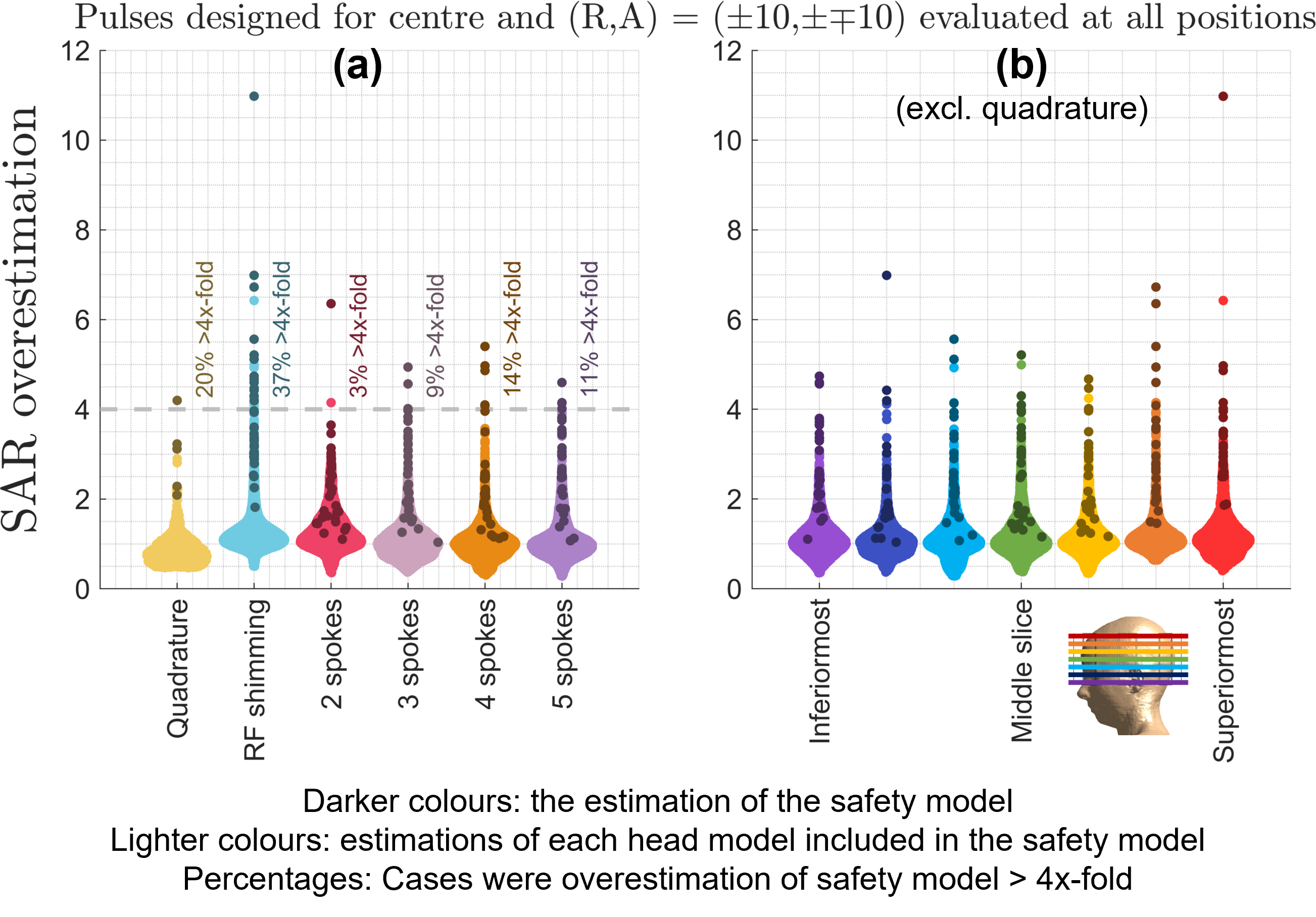
Figure 1: SAR overestimation is shown for different pulse types and target slice locations. (a) SAR overestimation is highest for RF shimming, with maximum overestimation being 11-fold. 2-/3-/4-/5-spokes pulses were overestimated by up to 6.5-fold. (b) The superiormost slice lead to the maximum overestimation across all pulses, while overestimation varied between 5- to 7-fold for other slices. Quadrature mode was excluded in panel b as SAR overestimation is slice-independent.
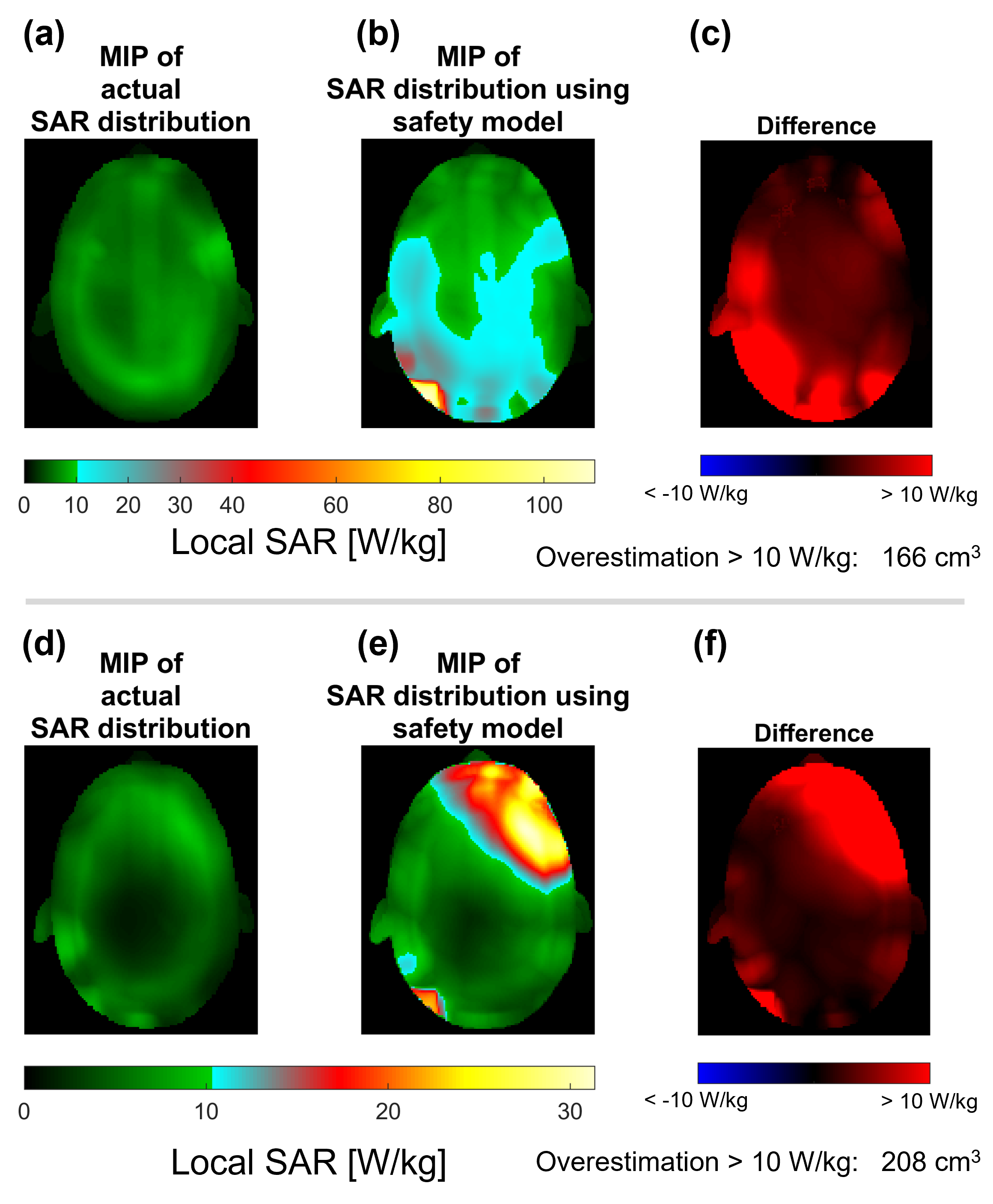
Figure 2: Maximum intensity projections of local SAR distributions (along superior-inferior). Pulse duration was adjusted so that actual peak local SAR is 10 W/kg. (a-c) RF Shimming, worst case SAR overestimation. (d-f) Example SAR overestimation for a 2-spoke pulse (not the worst-case 2-spokes overestimation). (a,d) Actual SAR distributions. (b,e) SAR distributions estimated using the safety model. (c,f) Difference between actual and estimated SAR (colour axes were saturated at ±10 W/kg).
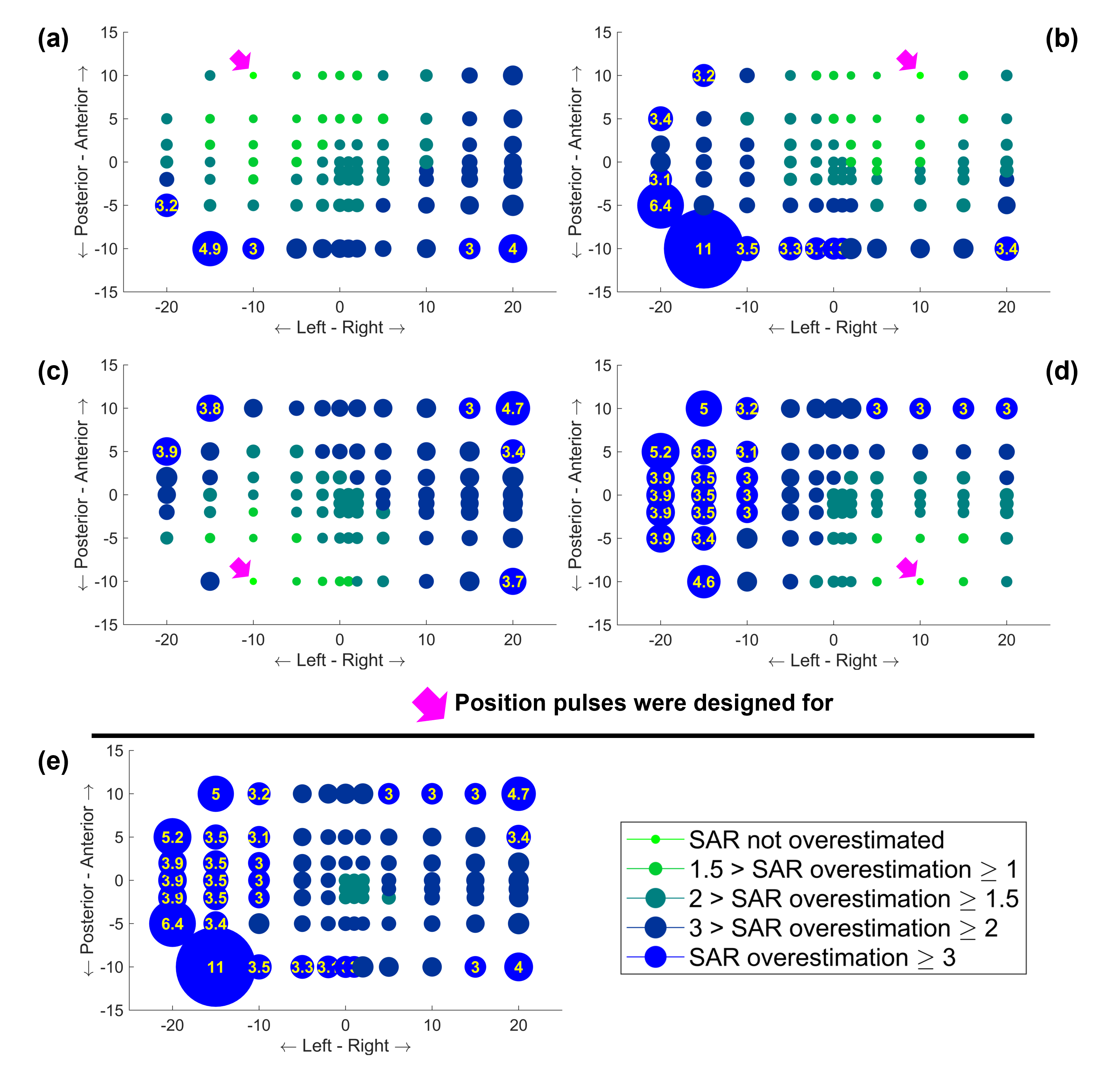
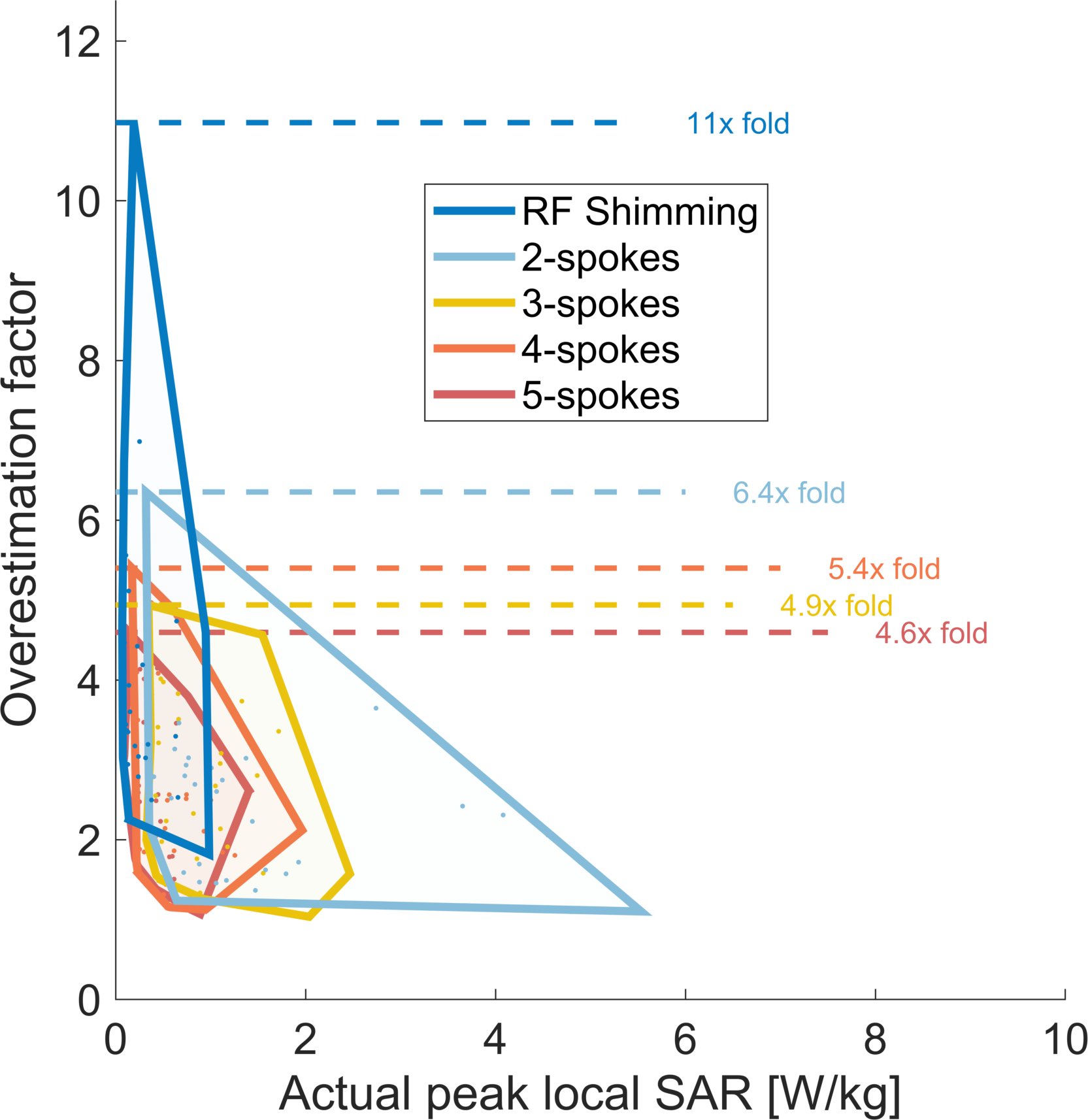
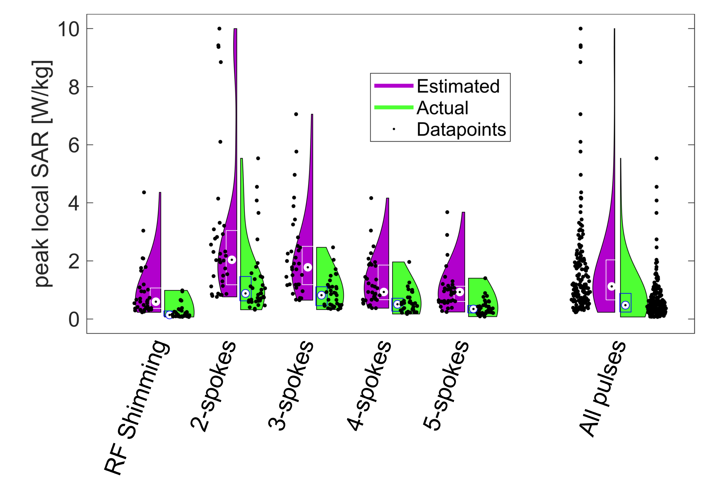
Figure 5: Estimated and actual peak local SAR values are shown for different types of pTx pulses. The range of actual peak local SAR was much smaller across the pulses designed for different slices and head positions, whereas the safety model yielded a much wider range of estimation values. The increased dynamic range of values would limit scanning performance due to the implied limitations on scan parameters (such as flip-angle or TR) especially for RF shimming which suffers the largest increase. Box plots show median, 25th and 75th percentile.