0592
Are fMRI findings on emotional aging affected by cerebrovascular aging?1Diagnostic Radiology and Nuclear Medicine, University of Maryland, Baltimore, Baltimore, MD, United States
Synopsis
Keywords: Neurodegeneration, fMRI (task based)
Age-related changes in emotional circuitry have been studied using BOLD fMRI and revealed age-related increases in the activation of the prefrontal cortex and inconsistent findings in the amygdala. Previous emotional aging studies did not account for vascular aging which causes a reduction in cerebrovascular reactivity (CVR). Using picture viewing task fMRI and gas inhalation MRI, the fMRI signals are calibrated by the vascular measures to improve the inference of neural activity. After accounting for vascular changes, age-invariant activity was seen in the amygdala, and increased age-related activation of prefrontal regions was observed compared to activation before vascular correction.INTRODUCTION
Functional MRI (fMRI) is an important tool to study neural circuit activity associated with emotional aging. Previous fMRI studies identified the fronto-amygdalar circuit that is involved in affective picture perception, revealing consistent age-related increases in the recruitment of prefrontal cortex and inconsistent age-effect in amygdala [1-3]. However, an important but under-studied issue in the interpretation of fMRI data in emotional aging is that the Blood-Oxygenation-Level-Dependent (BOLD) signal used in fMRI relies upon an indirect signal -- changes in oxygenation and blood flow -- to detect the intensity of neural activation. Given that aging is known to cause profound vascular changes such as reduction in cerebrovascular reactivity (CVR), which indicates the dilatory function of cerebral blood vessels [4], it is important to re-examine the emotional aging fMRI findings after accounting for vascular factors. Therefore, we conducted the present study to assess how the emotional circuit changes after accounting for age-related CVR changes.METHODS
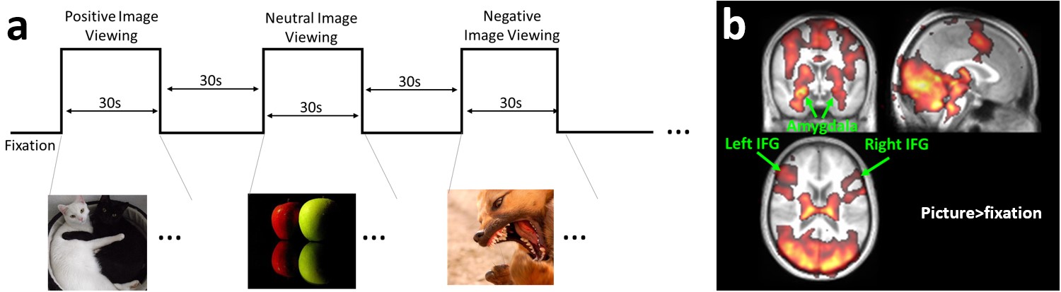
Subjects and imaging protocolsIn total, 27 subjects were studied on a Siemens 3T MRI scanner, including 16 young subjects (age range 23-38yrs, 8 Females) and 11 elderly subjects (age range 53-73yrs, 5 Females). Each subject underwent a 10min task-evoked fMRI scan and a 7min hypercapnia-inhalation MRI scan using identical HCP BOLD-MRI protocols (field-of-view=208x208x144mm3, 2mm isotropic voxels, TR/TE/FA=720ms/38ms/52º, SMS factor=8). The affective picture perception task paradigm began with a 30s fixation period, followed by 9 blocks of 30s picture blocks interleaved by 30s fixation periods (Figure 1a). The 9 picture blocks included 3 positive, 3 negative, and 3 neutral picture blocks in randomized order. Each block consisted of 10 IAPS pictures [5] each displayed for 3s. Subjects were instructed to press buttons with their left/right hand if they saw non-human/human objects in the pictures, in order to keep their attention. The hypercapnia inhalation paradigm included 5%CO2 gas inhalation following the MarkVCID CVR protocol [6]. A T1-MPRAGE scan (voxel=1x1x1mm3) was also performed for anatomic reference.
Data analysis
After motion correction, CVR BOLD images were coregistered to the task fMRI BOLD images, and then all BOLD images were normalized to MNI space via T1-MPRAGE. GLM analyses were performed for the task and CO2 data. For the task fMRI data, four contrasts were examined, including picture vs. fixation, positive vs. neutral pictures, negative vs. neutral pictures, and positive vs. negative pictures. CVR maps were also obtained following previous literature [7]. Three Region-of-interest (ROI) of the emotion circuit, amygdala, left and right inferior frontal gyrus (IFG), were used for quantitative analysis. CVR correction was performed as SfMRI,corr = SfMRI,uncorr/CVR. Two-sample t-tests were performed between the young and old groups before and after CVR correction.
RESULTS
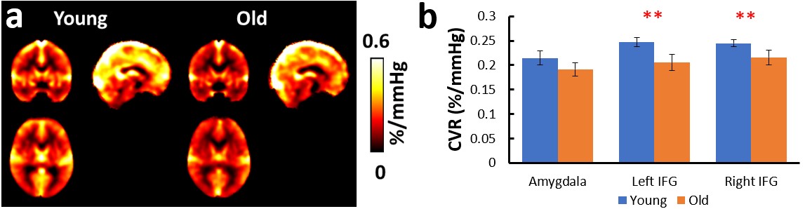
Figure 1b shows the group activation map across all subjects in viewing the affective pictures. It can be seen that amygdala, left and right IFGs were activated, confirming the involvement of the emotional circuit in our task, which is consistent with previous literature on emotion perception [1-3]. Visual, motor and thalamus areas were also activated as expected.Figure 2a shows the averaged CVR maps in young and old subjects. Visual inspection suggested reduced CVR in the older subjects, consistent with those reported previously. CVR values of the left and right IFGs, shown in Figure 2b, were significantly lower in the older group (p=0.018 and 0.044 for left and right IFG, respectively), although the difference was not significant in amygdala (p=0.14).
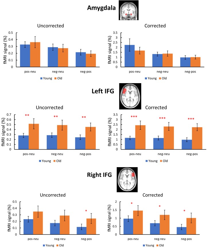
Figure 3 shows the group comparisons of fMRI signals between young and old in each ROI before and after CVR correction for 3 different contrasts. No significant group difference was found between young and old in amygdala in all contrasts both before and after correction. The older subjects had significantly higher fMRI signals than the young subjects in left IFG in all three contrasts (p=0.018, 0.036, and 0.019 for positive>neutral pictures, negative>neutral pictures, and negative>positive pictures, respectively), and these differences became more significant after accounting for CVR differences (p=0.004, 0.007, and 0.003 for positive>neutral pictures, negative>neutral pictures, and negative>positive pictures, respectively). In right IFG, before CVR correction, the older subjects showed a trend of higher fMRI signal only in the negative>positive picture contrast (p=0.080), but after CVR correction, the trend of higher fMRI signal in the older group was observed in all three contrasts (p=0.088, 0.071, and 0.061 for positive>neutral pictures, negative>neutral pictures, and negative>positive pictures, respectively), suggesting that frontal over-recruitment in emotional aging was present in both hemispheres.
DISCUSSION and CONCLUSION
Our findings are in agreement with previous literature on emotion perception [2-3]. Age-invariant activity in the amygdala was observed even after accounting for vascular factors in amygdala, suggesting the function of the amygdala remains intact in aging. The age-related increase in frontal lobe observed in this study was consistent with the emotional aging literature [1-2], but the degree of increased recruitment in the prefrontal cortex was much higher and consistent throughout all contrasts once the age-related CVR decrease was accounted for. This age-related increase in frontal recruitment is thought to represent a compensatory activation that occurs in the aging brain to accommodate the decreased volume of neural tissue and declining efficiency of neural circuitry [8]. Accounting for age-related changes in vascular function could improve the examination of fMRI responses in emotional aging.Acknowledgements
No acknowledgement found.References
1. Tessitore A, Hariri AR, Fera F, Smith WG, Das S, Weinberger DR, et al. Functional changes in the activity of brain regions underlying emotion processing in the elderly. Psychiatry Research. 2005;139:9–18.
2. Leclerc CM, Kensinger EA. Age-related differences in medial prefrontal activation in response to emotional images. Cognitive, Affective & Behavioral Neuroscience. 2008a;8:153–164.
3. Mather M, Canli T, English T, Whitfield S, Wais P, Ochsner K, et al. Amygdala responses to emotionally valenced stimuli in older and younger adults. Psychological Science. 2004;15:259–263.
4. Lu H, Xu F, Rodrigue KM, Kennedy KM, Cheng Y, Flicker B, Hebrank AC, Uh J, Park DC. Alterations in cerebral metabolic rate and blood supply across the adult lifespan. Cereb Cortex. 2011 Jun;21(6):1426-34.
5. Lang PJ, Bradley MM, Cuthbert BN. International affective picture system (IAPS) Gainesville, FL: University of Florida; 1997.
6. Lu H, Kashani AH, Arfanakis K, Caprihan A, DeCarli C, Gold BT, Li Y, Maillard P, Satizabal CL, Stables L, Wang DJJ, Corriveau RA, Singh H, Smith EE, Fischl B, van der Kouwe A, Schwab K, Helmer KG, Greenberg SM; MarkVCID Consortium. MarkVCID cerebral small vessel consortium: II. Neuroimaging protocols. Alzheimers Dement. 2021 Apr;17(4):716-725.
7. Liu P, De Vis JB, Lu H. Cerebrovascular reactivity (CVR) MRI with CO2 challenge: A technical review. Neuroimage. 2019 Feb 15;187:104-115.
8. Cabeza R, Anderson ND, Locantore JK, McIntosh AR. Aging gracefully: compensatory brain activity in high-performing older adults. Neuroimage. 2002;17:1394–1402.
Figures