0548
Fetal cardiac 3D cine MRI at low field - whole heart slice-to-volume reconstruction from real-time spiral SSFP at 0.55T1Translational Medicine, Hospital for Sick Children, Toronto, ON, Canada, 2University of Southern California, Los Angeles, CA, United States, 3Department of Biomedical Engineering, University of Southern California, Los Angeles, CA, United States, 4Division of Cardiology, Children's Hospital Los Angeles, Los Angeles, CA, United States, 5Department ofMedical Biophysics, University of Toronto, Toronto, ON, Canada
Synopsis
Keywords: Heart, Fetus, Cine, 4D, Whole Heart, Morphology
Spiral SSFP of the fetal heart at low-field has the advantage of low artifact and high sampling efficiency, compared to conventional systems. This work demonstrates the feasibility of spiral SSFP on a high-performance 0.55 T scanner to produce high spatiotemporal resolution real-time 2D and motion-corrected slice-to-volume super-resolution reconstructed 3D cine images of the heart and great vessels of the human fetus in utero. Patient comfort is improved with the use of a low-field scanner, while motion-robust slice-to-volume reconstruction allows for free-breathing acquisitions that do not require precise scan plane prescriptions during acquisition.Introduction
Imaging of the small, rapidly beating fetal heart in the presence of fetal and maternal movement is challenging. Volumetric imaging of the heart and great vessels in utero using slice-to-volume reconstruction (SVR) can overcome these challenges by using rapid MR imaging to capture movement and then correct for motion as images are combined as an image volume1,2. However the resolution of SVR output is governed by the spatiotemporal characteristics of the input MR images2.High-performance low field MR systems have many characteristics ideal for real-time steady state free precession (SSFP) spiral imaging of the fetal heart, including reduced artifact, higher sampling efficiency and increased patient comfort and safety3, compared to 1.5 T systems typically used for fetal cardiac MR. This work examines the feasibility of spiral SSFP on a high-performance 0.55 T scanner to produce high spatiotemporal resolution real-time 2D and motion-corrected SVR cine-resolved 3D images of the human fetal heart.
Methods
MRI Acquisition & Real-Time Image ReconstructionFive human subjects with third-trimester singleton pregnancies (gestational ages 31–39 weeks) and normal fetal cardiac anatomy were scanned with ethics board approval and written informed consent. Imaging was performed on a whole-body 0.55 T system (prototype Magnetom Aera, Siemens Healthineers) equipped with high-performance shielded gradients. Subjects were scanned in supine position and free breathing using a flexible body and table-integrated spine coil arrays.
Two-dimensional spiral SSFP MRI was acquired in three or more multi-slice stacks oriented axial, sagittal and coronal to the fetal trunk with the following parameters: spiral-out trajectory, golden angle sampling order, 240 mm supported field-of-view, 1.5 mm in-plane spatial resolution, 4 mm slice thickness, 90° flip angle, TE/TR 0.8–0.9/5.3–5.7 ms, acquisition duration 2.5–3.0 seconds/slice, 15–35 slices/stack with 2–3 mm overlap between slices, trajectory correction using gradient impulse response function4.
Real-time image reconstructions were performed with compressed sensing using nonuniform fast Fourier transforms with spatial and temporal total variation regularization5, and sensitivity maps estimated from time-averaged coil images. A larger reconstructed field of view was used to avoid aliasing artifact.
Cine-Resolved Slice-to-Volume Reconstruction
The Slice-to-Volume Reconstruction Toolkit was used to combine 2D real-time images as a 3D cine volume2,6. This is done by forward modeling of the real-time image acquisition process from an underlying high-resolution beating fetal heart, with cine SVR formulated as the solution to the inverse problem. The framework consisted of an initial motion correction stage using temporal mean images for stack-stack and then slice-volume alignment, followed by per-slice heart rate estimation and between-slice cardiac synchronization and, finally, a full volumetric cine reconstruction, including outlier rejection, interleaved with further motion-correction for each real-time image frame. The entire process was automated aside from initial user-specified regions of interest of the fetal heart and chest. The image acquisition model was modified for the real-time spiral images, employing jinc spatial in-plane (Gaussian approximation of main lobe) and Gaussian temporal point spread functions. In addition, temporal smoothing of spatial transformations between real-time image frames was implemented. SVR was performed using time-average images reconstructed with spatial regularization parameter 0.0005 relative to the highest pixel intensity and real-time images reconstructed with temporal resolution ~40 ms and spatial and temporal regularization parameters 0.002 and 0.02, respectively.
Results
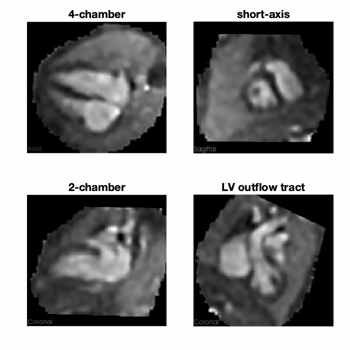
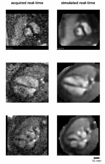
Three to five stacks of multi-slice real-time spiral SSFP were acquired in each subject (80–145 slices total). Cine volume reconstruction (Fig.1) was successful in 4 of 5 cases. Stacks were acquired within a window of 10 minutes in all successful cases, and 15 minutes in the unsuccessful case. No manual intervention of the 3D cine SVR process was attempted, e.g., forcing exclusion of motion-corrupted images or applying manual stack transformations to improve initial alignment.Fetal motion and cardiac cycle were effectively tracked from the input real-time images (Fig.2) and combined as a 3D cine that could be viewed in arbitrary planes, including views not available in the acquired real-time stacks (Fig.1). Mean displacement was 6.3–14.8 mm and outlier rejection excluded 9–19% of all real-time images used in SVR for each subject. The mean heart rate was 129–152 beats per minute.
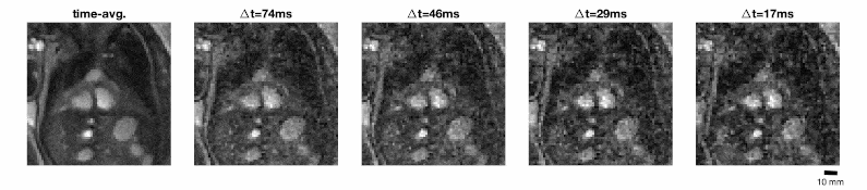
Real-time images could be reconstructed for a range of temporal resolutions (Fig.3). Image noise increased at higher temporal resolutions, but the underlying anatomical detail was not lost.
Discussion
These initial results demonstrate that 3D cine images of the fetal heart can be generated by SVR of real-time spiral images acquired on a high-performance 0.55 T scanner. Comfort and safety are improved with the use of a low-field scanner, while the motion-robust volume reconstruction allows for free-breathing acquisitions. Furthermore, the retrospective volume reconstruction does not require precise scan plane prescription during acquisition, and still produces a 3D cine that can be viewed in any slice.This work establishes the feasibility of 3D cine SVR with 0.55 T MRI. Further work that should improve the reconstructed volumes include better modeling of point spread functions of the acquisition process in the SVR algorithm and use of separate real-time image reconstructions for cardiac synchronization, motion-correction and volume reconstruction.
The high spatiotemporal resolutions that can be obtained with 0.55 T real-time spiral SSFP opens exciting possibilities for motion-tolerant SVR 3D cine imaging of the fetal heart and great vessels, particularly functional cardiac assessment and 4D flow quantification7,8.
Acknowledgements
Krishna Nayak and Chris Macgowan contributed equally as joint senior authors.The work described in this abstract was built on existing code repositories for spiral image reconstruction (https://github.com/usc-mrel/spiral_aliasing_reduction), the Slice-to-Volume Reconstruction Toolkit (https://github.com/SVRTK/SVRTK) and fetal whole-heart cine reconstruction using multiple real-time non-coplanar balanced SSFP stacks (https://github.com/mriphysics/fetal_cmr_4d).
This work was supported by a University of Southern California (USC) Provost’s Strategic Direction for Research Award and Keck School of Medicine of USC Dean’s Pilot Grant.
References
1. DFA Lloyd, K Pushparajah, JM Simpson, JFP van Amerom, MPM van Poppel, A Schulz, B Kainz, M Deprez, M Lohezic, J Allsop, S Mathur, H Bellsham-Revell, T Vigneswaran, M Charakida, O Miller, V Zidere, G Sharland, MA Rutherford, JV Hajnal, R Razavi. Three-dimensional visualisation of the fetal heart using prenatal MRI with motion-corrected slice-volume registration: a prospective, single-centre cohort study. Lancet. 2019;393(10181):1619–27. doi:10.1016/S0140-6736(18)32490-5
2. JFP van Amerom, DFA Lloyd, M Deprez, AN Price, SJ Malik, K Pushparajah, MP van Poppel, MA Rutherford, R Razavi, JV Hajnal. Fetal whole-heart 4D imaging using motion-corrected multi-planar real-time MRI. Magn Reson Med. 2019;82(3):1055–72. doi:10.1002/mrm.27798
3. KS Nayak, Y Lim, AE Campbell-Washburn, J Steeden. Real-Time Magnetic Resonance Imaging. J Magn Reson Imaging. 2020; doi:10.1002/JMRI.27411
4. AE Campbell-Washburn, H Xue, RJ Lederman, AZ Faranesh, MS Hansen. Real-time distortion correction of spiral and echo planar images using the gradient system impulse response function. Magn Reson Med. 2016;75(6):2278-85. doi:10.1002/mrm.25788
5. Y Tian, Y Lim, Z Zhao, D Byrd, S Narayanan, KS Nayak. Aliasing Artifact Reduction in Spiral Real-Time MRI. Magn Reson Med. 2021;86(2):916–25. doi:10.1002/mrm.28746
6. M Kuklisova-Murgasova, G Quaghebeur, MA Rutherford, JV Hajnal, JA Schnabel. Reconstruction of fetal brain MRI with intensity matching and complete outlier removal. Medical Image Analysis. 2012;16(8):1550–64. doi:10.1016/j.media.2012.07.004
7. TA Roberts, JFP van Amerom, A Uus, DFA Lloyd, MPM van Poppel, AN Price, JD Tournier, CA Mohanadass, LH Jackson, SJ Malik, K Pushparajah, MA Rutherford, R Rezavi, M Deprez, JV Hajnal. Fetal whole heart blood flow imaging using 4D cine MRI. Nat Commun. 2020;11(1):4992. doi:10.1038/s41467-020-18790-1
8. DS Goolaub, J Xu, EM Schrauben, D Marini, JC Kingdom, JG Sled, M Seed, CK Macgowan. Volumetric Fetal Flow Imaging with Magnetic Resonance Imaging. IEEE Trans Med Imaging. 2022;41(10)2941–52. doi:10.1109/TMI.2022.3176814
Figures