0545
REACT-QUANT: simultaneous non-contrast-enhanced subclavian MRA and vessel-wall imaging with multi-parametric quantitative mapping1Philips Japan, Tokyo, Japan, 2Department of Radiological Services, Tokyo Women's Medical University, Tokyo, Japan, 3Department of Diagnostic Imaging and Nuclear Medicine, Tokyo Women’s Medical University, Tokyo, Japan, 4Philips Healthcare, Hamburg, Germany, 5Philips Healthcare, Best, Netherlands
Synopsis
Keywords: Atherosclerosis, Atherosclerosis
Clinical imaging procedure for vascular evaluation and tissue characterization often needs separate MR scans. Both scenarios require multiple imaging sequences with different image contrasts, which results in long exam times and image misalignment due to possible motion between the scans that needs to be corrected in post-processing. In this work, the recently proposed REACT (Relaxation-Enhanced Angiography without ContrasT) technique was further developed and combined with multi-echo data acquisitions and Dixon-based chemical-shift-based water-fat separation. Initial results in patients showed great promise in detection and assessment of vascular and vessel wall lesions in one single scan.INTRODUCTION
Typical clinical imaging procedure for vascular evaluation including vessel wall lesions, such as atherosclerotic plaque imaging, needs separate MR scans1,2, it requires multiple imaging sequences with different image contrasts. This results in 1) long exam times, 2) image datasets misalignment due to possible motion between the scans that need to be corrected in post-processing. Furthermore, existing quantitative multi-parametric measurements do not provide angiographic information, in which case a complete diagnosis is compromised.REACT (Relaxation-Enhanced Angiography without ContrasT)3 with Multiple Delays (REACT-MD)4 simultaneously provides non-contrast-enhanced MR angiogram and MPRAGE (magnetization prepared rapid gradient echo) type images with uniform background tissue suppression over a large field of view (FOV) for the direct visualization of the plaques and vessel wall lesion in addition to luminal changes. Initial results in patients showed great promise in the detection of luminal changes and plaques for the assessment of systemic atherosclerosis in one single scan4.
In this study, we attempted to develop simultaneous non-contrast MRA, black-blood imaging and quantitative maps by extending the technique with combination of multi-echo DIXON chemical-shift-based water-fat separation5,6. The new REACT-Quant method was tested in volunteers for feasibility, compared with the original technique, and subsequently applied in patients for assessment of vascular lesions in the subclavian region.
METHODS
A schematic overview of the REACT-Quant sequence is shown in Figure 1. It basically consists of two signal readouts by 3D Dixon multi-echo turbo field-echo (TFE) preceded by a T2prep pulse and a non-selective IR pulse. Magnetization preparation pulses were implemented to suppress signal from tissues such as muscles, nerves and organs, exploiting their difference in relaxation times. In detail, immediately after the T2prep pulse, an IR pulse was applied with a short inversion time (TI), enhancing the blood-to-tissue contrast during the first signal readout for MRA. Thereafter, at the time of the blood null point, the second signal readout was carried out additionally to obtain the MPRAGE images. Consequently, REACT-Quant provides MRA (phase-1) and MPRAGE (phase-2) images in one single scan without prolonging the total acquisition time. In addition, multi-echo signal readout not only allows water-fat separation based on chemical-shift Dixon reconstruction, but also captures T2* signal delay. The latter helps to derive multiple quantitative parametric maps at the end of the same single scan, including fat fraction (FF) map, T2*/R2* maps and T1 map [Figure 2]. Since each TFE readout phase has multiple echoes, mDIXON reconstruction framework generates both bright-blood (phase-1) and black-blood (phase-2) fat fraction maps, T2* and R2* maps respectively, these might be helpful in assessment around and/or inside the blood vessels. Furthermore, a T1 map is also generated by using dual-phase readouts because these images have different TIs.As all datasets including multi-contrast images and quantitative maps are inherently spatially aligned from one single scan, no post-processing for misalignment is needed.
A total of 5 volunteers and 6 patients were examined on a 3.0T whole-body clinical system (Ingenia, Philips Healthcare, Best, the Netherlands). The study was approved by the local IRB, and written informed consent was obtained from all subjects.
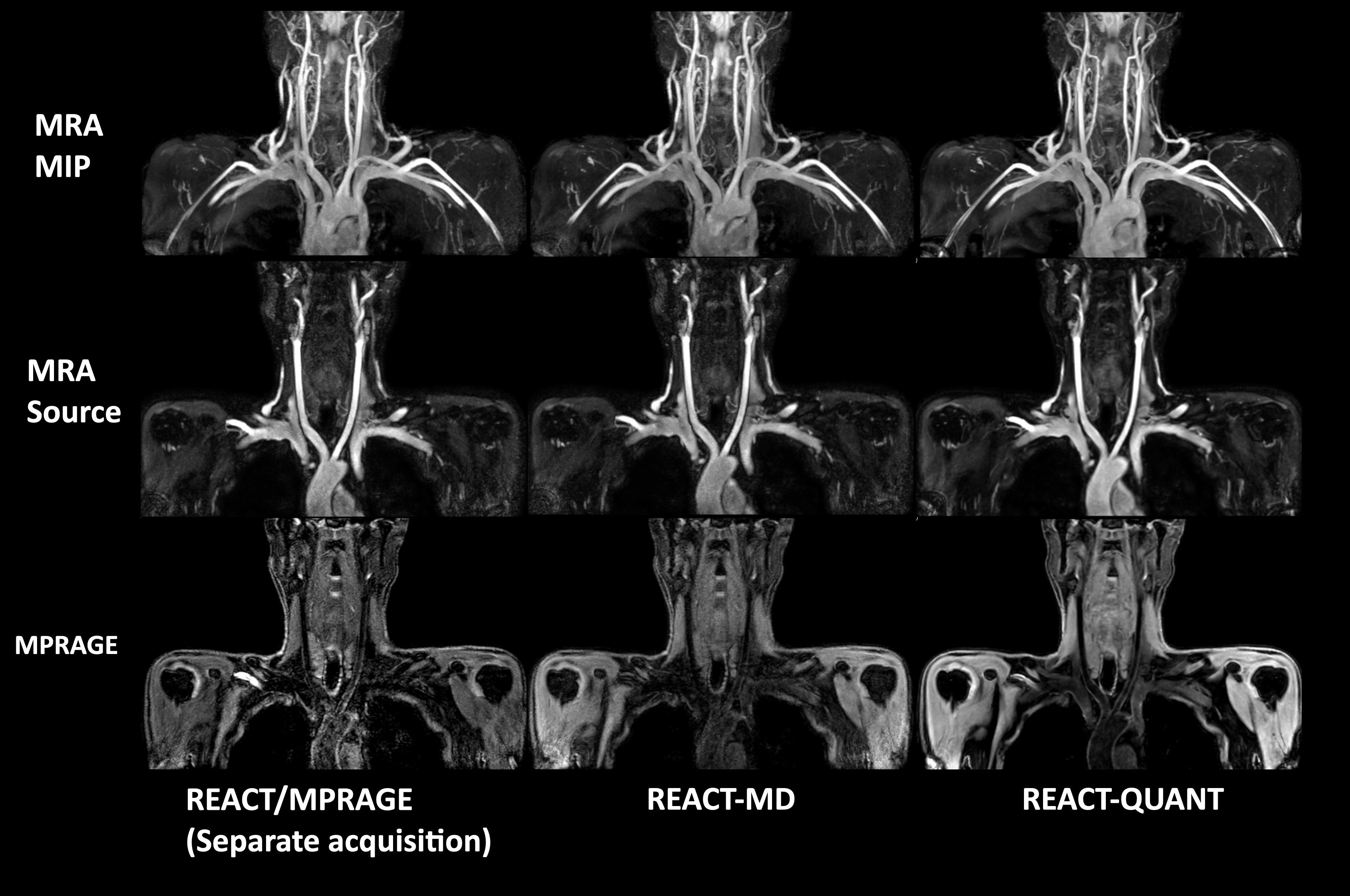
REACT-Quant images were compared with conventional REACT/MPRAGE and REACT-MD images for image quality, especially for the overall SNR and the presence of artifacts. Quantitative maps provided by REACT-Quant were also evaluated in the patients with atherosclerotic plaque.
Imaging parameters for REACT-Quant were; Coronal, voxel size=1.4x1.40 x3.0(1.5)mm3, TFE shot-interval=3000ms, TR=7.9ms, 6echoes, TE1/delta TE=1.3/1.0ms, flip angle=12°, NSA=1, TFE factor=40, Compressed SENSE reduction factor=3.5, and total acquisition time=4m30s.
RESULTS and DISCUSSION
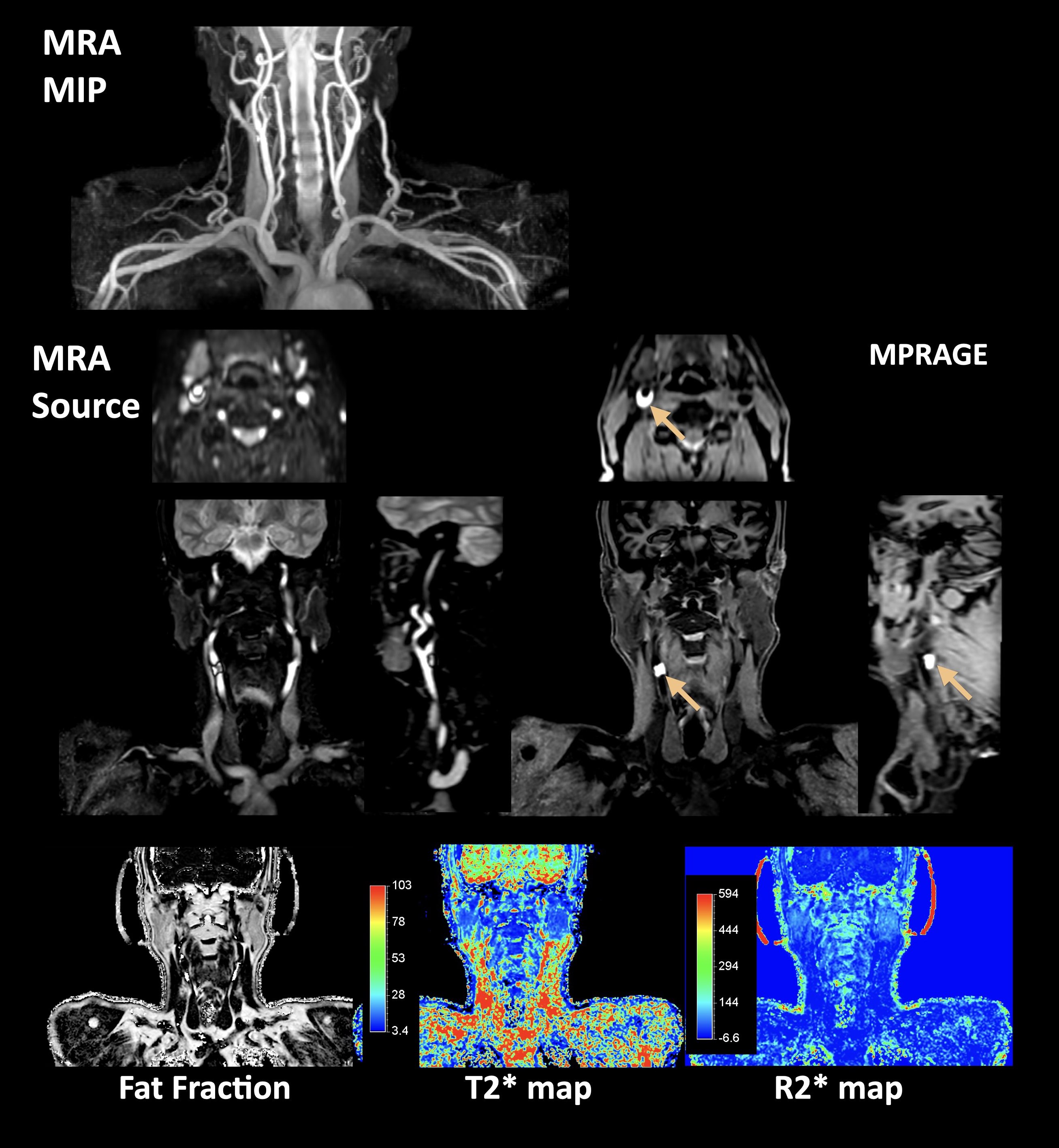
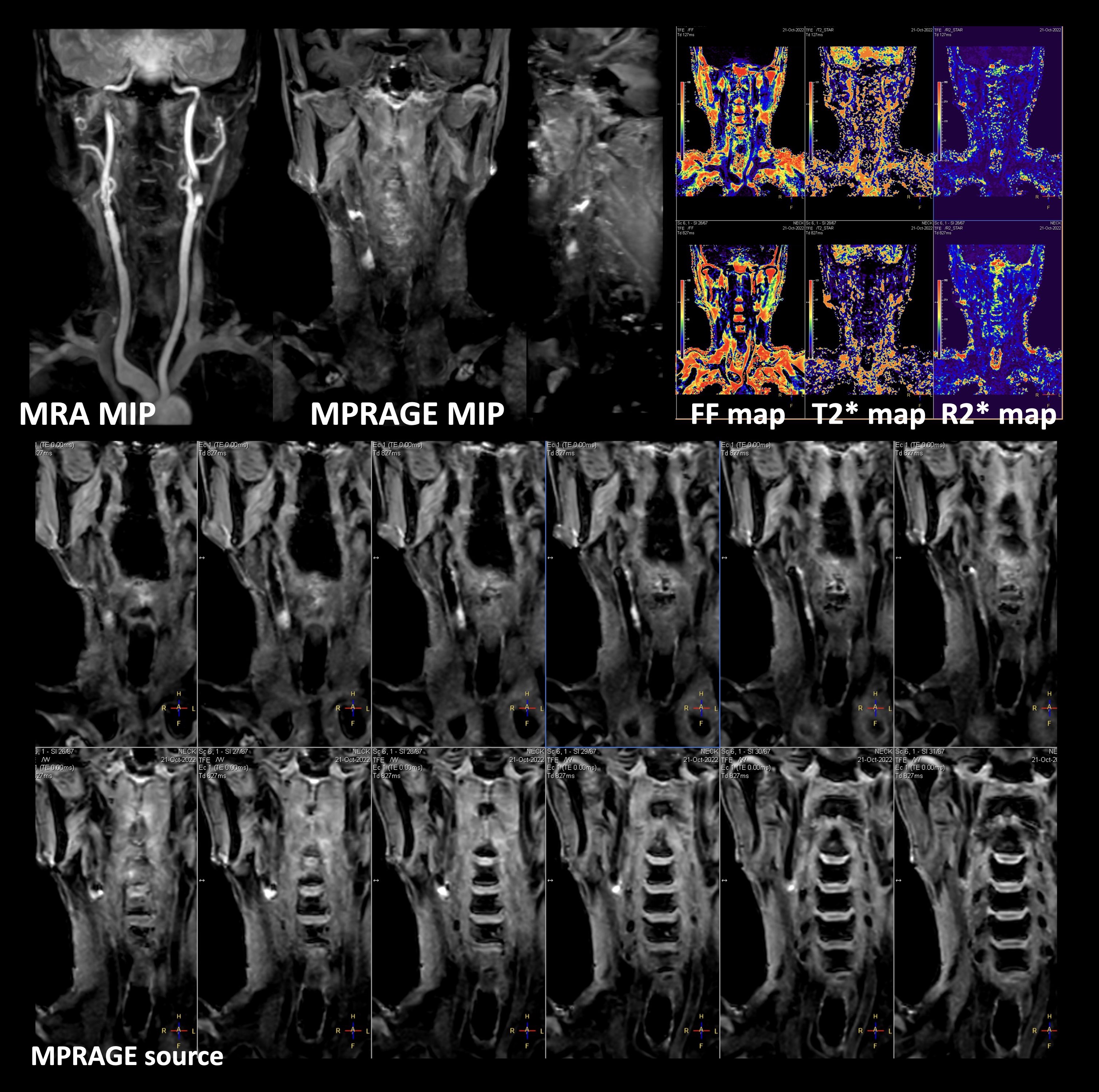
The quality of REACT-Quant MRA and MPRAGE images was considered comparable to that of the conventional REACT MRA/mDIXON-MPRAGE and REACT-MD, respectively. Representative images from all these techniques are shown in Figure 3. It is noteworthy that REACT-Quant provided REACT-MRA and REACT-MPRAGE images in one single scan and allowed for imaging over a large FOV at 3.0T with homogenous fat suppression without obvious artifacts.Two clinical cases with patients with right common carotid artery occlusion due to the presence of thrombus [Fig.4] and systemic atherosclerotic plaques [Fig.5] demonstrated that multiple plaques with IPH may be clearly visualized in REACT-Quant with complementary MRA on vascular morphology. Almost all plaques, which showed significant high signal intensity on REACT-MPRAGE, indicated low fat content on FF map and shorter T2* values (20.9±5.8ms) compared with that of the sternocleidomastoid muscle (39.0±6.4ms). Such quantitative information may present additional value to readily available multiple contrasts, though its clinical usefulness would need to be investigated more systematically.
CONCLUSION
REACT-Quant enables both morphological images, including non-contrast MR angiography and MPRAGE direct thrombus imaging, and quantitative mapping, including fat fraction, T2*/R2* maps and T1map simultaneously, with uniform fat suppression over a large FOV, in one single scan. It holds promise for assessment of systemic atherosclerosis, but further clinical studies in comparison to the other state-of-the-art methods7 are warranted.Acknowledgements
No acknowledgement found.References
1. Saba L, et al. Carotid artery wall imaging: perspective and guidelines from the ASNR vessel wall imaging study group and expert consensus recommendations of the American Society of Neuroradiology. AJNR Am J Neuroradiol. 2018;39(2):E9-E31.
2. Yuan C, et al. MRI of atherosclerosis. J Magn Reson Imaging. 2004;19(6):710-9.
3. Yoneyama M, et al. Free-breathing non-contrast-enhanced flow-independent MR angiography using magnetization-prepared 3D non-balanced dual-echo Dixon method: A feasibility study at 3 Tesla. Magn Reson Imaging. 2019:16;63:137-146.
4. Yoneyama M, et al. REACT-MD: simultaneous non-contrast-enhanced subclavian MRA and fat suppressed direct thrombus imaging with a large field-of-view, Proc Intl Soc Mag Reson Med. 2020;28:1320.
5. Pedrosa I, et al. mDIXON Quant non-invasively aids in high quality assessment of fatty liver disease. FieldStrength issue 2014;50(1):16-19.
6. Yokoo T, et al. Fat and Iron Quantification in the Liver Past, Present, and Future. Top Magn Reson Imaging 2014;23:73–94.
7. Wang J, et al. Simultaneous noncontrast angiography and intraplaque hemorrhage (SNAP) imaging for carotid atherosclerotic disease evaluation. Magn Reson Med 2013;69:337-345.
Figures
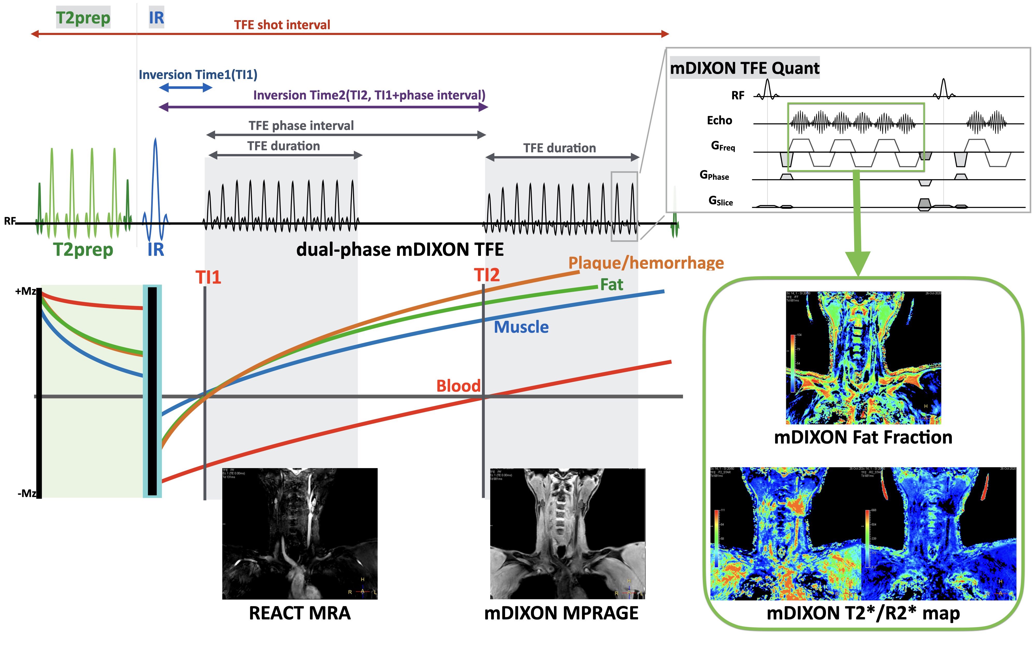
Figure 1. Scheme overview of REACT-Quant sequence.
REACT-Quant consisting of dual-phase 3D DIXON-TFE with T2prep and IR pulse. Immediately after the T2prep pulse, an IR pulse was applied with a short TI to suppress muscle and fat. Thereafter, an additional DIXON-TFE shot at the timing with the null point of the blood is also applied. REACT-Quant provides MRA (phase-1) and MPRAGE (phase-2) images in one single scan. Multi-echo signal readout is used to derive multiple quantitative parametric maps at the end of the same single scan, including fat fraction (FF) map, T2*/R2* maps.
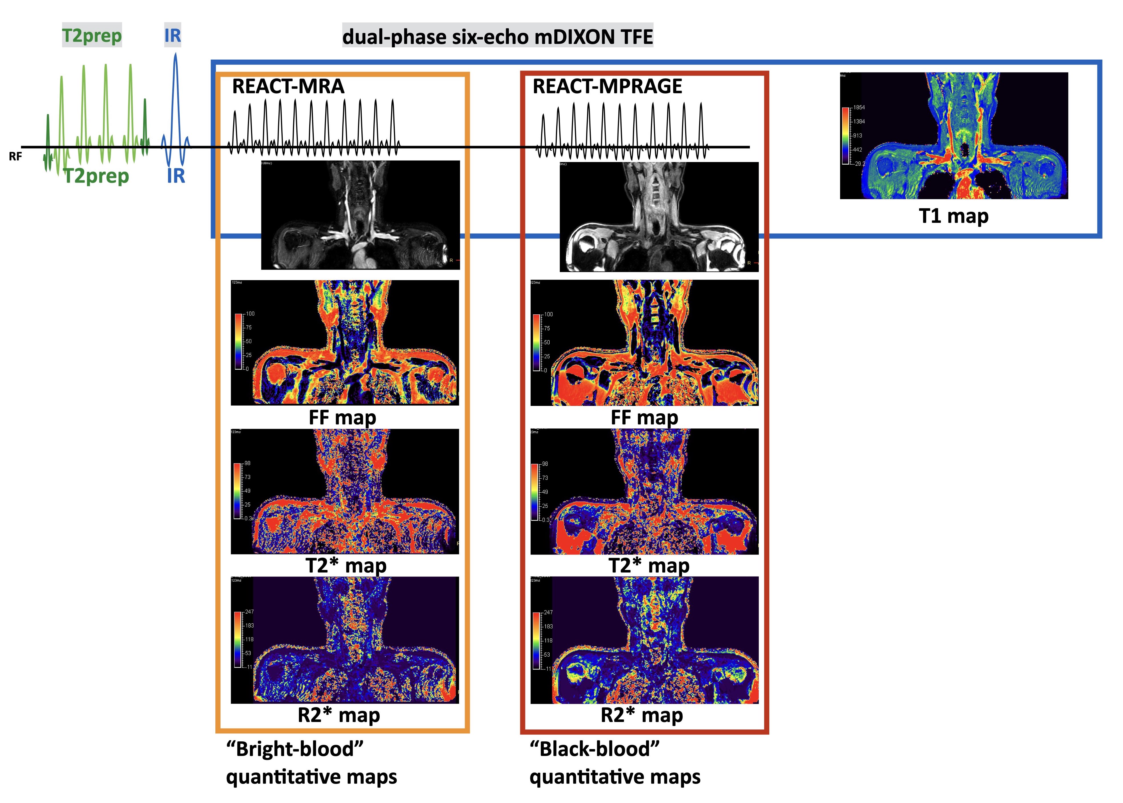
Figure 2. Quantitative maps delivered from RECAT-Quant dataset.
Since each mDIXON-TFE readout phase has multiple echoes, mDIXON reconstruction framework generates both bright (phase-1) and black-blood (phase-2) fat fraction maps, T2* and R2* maps respectively, these might be helpful in assessment around and/or inside the blood vessels. Furthermore, T1 map is also generated by using dual-phase readouts because these images have different TIs.