0505
Real-time MRI using deep learning in gastroesophageal reflux disease: a feasibility study1Radiology, Beichen Hospital, Tianjin, China, 2UIH America, Inc., Houston, TX, United States, 3United Imaging Intelligence, Cambridge, MA, United States, 4Radiology, Tianjin Medical University General Hospital, Tianjin, China
Synopsis
Keywords: Digestive, Digestive
A real-time MRI technique is developed using deep-learning reconstruction. Its feasibility is evaluated in both healthy subjects and patients with gastroesophageal reflux diseases.Introduction
Gastroesophageal reflux disease (GERD) is a common disease caused by motility disorder of the gastroesophageal junction, which regulates flow of food and fluid between the esophagus and the stomach. The medial lower esophageal sphincter (LES) and the lateral diaphragm, together with the gastric cardia ring and the chordae tendinous fibers, form a complete sphincter mechanism [1]. Among of them, relaxation of the LES is mostly relevant to reflux symptoms.Magnetic resonance imaging (MRI) has advantages in GERD evaluation due to its ability to monitor dynamic physiological processes non-invasively with high resolution and soft-tissue contrast. Prior researchers have developed a super-fast technique, so called real-time MRI, with a temporal resolution up to 20 ms per frame. It used motion-insensitive spatial encoding with a high degree of data under-sampling, combined with iterative, regularized nonlinear inversion image reconstruction [2,3]. This non-invasive approach has shown promises in cardiovascular imaging and imaging of more complex movements such as swallowing and speech [4,5].
Deep learning (DL) has gained momentum in MRI image reconstruction in recent years due to its faster reconstruction speed and potentially better signal-to-noise ratio than conventional methods [6]. Real-time MRI using DL has been demonstrated in cardiac cine imaging. For example, a cascade of 2D CNN model with data-sharing layers for dynamic image reconstruction was developed [7]. Chen et al. proposed a Res-RNN model and evaluated it in real-time cine reconstruction [8]. Despite these developments, there has been no attempt in imaging GERD using DL.
This study aims to show the feasibility of using DL-based real-time MRI to image the swallowing and reflux processes, in both healthy volunteers and GERD patients.
Methods
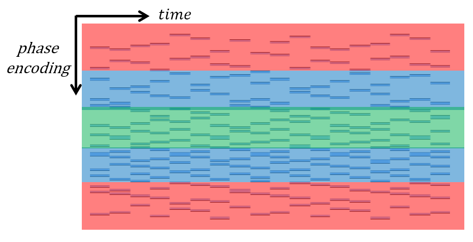
Sequence design: A 2D spoiled-GRE pulse sequence was developed for high spatial- and temporal- resolution imaging. The acquisition pattern features high under-sampling in the spatiotemporal domain by using Latin hypercube designs and echo-sharing. The k-space is roughly divided into five regions: a central region having 4-fold acceleration, two middle regions having 6-fold acceleration, and two outer regions having 14-fold acceleration [9]. Data sharing is used in the outer regions during reconstruction so the effective accelerates lowers to 7-fold.MRI: After localizer scans to identify anatomical landmarks, real-time imaging was performed using the above sequence, on a clinical 3.0T scanner (uMR 790, United Imaging, Shanghai, China). A 2D coronal slice covering the esophagogastric junction was used. 33 healthy volunteers and 7 GERD patients were recruited after written approval. The subjects were instructed to swallow pineapple juice during scan. Imaging parameters were: FOV = 300 mm × 300 mm, resolution = 1.56 mm × 1.56 mm, slice thickness = 8 mm, flip angle = 10 deg, TE/TR = 1.51/3.28 ms, bandwidth = 800 Hz/px. Each temporal frame contains 15 phase-encoding lines, resulting in a temporal resolution of 49.2 ms. A total of 400 temporal frames were scanned leading to a scan time of 19.7 s.
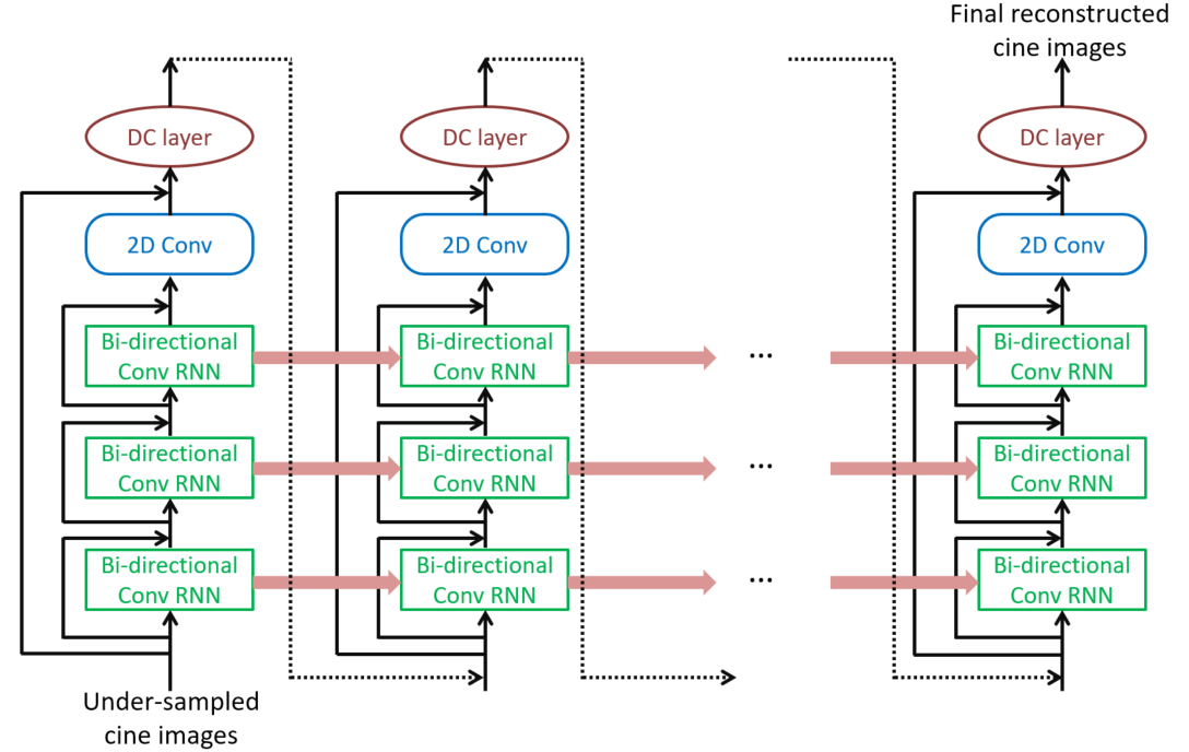
Reconstruction: DL reconstruction uses a Res-CRNN neural network [8], including 3 bi-directional ConvRNN layers, data consistency layers, and 2 levels of residual connections. Images of different coils are simultaneously reconstructed and then combined using sum-of-squares. The model was trained on simulated under-sampled data from 1,610 retrospectively gated, balanced-steady-state-free-procession cardiac cines from healthy volunteers and trained on a Nvidia Tesla V100 graphics processing unit (GPU). Mean square error (MSE) + 0.1x structural similarity (SSIM) as the training loss and a learning rate of 0.0001 along with the Adam optimizer for total 100 epochs were employed. The use of extra 2D Conv layers allows this neural network to learn high-frequency details while reducing memory consumption and accelerating reconstruction. Reconstruction was performed inline on the scanner and reconstruction time was < 3s in all cases.
Results
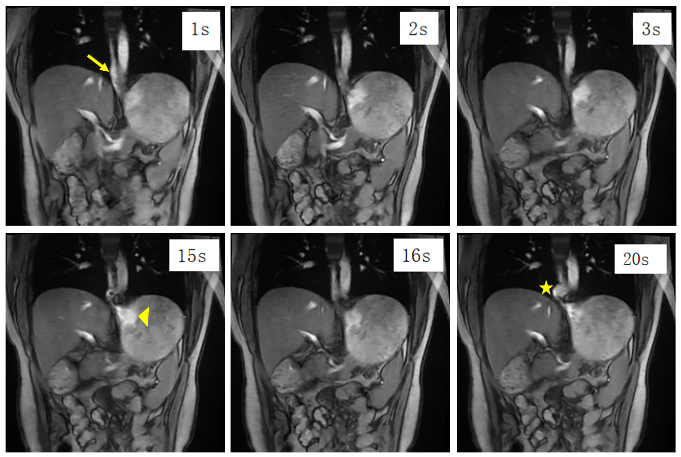
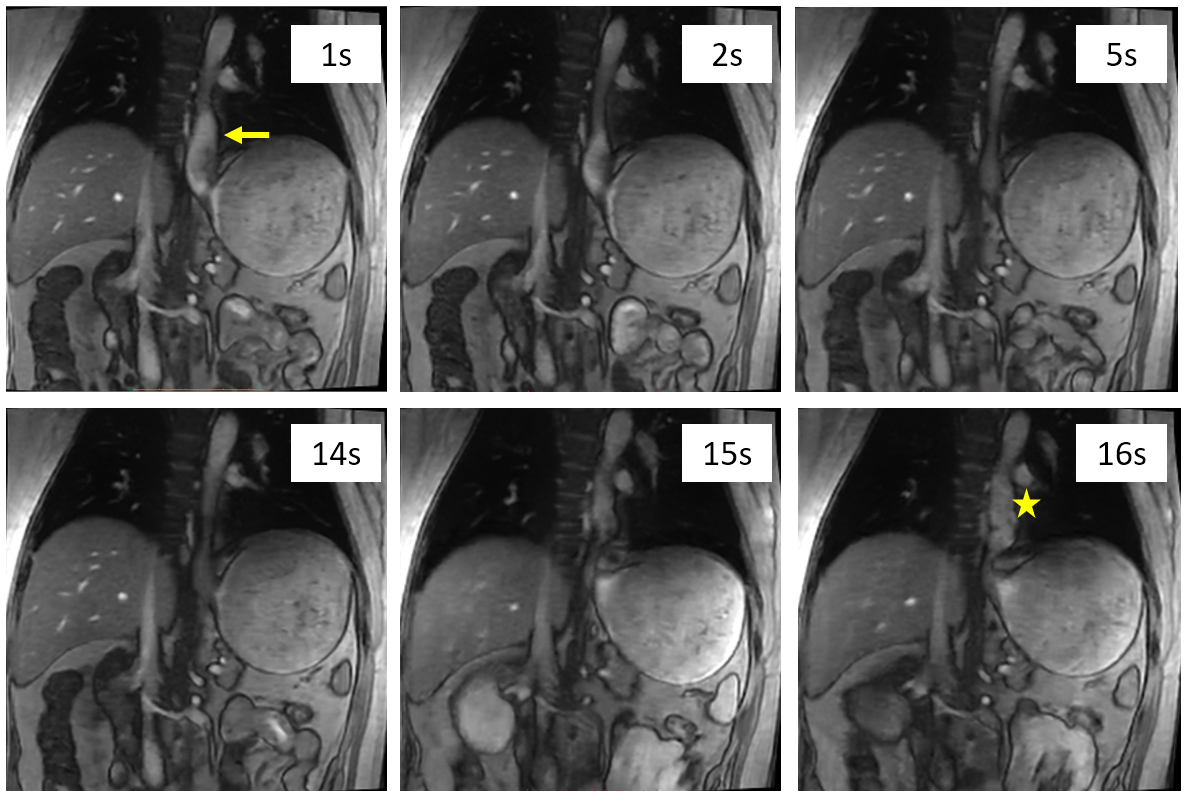
In healthy volunteers, normal swallowing dynamics were observed. Real-time MRI clearly visualized the lower esophageal sphincter transiently relaxed, with the pineapple juice appeared as bright fluid flowing from the lower esophageal to the cardia region of the stomach.In GERD patients, normal swallowing dynamics were first observed and then they were asked to perform the Valsalva maneuver to induce reflux. 4 out of the 7 patients demonstrated gastric juice reflux, with the lower esophageal sphincter kept relaxed and a small volume of the pineapple juice seen flowed back to the lower esophagus. Figures 3 & 4 show images from 2 typical patients.
Conclusion
Real-time visualization of swallowing and reflux process by DL-based MRI is feasible. It is a promising technique for clinical management in gastroesophageal reflux diseases. Compared to prior techniques, our results had higher spatial resolution and similar temporal resolution [2].Acknowledgements
No acknowledgement found.References
[1] Miller L, Vegesna A, Kalra A, et al. New observations on the gastroesophageal antireflux barrier[J]. Gastroenterol Clin North Am, 2007, 36 (3):601-617. DOI: 10.1016/j.gtc.2007.07.008.
[2] Zhang S, Joseph AA, Gross L, Ghadimi M, Frahm J, Beham AW. Diagnosis of Gastroesophageal Reflux Disease Using Real-time Magnetic Resonance Imaging. Sci Rep. 2015;5:12112.
[3] Uecker M, Zhang S, Voit D, et al. Real-time MRI at a resolution of 20 ms[J]. NMR Biomed, 2010, 23(8):986-994. DOI: 10.1002/nbm.1585.
[4] Zhang S, Joseph AA, Voit D, et al. Real-time MRI of cardiac function and flow-recent progress[J]. Quant imaging Med Surg, 2014, 4(5):313-329. DOI: 10.3978/j.issn.2223-4292.2014.06.03.
[5] Iltis PW, Frahm J, Voit D, et al. High-speed real-time magnetic resonance imaging of fast tongue movements in elite horn players. Quant imaging Med Surg, 2015, 5(3):374-381. DOI: 10.3978/j.issn.2223-4292.2015.03.02.
[6] Lin DJ, Johnson PM, Knoll F, Lui YW. Artificial Intelligence for MR Image Reconstruction: An Overview for Clinicians. J Magn Reson Imaging. 2021;53:1015-1028.
[7] Schlemper J, Caballero J, Hajnal JV, Price AN, Rueckert D. A deep cascade of convolutional neural networks for dynamic MR image reconstruction. IEEE Trans. Med. Imag. 2017;37:491–503.
[8] Chen EZ, Chen X, Lyu J, Zheng Y, Chen T, Xu J, Sun S. Real-time cardiac cine MRI with residual convolutional recurrent neural network. In: Proceedings of the 28th annual meeting of ISMRM; 2020. Abstract 3588.
[9] Lyu J, Ding Y, Zhong J, Zhang Z, Zhao L, Xu J, Liu Q, Peng R, Zhang W. Toward single breath-hold whole-heart coverage compressed sensing MRI using VAriable spatial-temporal LAtin hypercube and echo-Sharing (VALAS). In: Proceedings of the 27th annual meeting of ISMRM; 2019. Abstract 4752.
Figures