0486
Detecting the role of hippocampal subfields in pattern separation and completion task using 7T object memory task fMRI1Lou Ruvo Center for Brain Health, Cleveland Clinic, Las Vegas, NV, United States, 2Cleveland Clinic, Las Vegas, NV, United States, 3Cleveland Clinic, Cleveland, OH, United States
Synopsis
Keywords: Data Analysis, fMRI, Task fMRI; reigstration
In this study, we have established a reliable hippocampal subfield segmentation and activation analysis pipeline to probe the role of hippocampal subfields in pattern separation task by using 7T fMRI data, which could be able to identify the role of subfield dysfunction in cognitive impairment in prodromal AD.Introduction
Reduced episodic memory ability is a hallmark symptom of prodromal Alzheimer’s disease, which implicates the dysfunction in the medial temporal lobe. Hippocampal subfields respond differentially in processing episodic memories and their activation profiles differ in the pattern separation and completion memory tasks in cognitively normal subjects. The differentiated roles of subfields suggest that the cognitive impairment in AD may be driven by subfield-specific dysfunction. Identifying the relevance of hippocampal subfields in the pattern separation and completion memory tasks in subjects with stratified amyloid status before cognitive onset could provide valuable insight to the pathological mechanism in AD, which may be crucial for developing therapeutic target for treating or delaying clinical syndromes. Due to low signal-to-noise ratio, capturing activation in hippocampal subfield with 3 tesla MRI scanner is challenging. To address this issue, we collected object memory task fMRI data at 7 tesla MRI scanner among a group of cognitively normal participants with stratified amyloid status and comprehensive cognitive assessments.Methods
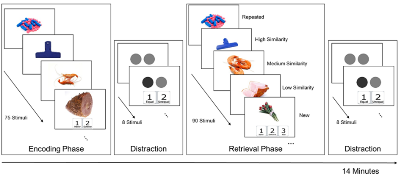
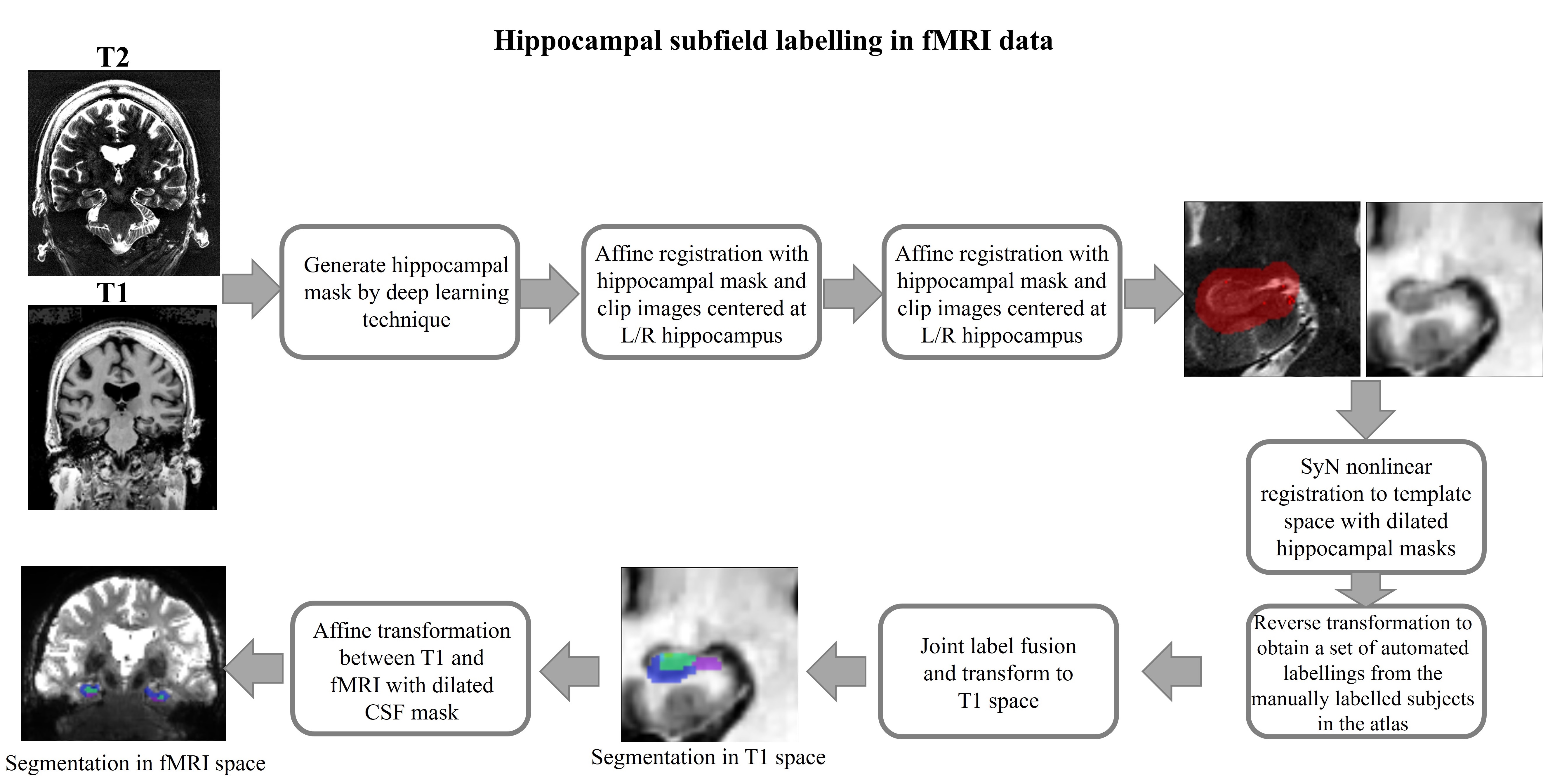
Fifteen normal subjects have been enrolled in the study with amyloid status determined from cerebrospinal fluid. The task fMRI data and structural MRI data were collected on a single 7T Siemens MRI scanner. The fMRI task examines pattern separation performance using similarities in the object as the lure type level measurements. A modified Mnemonic Similarity Task (MST) is used, which was originally designed by Stark et al. (2013) and Yassa & Stark (2011). Fig. 1 depicts the detailed fMRI task design. Three fMRI runs were collected during each scan session, and the object arrangement during the retrieval phases were optimized to have the maximum detection power for the pattern separation contrast (response “different” given lure stimuli minus response “different” given new stimuli). The whole brain three-dimensional T1-weighted images with isotropic voxel size 0.83mm were collected for all subjects. Thirteen subjects had high-resolution T2 structural MRI data collected along the long axis of the hippocampus with voxel size 0.44x0.44x1mm3. The automated hippocampal subfield segmentation pipeline was implemented by taking advantage of various machine learning and deep learning methods. The schematic diagram for the segmentation pipeline is shown in Fig. 2. Briefly, a deep-learning based approach was first used to obtain hippocampal mask for a new subject. Then nonlinear registration with dilated hippocampal mask using ANTS-SyN was carried out for aligning the new subject’s structural image(s) to the manually-labelled individuals in the atlas, achieving a set of automated subfield labelling. Finally, a joint label fusion technique was applied to obtain the final automated labeling for the subject. For the subjects having T1 and T2 structural images, both modalities were used for automated segmentation. In the standard affine registration between fMRI and structural MRI images, the voxels in fMRI data could be a few slices away from its actual anatomical location after the transformation, which is problematic for analyzing the activation of fine-structure region such as hippocampus, not to mention its subfields. To address the issue, we took advantage of the evident contrast between cerebrospinal fluid (CSF) voxels and non-CSF voxels in both fMRI and structural MRI data and conducted the affine transformation with a dilated CSF mask, which substantially improved the registration quality.Results
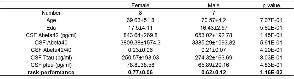
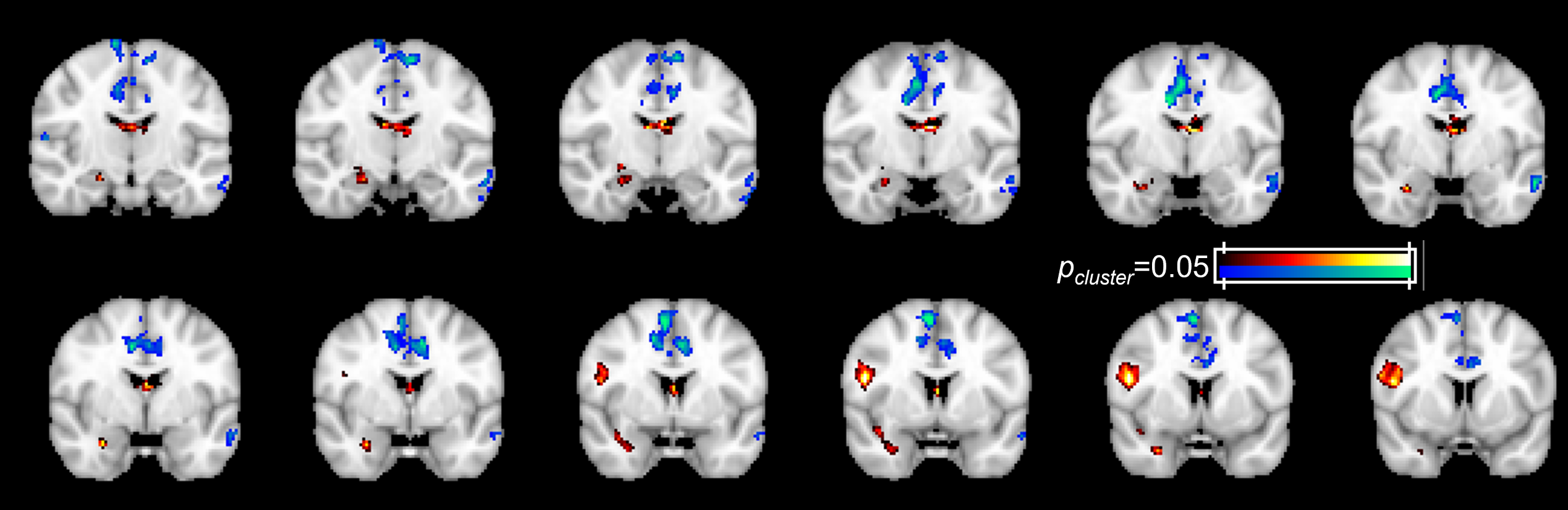
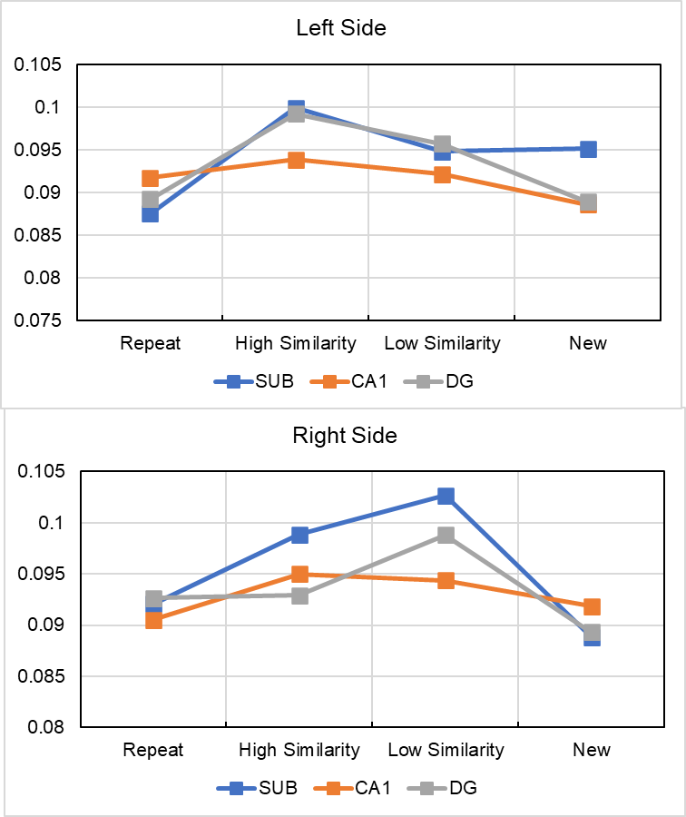
Table 1 shows the demographic information for the 15 enrolled normal subjects. As listed in Table 1, women performed significantly better than men during the object lure fMRI tasks. Fig. 3 is the group activation map for the pattern separation contrast. As shown in Fig. 3, hippocampal activation were detected during this contrast. Fig. 4 plots the beta value for each stimuli type in the fMRI analysis, and as shown in Fig. 4, bilateral subiculum and left dentate gyrus were activated in recognizing subtle differences in the stimuli.Discussion
In this study, we have established a reliable automated pipeline to categorize voxels in fMRI data to various hippocampal subfields and demonstrated the feasibility of using task fMRI data at 7 tesla MRI scanner to probe hippocampal subfield activation, which could potentially be used to detect the alteration of hippocampal subfield activation among the patients with prodromal AD.Acknowledgements
This study was funded by NIH-RF1AG071566.References
Reagh, Z. M., Noche, J. A., Tustison, N. J., Delisle, D., Murray, E. A., & Yassa, M. A. (2018). Functional Imbalance of Anterolateral Entorhinal Cortex and Hippocampal Dentate/CA3 Underlies Age-Related Object Pattern Separation Deficits. Neuron, 97(5), 1187-1198.e4. https://doi.org/10.1016/j.neuron.2018.01.039
Reagh, Z. M., & Yassa, M. A. (2014). Object and spatial mnemonic interference differentially engage lateral and medial entorhinal cortex in humans. Proceedings of the National Academy of Sciences, 111(40), E4264–E4273. https://doi.org/10.1073/pnas.1411250111
Stark, S. M., Yassa, M. A., Lacy, J. W., & Stark, C. E. L. (2013). A task to assess behavioral pattern separation (BPS) in humans: Data from healthy aging and mild cognitive impairment. Neuropsychologia, 51(12), 2442–2449. https://doi.org/10.1016/j.neuropsychologia.2012.12.014
Figures