0465
Distortion-corrected cervical spine diffusion-weighted imaging at 3.0T1Shanghai Sixth People's Hospital, Shanghai, China, 2Philips Healthcare, Shanghai, China, 3Philips Healthcare, Guangzhou, China
Synopsis
Keywords: Head & Neck/ENT, Spinal Cord
Diffusion-weighted imaging (DWI) could benefit the detection and evaluation of the spine and spinal cord pathologies. Traditional single-shot EPI (SS-EPI) DWI suffers from a large geometry distortion due to the inhomogeneous B0 field and the low bandwidth in the phase encoding direction. This work demonstrated the application of an EPI geometry correction method for the cervical spine which can improve the geometry accuracy of the DWI images.
Introduction
Diffusion-weighted imaging (DWI) can reveal the underlying random Brownian motion of the water molecules, which could be altered in different pathologies. Traditional DWI based on echo planner imaging (EPI-DWI) may suffer from geometry distortion, this is even worse for the cervical spine since the field is extremely inhomogeneous.Several acquisition methods have been proposed to shorten the EPI echo train and overcome this issue. Such as parallel imaging (PI)1, multi-shot EPI (MS-EPI)2,3, reduced field-of-view (rFOV)4, and turbo spine echo DWI (TSE-DWI)5, etc. PI and rFOV can reduce the distortion for cervical spine DWI, but the improvement is limited. MS-EPI could decrease the distortion, at the cost of an increased scan time. TSE-DWI could provide distortion-free images, but should compromise on scan speed, image sharpness, and signal-to-noise ratio (SNR)6.
Besides the above-mentioned acquisition methods, two kinds of distortion correction methods, namely field-mapping7 and the top-up8 methods can also be used here. The field-mapping method relies on an accurate calculation of the field map and may be prone to unwrapping errors. The top-up method can correct the signal pile-up, but the result is also dependent on the estimated displacement map.
Here we demonstrated the utilization of a combination of the above-mentioned two distortion correction methods on the cervical spine at a 3T system.
Methods
The scan was conducted on a Philips 3.0T scanner (Ingenia Elition, Philips Healthcare, Best, The Netherlands) with a healthy volunteer. Informed consent was obtained from the volunteer.For the distortion-corrected SS-EPI DWI scan, one B0 prescan (scan duration: 10 seconds) was inserted automatically to generate the field inhomogeneity based on the Dixon method. For each b0 DWI scan, an extra blip-down scan (traversing the kspace in an opposite way compared with the conventional scan) was acquired, like the top-up method. The calculated B0 map, conventional b0 images, and blip-down b0 images were used to calculate the distortion and intensity correction maps, which were used to correct b0 and high-b images later (Figure 1). Imaging parameters could be found in Table 1. It should be noticed that if no blip-down images were acquired, the total scan duration was 1 minute 48 seconds.
For comparison purpose, T2w SPectral Attenuated Inversion Recovery (SPAIR) images, TSE-DWI, MS-EPI DWI (combined with rFOV) are also acquired. Imaging parameters were shown in Table 1.
Results
Figure 2 shows the comparison between the results of the above-mentioned sequences. After distortion correction, SSH-EPI DWI (Figure 2B) has nearly no distortion, and has a better image quality (higher SNR, sharper boundary) compared with TSE DWI (Figure 2E). Although MSH-EPI DWI (Figure 2D) can reduce the distortion, the improvement is limited.Discussion
We demonstrated the feasibility of distortion-corrected SSH-EPI DWI for the cervical spine. It can improve the geometry accuracy of the traditional SSH-EPI DWI and have a comparable distortion level with TSE DWI. And the SNR and image sharpness was better than TSE DWI.Since an extra blip-down b0 image should be acquired for each slice, the total scan duration could prolong a little bit (12 seconds in this case). But this time increment was negligible compared with the total scan time (2 minutes in this case). What’s more, the extra B0 prescan, which cost 10 seconds, could be reused by different scans with the same scan location, therefore it could be acquired only once for all the sequences.
For MSH-EPI DWI, the distortion level could be further reduced by increasing the half-scan factor or increasing the shot number, at the cost of a decreased SNR and an increased scan time. But again, the distortion could never be eliminated due to the intrinsic characteristic of the EPI acquisition.
Conclusion
Distortion-corrected SSH-EPI DWI could be used for the cervical spine for improved geometry accuracy.Acknowledgements
No acknowledgement found.References
[1] Cercignani M, Horsfield MA, Agosta F, Filippi M. Sensitivity-encoded diffusion tensor MR imaging of the cervical cord. AJNR Am J Neuroradiol. 2003 Jun-Jul;24(6):1254-6.
[2] Jeong HK, Gore JC, Anderson AW. High-resolution human diffusion tensor imaging using 2-D navigated multishot SENSE EPI at 7 T. Magn Reson Med. 2013 Mar 1;69(3):793-802.
[3] Holdsworth SJ, Skare S, Newbould RD, Guzmann R, Blevins NH, Bammer R. Readout-segmented EPI for rapid high resolution diffusion imaging at 3 T. Eur J Radiol. 2008 Jan;65(1):36-46.
[4] Andre JB, Zaharchuk G, Saritas E, Komakula S, Shankaranarayan A, Banerjee S, Rosenberg J, Nishimura DG, Fischbein NJ. Clinical evaluation of reduced field-of-view diffusion-weighted imaging of the cervical and thoracic spine and spinal cord. AJNR Am J Neuroradiol. 2012 Nov;33(10):1860-6.
[5] Tsuchiya K, Katase S, Fujikawa A, Hachiya J, Kanazawa H, Yodo K. Diffusion-weighted MRI of the cervical spinal cord using a single-shot fast spin-echo technique: findings in normal subjects and in myelomalacia. Neuroradiology. 2003 Feb;45(2):90-4.
[6] Yoshida T, Urikura A, Shirata K, Nakaya Y, Terashima S, Hosokawa Y. Image quality assessment of single-shot turbo spin echo diffusion-weighted imaging with parallel imaging technique: a phantom study. Br J Radiol. 2016 Sep;89(1065):20160512.
[7] Jezzard P, Balaban RS. Correction for geometric distortion in echo planar images from B0 field variations. Magn Reson Med. 1995 Jul;34(1):65-73.
[8] Chang H, Fitzpatrick JM. A technique for accurate magnetic resonance imaging in the presence of field inhomogeneities. IEEE Trans Med Imaging. 1992;11(3):319-29.
Figures
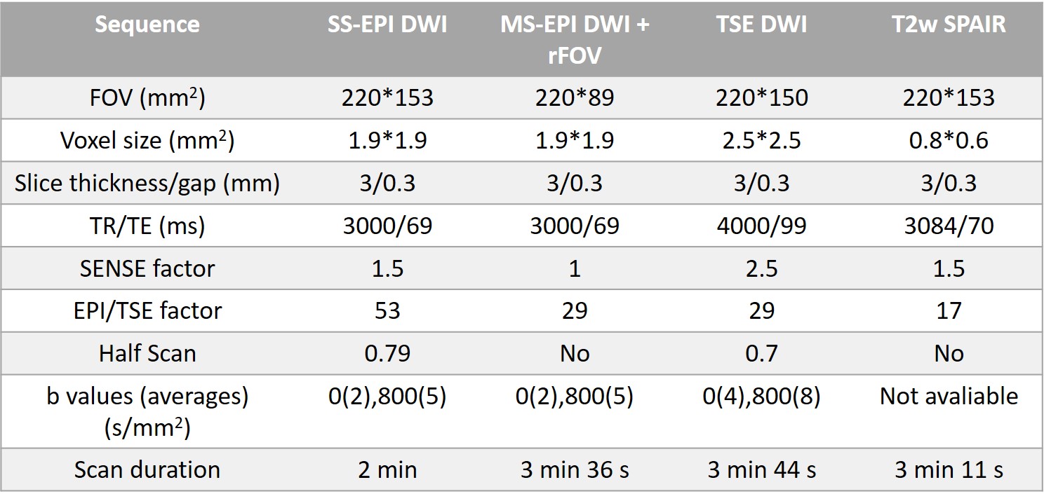
Table 1. Imaging parameters for SS-EPI DWI, MS-EPI DWI, TSE DWI and T2w SPAIR.
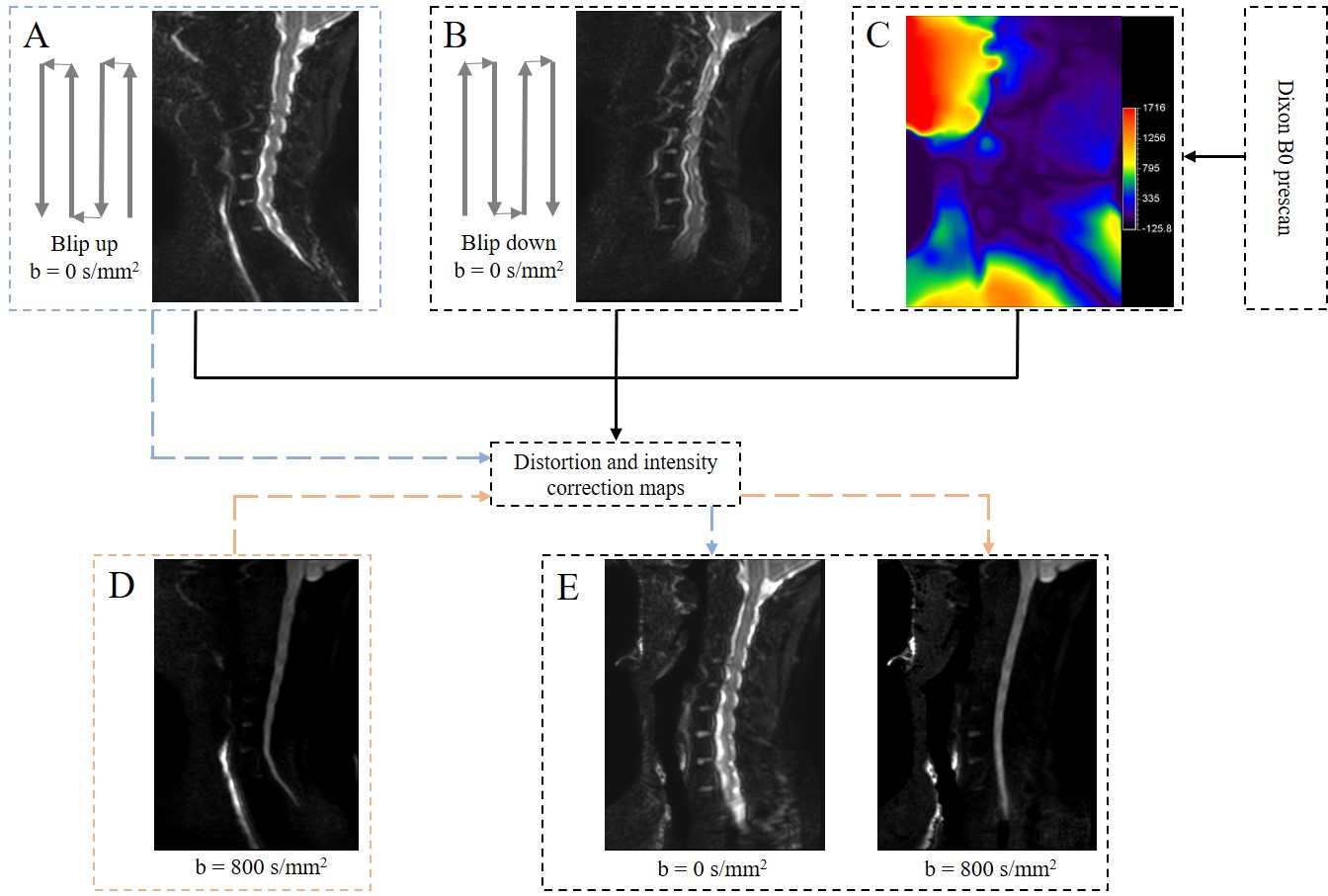
Figure 1. Flow chart of the distortion correction. One Dixon B0 prescan was inserted automatically to generate a (C) B0 map. For each slice, an extra blip-down b0 scan (B) was added. The B0 map, blip-down b0 scan, together with the original blip-up b0 scan (A), were used to estimate distortion and intensity correction maps. Finally, original distorted b0 (A) and high b-value images (D) were corrected with the distortion and intensity correction maps respectively to generate distortion-corrected images (E).
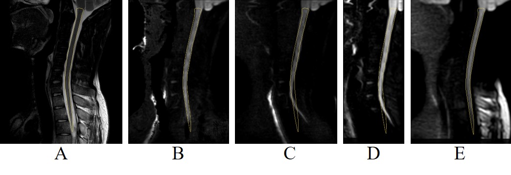
Figure 2. Comparison between different acquisition sequences. (A) T2w SPAIR. (B) SSH-EPI DWI with distortion correction. (C) SSH-EPI DWI without distortion correction. (D) MSH-EPI with rFOV acquisition. (E) TSE DWI. The spinal cord region is extracted from the T2w SPAIR image and copied to all the other images (marked with yellow polygons).