0461
Influence of White Matter Microstructure on Interhemispheric Processing Speed After Mild Traumatic Brain Injury Using Advanced Diffusion MRI1Center for Advanced Imaging Innovation and Research (CAI2R), Department of Radiology, New York University Grossman School of Medicine, New York, NY, United States, 2Bernard and Irene Schwartz Center for Biomedical Imaging, Department of Radiology, New York University Grossman School of Medicine, New York, NY, United States, 3Department of Neurology, New York University Grossman School of Medicine, New York, NY, United States, 4Department of Rehabilitation Medicine, New York University Grossman School of Medicine, New York, NY, United States
Synopsis
Keywords: Traumatic brain injury, Diffusion/other diffusion imaging techniques
The corpus callosum (CC) is especially vulnerable to mild traumatic brain injury (MTBI). Since it connects left and right cerebral hemispheres, damage to the CC or neighbor white matter (WM) pathways may specifically disrupt interhemispheric communication. Here we employ a mediation framework to study the collaboration between tissue microstructure of the CC and neighbor WM pathways that may influence interhemispheric processing. Our results show different patterns in individuals with recent MTBI compared to healthy controls in terms of the relationships between callosal microstructure and interhemispheric communication mediated by other pathways, highlighting tracks specifically related to primary visual and language processes.
Introduction
Mild traumatic brain injury (MTBI) is a common injury with potentially serious clinical sequelae. Primary stretch and torsion injuries of white matter (WM) following MTBI have been previously shown and the corpus callosum (CC) is known to be particularly at risk.1-4 The CC is a critical pathway for interhemispheric communication and information transfer5 and also connects a variety of WM bundles across the brain in complex ways. Here, we use an adapted form of a visual stimulus-response test, termed the Interhemispheric Speed of Processing Task (IHSPT),6 to assess interhemispheric processing speed in MTBI patients. IHSPT is based on measuring the latency between words presented to the right (RVF) and left visual fields (LVF). Subject’s reaction time between visual presentation and articulation of words is expected to be longer when presented to the LVF (crossed reaction time) compared with the RVF (uncrossed reaction time) assuming left hemisphere language dominance. Words presented to the RVF project directly to the left cerebral hemisphere where language functions are lateralized in such individuals, while words presented to the LVF project to the posterior right hemisphere and must then cross the corpus callosum to reach Broca’s area, the center for expressive language (Fig.1). Here we study the indirect effects of callosal microstructure on interhemispheric processing by passing through neighbor WM pathways using mediation analysis.7 WM microstructure was assessed by using DTI, DKI, and the standard model (SM) of diffusion in WM.8 In particular, the SM is a unifying framework of WM diffusion models and provides biophysically meaningful compartment-specific measures such as axonal water fraction (ƒ), intra-axonal diffusivity (Daxon), extra-axonal axial (De||) and radial (Deㅗ) diffusivities.Methods
We studied 24 MTBI patients (35±14 years old) within a month of injury and 19 normal controls (NC) (35±14 years old). MRI was performed on 3T MR scanners (Skyra/Prisma, Siemens). Multi-shell diffusion imaging was performed with 5 b-values (0.25, 1, 1.5, 2, 2.5 ms/µm2) along with total 137 diffusion-encoding-directions using multiband (factor=2) (FOV=220×220mm2, 2.5mm-isotropic resolution, slices=56, TR/TE=4.9s/95ms, GRAPPA factor=2). We calculated 11 diffusion maps of DTI (fractional anisotropy [FA], mean/axial/radial diffusivity [MD/AD/RD]), DKI (mean/axial/radial kurtosis [MK/AK/RK]), and SM metrics (ƒ, Daxon, De||, Deㅗ)9. 23 major cerebral WM ROIs were identified,7 including corpus callosum (genu/body/splenium), right and left anterior/posterior/ retrolenticular limb of internal capsule (aIC/pIC/rIC), anterior/superior/posterior corona radiata (aCR/sCR/pCR), posterior thalamic radiation (pTR), external capsule (EC), superior longitudinal fasciculus (SLF), superior fronto-occipital fasciculus (SFOF), respectively. Inter-scanner harmonization was performed using ComBat.10 For each ROI, mean value of each diffusion metric was calculated only in voxels with FA ≥ 0.3 to restrict the analysis to WM. The IHSPT test: Each trial consisted of a three-letter word randomly presented for 150ms to either the LVF or RVF (Fig.1), and subjects were required to speak each word as quickly as possible. A total of 80 trials were assigned for each subject and reaction time (RT) was recorded for each trial. The IHSPT score (%) was calculated by the absolute difference between median RTs for LVF and RVF word presentations, divided by the average RT between them, and then multiplied by 100 to obtain a percentage score. Mediation analysis was conducted by using PROCESS in the SPSS framework with 5000 bootstrap resamples adjusted for age and sex (Fig.2).Results
Several WM pathways were found to be significant mediators of associations between callosal microstructure and IHSPT performance, showing different patterns in MTBI compared to NC (Fig.3). The MTBI group demonstrated significant mediators mostly in the anterior and posterior portion of the brain (aCRL, sCRR, SLFR, SLFL, SFOFR), whereas in normal controls, significant mediators were present mainly in the anterior and middle portion of the brain (aICL, aICR, pICR, aCRL, sCRL, pCRL, ECR, ECL, SFOFL). Specifically, dominant diffusion metrics having significant mediators were Daxon in the capsular areas and RD in the SLF.Discussion
Our study shows different patterns of significant WM pathways that mediate the relationship between callosal microstructure and IHSPT performance in MTBI compared to normal controls. Dissecting brain mechanisms of the effect of callosal microstructure on the interhemispheric processing through neighbor WM pathways is important for understanding the underlying mechanisms of interhemispheric processing particularly associated with language and visual pathways. Our findings show significant mediators mainly in capsular WM in NC, whereas the MTBI group demonstrates no such relationship. Poorer performance on IHSPT is mediated by lower Daxon in the capsular WM which has been previously reported in conditions of intra-axonal injury1. In MTBI, significant mediators are present mainly in the SLF which is critical for attention, memory, emotion, and language, showing poorer performance on IHSPT with lower RD which has been previously shown with the presence of cytotoxic edema which is often observed in contusive or relative severe injury in the acute stage.11Conclusion
This work identifies novel patterns of altered relationships between callosal microstructure and interhemispheric communication in MTBI, highlighting that not only the corpus callosum but the indirect mediation through neighbor WM pathways is important. These patterns may shed new and needed light on the relationship between WM microstructural injury and altered functional organization in MTBI.Acknowledgements
This work was supported in part by grant funding from the National Institute of Health (NIH): R01 NS119767-01A1, R01 NS039135-11, and R56 NS119767. This work was also performed under the rubric of the Center for Advanced Imaging Innovation and Research (CAI2R), a NIBIB Biomedical Technology Resource Center (NIH P41 EB017183).
References
1. Chung S, et al. White Matter Tract Integrity: an indicator of axonal pathology after mild traumatic brain injury. J Neurotrauma 35:1015-1020, 2018.
2. Chung S, et al. Investigating brain white matter in football players with and without concussion using a biophysical model from multishell diffusion MRI. AJNR 43:823-828, 2022.
3. McAllister TW, et al. Maximum principal strain and strain rate associated with concussion diagnosis correlates with changes in corpus callosum white matter indices. Ann Biomed Eng 40:127-140, 2012.
4. Quigley M, et al. Role of the corpus callosum in functional connectivity. AJNR 24:208-212, 2003.
5. Bloom JS and Hynd GW. The role of the corpus callosum in interhemispheric transfer of information: excitation or inhibition? Neuropsychol Rev 15:59-71, 2005.
6. Bacon JH, et al. Posterior corpus callosum atrophy is correlated with worse performance on inter-hemispheric speed of processing task in unimpaired MS patients. AAN 2015 Annual Meeting, Washington DC, 2015.
7. Baron RM, et al. The moderator-mediator variable distinction in social psychological research: conceptual, strategic, and statistical considerations. J Pers Soc Psychol 51:1173-1182, 1986.
8. Novikov D, et al. Quantifying brain microstructure with diffusion MRI: Theory and parameter estimation. NMR Biomed 32:e3998, 2019.
9. Coelho S, et al. Reproducibility of the Standard Model of diffusion in white matter on clinical MRI systems. Neuroimage 257:119290, 2022.
10. Fortin J., et al. Harmonization of multi-site diffusion tensor imaging data. Neuroimage 161:149-170, 2017.
11. Marmarou A, et al. Brain Edema. XIII. Vienna: Springer; Traumatic brain edema in diffuse and focal injury: cellular or vasogenic? Pp 24-29, 2006.
Figures
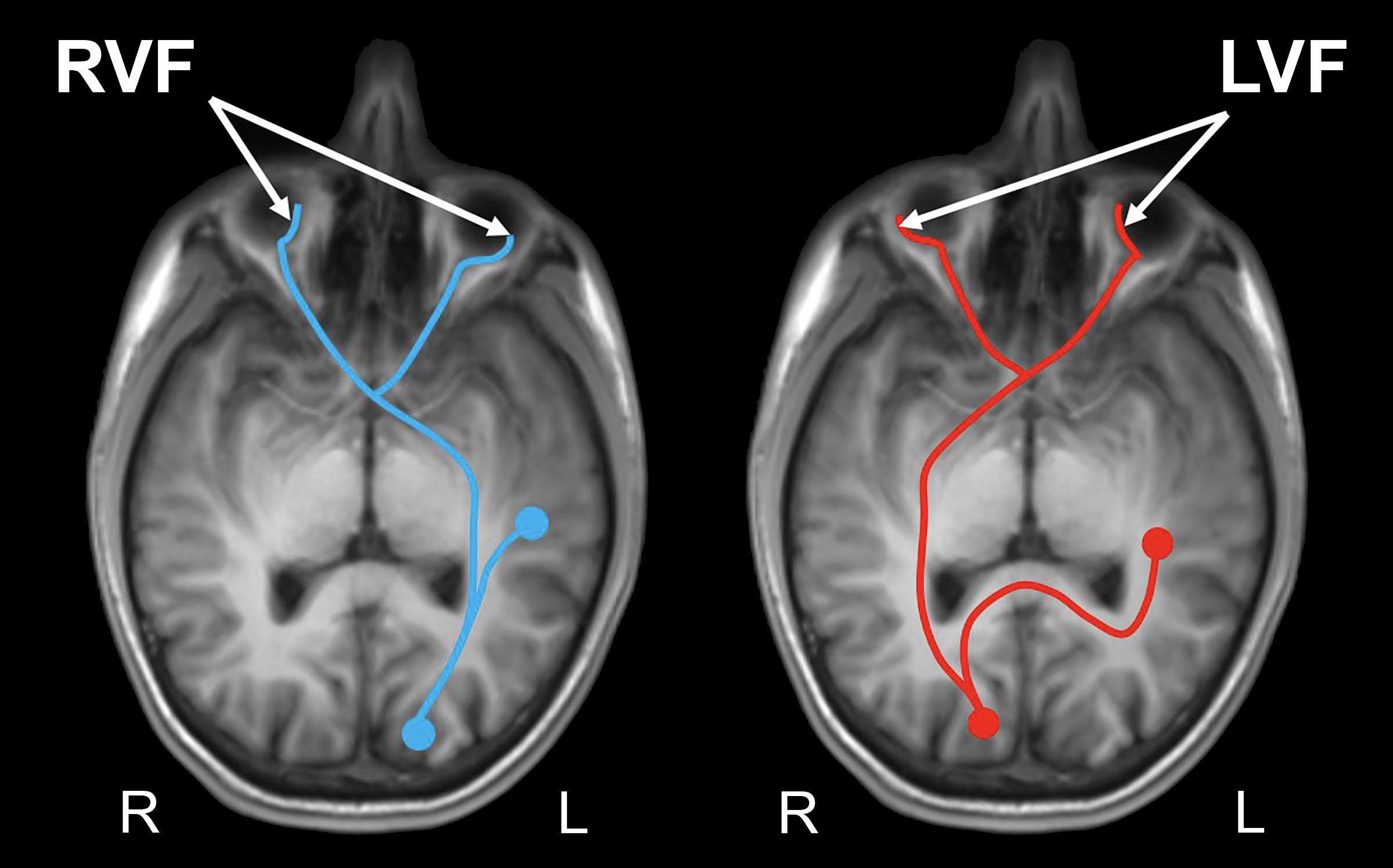
Figure 1. Interhemispheric Speed of Processing Task probes hemispheric and callosal microstructure by measuring processing speed and latency in articulation of words presented to the left and right visual fields (LVF and RVF) in individuals with left hemisphere language dominance. Visual information from LVF projects to contralateral (right) primary visual cortex, then should cross the midline to access core language centers (Red), whereas information presented to RVF projects to left primary visual cortex with access to primary language centers in the same hemisphere (Blue).
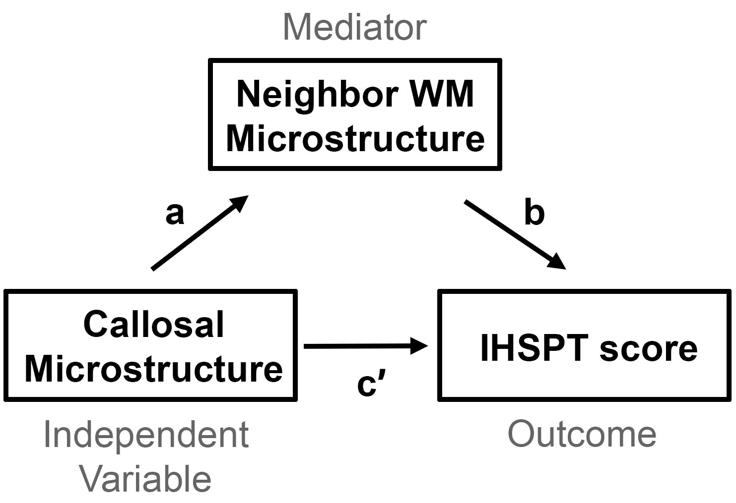
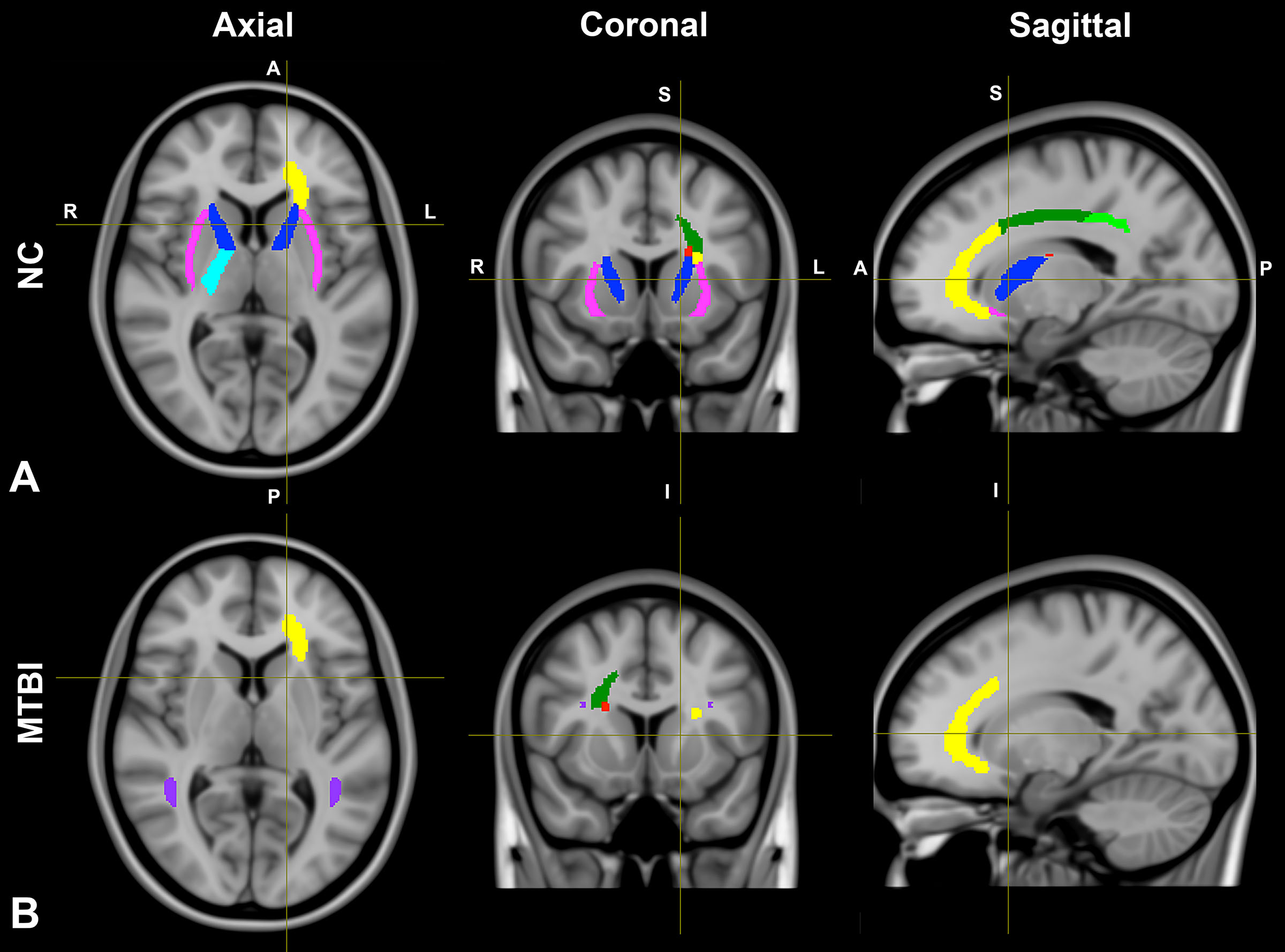
Figure 3. White matter regions of interest (ROIs) showing significant indirect effects between the corpus callosum (gene, body, splenium) microstructure as measured by diffusion MRI and interhemispheric processing speed as measured by IHSPT score (A) in healthy controls (aICL, aICR, pICR, aCRL, sCRL, pCRL, ECR, ECL, and SFOFL); and (B) in subjects with MTBI (bottom row) (aCRL, sCRR, SLFR, SLFL, and SFOFR).