0458
MRI Detection of Glymphatic Function in athletes after Sports-related Concussion1Department of Magnetic Resonance, Lanzhou University Second Hospital, Lanzhou, China, 2MR Research, GE Healthcare MR Research, Beijing, China
Synopsis
Keywords: Traumatic brain injury, Diffusion/other diffusion imaging techniques
Diffusion tensor image analysis along the perivascular space (DTI-ALPS) as a non-invasive method for evaluating the activity of the glymphatic system in human brain by using diffusion images. In this study, DTI-ALPS was used to evaluate the activity of the human lymphatic system in patients with Sports-related concussion (SRC). We found SRC has a higher ALPS index compared to the healthy controls. Our findings suggested that abnormal glymphatic function in brain might be a potential biomarker for explaining the cognitive function decline of SRC.Introduction and purpose
SRC is a complex and heterogeneous injury, which was often accompanied by cognitive dysfunction, but the neural basis is unclear1. The glymphatic system is a recently discovered waste drainage system in the brain that involves movement of the cerebrospinal fluid (CSF) along the perivascular space. This system promotes elimination of soluble proteins including amyloid-β (Aβ) and metabolites, also facilitates the distribution of glucose, lipids, amino acids, and neuromodulators. It has been suggested that glymphatic dysfunction may be associated with cognitive dysfunction2. Diffusion tensor image analysis along the perivascular space (DTI-ALPS) as a non-invasive method for evaluating the activity of the glymphatic system in human brain by using diffusion images3. Therefore, the aim of this study was to apply DTI-ALPS to compare the glymphatic system differences between SRC and NSRC groups and to study their relationship with the neurocognitive impairment of SRC.Materials and Methods
Participants:Thirty athletes (boxing and taekwondo) with a history of concussion (HoC) and 30 age, gender, and education-matched health controls (HCs) were recruited. Athletes were interviewed to determine their years in sports, the number of prior concussions, and the duration since the last SRC.
Neuropsychological testing:
All subjects underwent the same cognitive and affective measures before the MRI scan: (i) Montreal Cognitive Assessment (MoCA) assesses several categories of function, including the categories of visuospatial/executive,et al; (ii)Hamilton Depression Scale (HAMD); and (iii) Hamilton Anxiety Scale (HAMA). The additional assessment included the Rivermead Postconcussion Symptoms Questionnaire (RPQ) for contact-sport athletes.
MRI Acquisition and Processing:
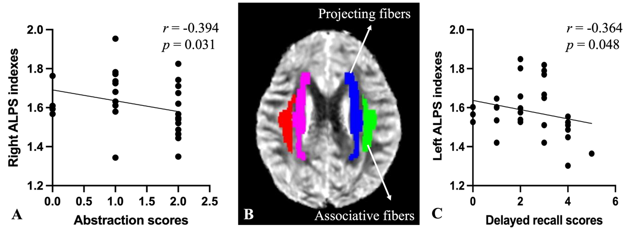
Diffusion imaging was acquired by using a 3T MR scanner (SIGNATM Premier; GE Healthcare, Waukesha, WI, USA) with a 48-channel head coil. A whole Brain diffusion data was performed with a multi-slice single-shot spin-echo echo-planar image (SE-EPI, TR = 5705 ms, TE = 68.8 ms, MPG = 120 directions, FOV = 240 mm, matrix = 120 × 120, 78 slices, slice thickness = 2 mm) set in 20 directions at b = 1000 s/mm2, 40 directions at b = 1800 s/mm2, and 60 directions at b = 2500 s/mm2. Sagittal three-dimensional (3D) T1-weighted images (T1WI) were acquired using the MP-RAGE sequence with TR = 2632 ms, TE = 3.0 ms, TI = 1000 ms, acquisition matrix = 256, 8° flip, 392 slices, and 1-mm isotropic resolution. Diffusion MRI data analysis was performed by using FSL (FSL; www.fmrib.ox.ac.uk/fsl). After calculating the fractional anisotropy (FA) map and co-registering ICBM DTI-81 Atlas, we extracted the periventricular projection (superior and posterior corona radiata) and association fibers (superior longitudinal fasciculus) (Fig. 2B). ALPS indexes from projection and association fibers derived from the ICBM DTI-81 Atlas were calculated.
Statistical analysis:
An independent samples t-test was applied to find the ALPS indexes differences between the two groups. Relationships between ALPS indexes and clinical characteristics were determined using Spearman correlation.
Results
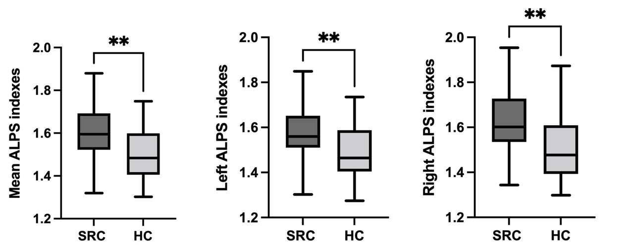
There were no group differences in age, sex composition, and years of education. Compared to the NSRC group, the SRC group had more years of training. For the neuropsychological testing, the concussed group performed more poorly than the control groups on cognitive measures. Depression and anxiety scores were not significantly different between the two groups (Table 1). The average ALPS indexes, the left ALPS indexes, and the right ALPS indexes showed significant differences between SRC and HC (P = 0.003, P = 0.003, and P = 0.009, respectively) (Fig.1). The spearman analysis showed that the right ALPS indexes were negatively correlated with abstraction scores in the SRC group (Fig. 2A). Furthermore, correlations were found between the left ALPS indexes and delayed recall scores (Fig.2C).Discussion
This study showed a higher ALPS index in the SRC group compared with the NSRC group. SRC is a form of mild traumatic brain injury that has been the leading cause of dementia. Research has linked dementia to abnormal deposits of harmful substances such as Aβ in the body. As one of the brain's clearance mechanisms, the glymphatic system can clear Aβ. An animal study found a widespread increase in phosphorylated tau in the brain after traumatic brain injury4-5. Meanwhile, a higher ALPS index represented better glymphatic activity. SRC has a higher ALPS index, we speculate that it may be related to its compensatory mechanism.In addition, we found longer years of training in the SRC group compared to the NSRC group. On neuropsychological tests, the concussion group performed worse on cognitive tests than the control group. This indicates that the SRC group already has some cognitive impairment. The APLS index was negatively correlated with the scores of some cognitive scales, indicating that the lower the score was, the worse the cognitive function was, and the higher the lymphatic-like system vitality was required.
Conclusion
The DTI-APLS method can be used for evaluating the activity of the glymphatic system. A higher ALPS index represented better glymphatic activity. SRC has a higher ALPS index, which may be related to its compensatory mechanism. ALPS-index may provide reveal potential biomarkers of an athlete's recovery trajectory.Summary of Main Findings
In this study, we used the DTI-ALPS method to assess lymphatic system activity in patients with sports-related concussion (SRC). Abnormalities in the ALPS index may provide potential biomarkers to explain cognitive decline in SRC.Acknowledgements
We would like to thank all participants in this study.References
1. Major B, Symons GF, Sinclair B, et al. White and Gray Matter Abnormalities in Australian Footballers With a History of Sports-Related Concussion: An MRI Study. Cereb Cortex. 2021;31(12):5331-5338. doi:10.1093/cercor/bhab161.
2. Fischer C, Schaub S, Büttner K, Hartmann K, Schmidt MJ. Dilated perivascular spaces can present incidental CSF-isointense foci within the ventral forebrain of dogs and cats in transverse MR images. Front Vet Sci. 2022;9:1002836. Published 2022 Oct 10. doi:10.3389/fvets.2022.1002836.
3. Taoka T, Masutani Y, Kawai H, et al. Evaluation of glymphatic system activity with the diffusion MR technique: diffusion tensor image analysis along the perivascular space (DTI-ALPS) in Alzheimer's disease cases. Jpn J Radiol. 2017;35(4):172-178. doi:10.1007/s11604-017-0617-z.
4. Iliff JJ, Chen MJ, Plog BA, et al. Impairment of glymphatic pathway function promotes tau pathology after traumatic brain injury. J Neurosci. 2014;34(49):16180-16193. doi:10.1523/JNEUROSCI.3020-14.2014.
5. Naganawa S, Taoka T. The Glymphatic System: A Review of the Challenges in Visualizing its Structure and Function with MR Imaging. Magn Reson Med Sci. 2022;21(1):182-194. doi:10.2463/mrms.rev.2020-0122.
Figures
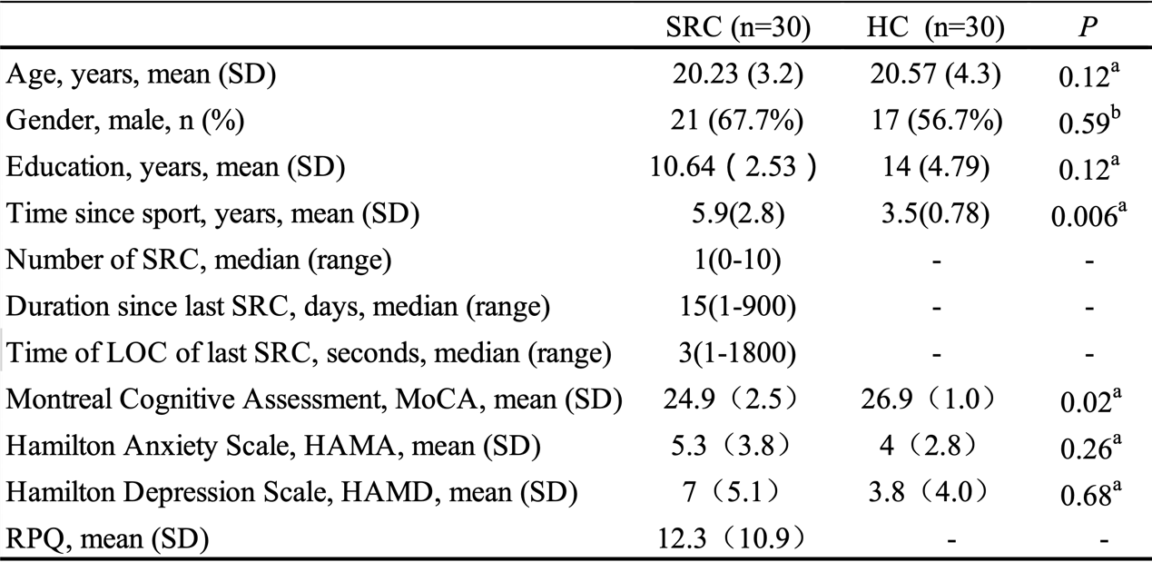
Table 1 Demographic and clinical characteristics of all participants.
Note: SRC = Sport-Related Concussion; HC = Health Control; LOC = Loss of consciousness; RPQ = Rivermead Postconcussion Symptoms Questionnaire; a two-sample t-test; b chi-square t-test; Values are mean ± standard deviations (SD).